
Article Figures & Data
Figures
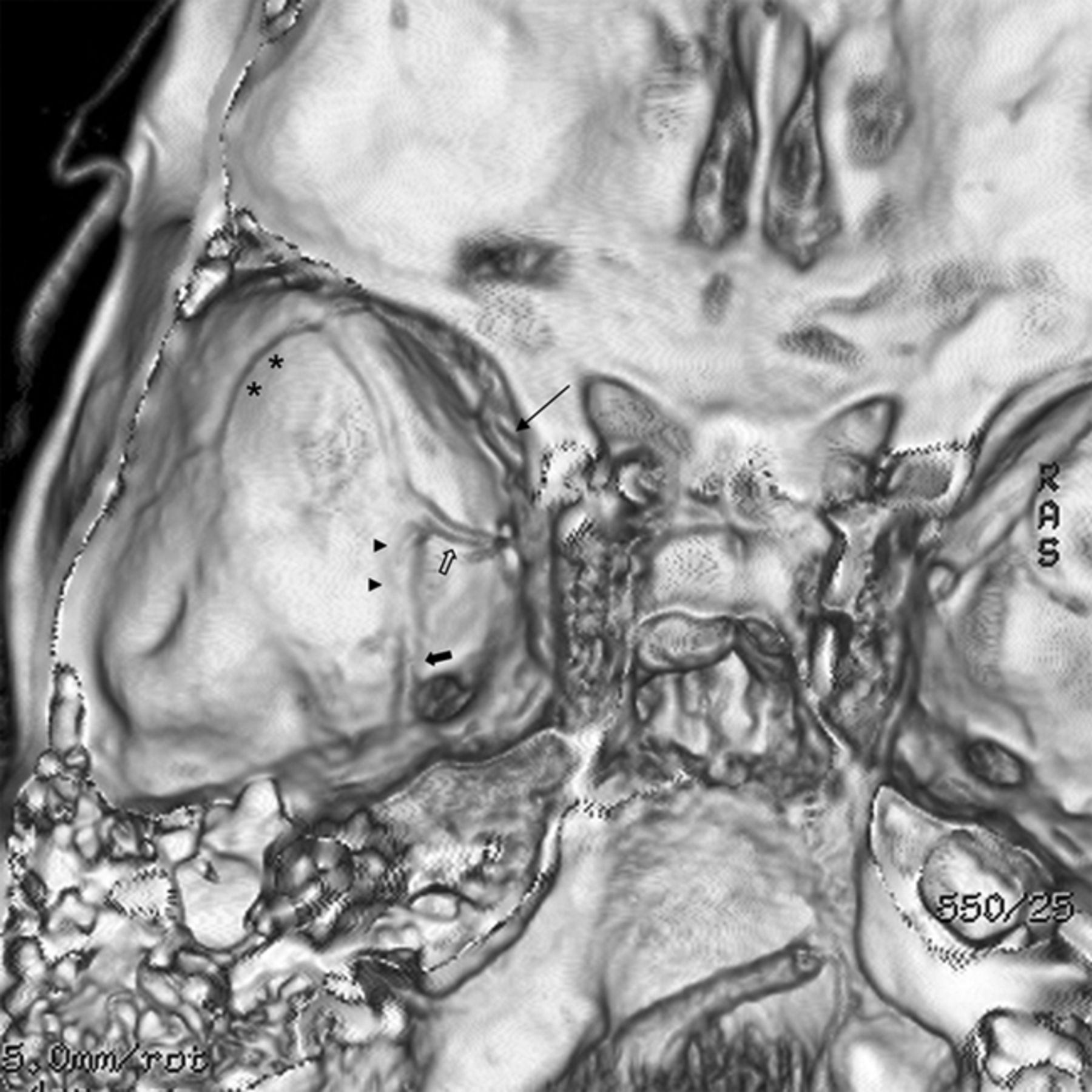
- Fig 1.
CT venogram reconstructon shows the typical locations of various sphenoidal group sinuses in the middle cranial fossa. The sinus is found along the lesser wing of sphenoid (long arrow). The sphenobasal sinus (arrowheads) and sphenopetrosal sinuses (asterisk) travel medially and laterally on the middle cranial fossa floor, respectively. They also have connections to the emissary veins of the foramen rotundum (solid arrow) and foramen ovale (open arrow).
- Fig 2.
Patient 8. Cerebral angiograms show a fistula (arrow) along the lesser sphenoid wing, primarily fed by the right MMA and draining into the right cavernous sinus, bilateral inferior petrosal sinuses, and the right superior ophthalmic vein. A, Anteroposterior view of the right ECA angiogram. B, Lateral view of the right ECA angiogram.
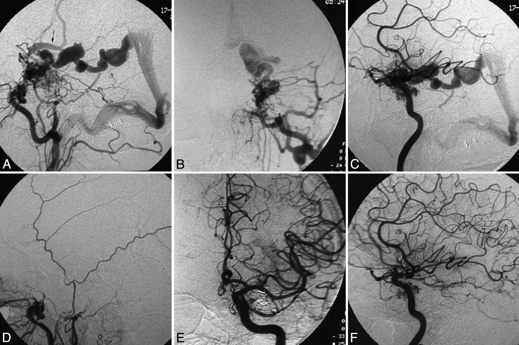
- Fig 3.
Patient 5. The ECA (A, lateral view; B, anteroposterior view) and ICA (C, lateral view) angiograms show that the high-flow fistula is fed by multiple branches of the ICA and the ECA, and there is venous drainage into the basal vein of Rosenthal and subsequently the vein of Galen, as well as into the Sylvian and Trolard veins (arrow) into the superior sagittal sinus. A triaxial system with a guide catheter inserted into the internal jugular vein, an intervening Tracker-38 catheter (Boston Scientific) placed in the transverse sinus near the torcula—and, in this, a longer microcatheter was navigated to the exact fistula site through the straight sinus, the vein of Galen, and the basal vein of Rosenthal—and transvenous embolization of the fistula was performed using coils. Postembolization angiograms of the ECA (D, lateral view) and ICA (E, anteroposterior view; F, lateral view) show complete obliteration of the fistula.
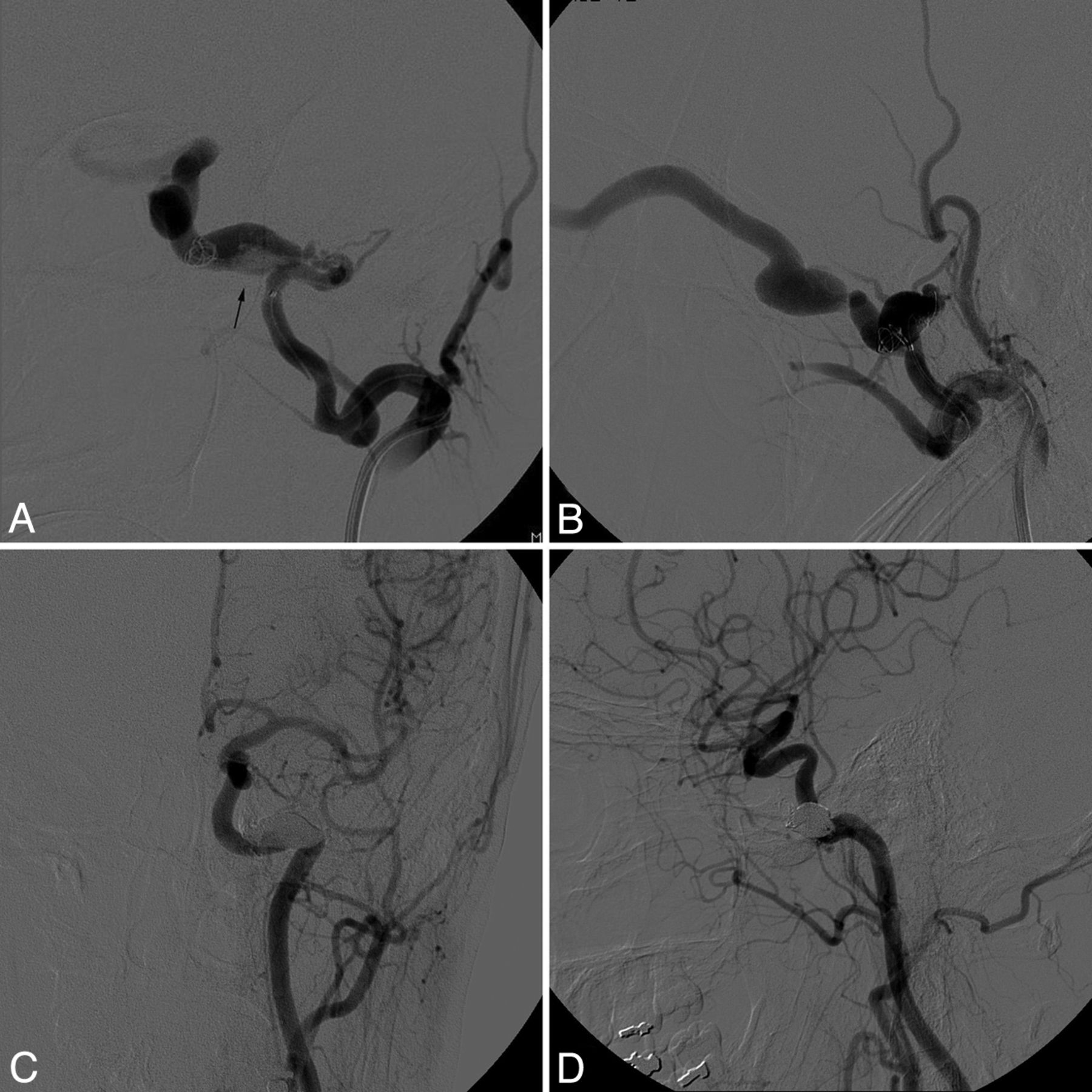
- Fig 4.
Patient 3. Anteroposterior (A) and lateral (B) view of the left ECA angiograms during transarterial coil embolization show that the fistula site involves the sphenobasal sinus (arrow) draining to the left cavernous sinus and SOV. Five-month follow-up angiogram of the left common carotid artery (C, anteroposterior view; D, lateral view) demonstrates durable complete obliteration of the fistula by Onyx and coils.
- Fig 5.
Patient 11. Lateral angiographic view of the right ICA (A) shows primary feeding arteries from the recurrent meningeal branch of the ophthalmic artery as well as the inferolateral trunk of the ICA, with resultant venous drainage into an enlarged vein of Labbe. Anteroposterior (B) and lateral (C) view of the right internal maxillary artery angiogram showing a fistula at the lesser sphenoid wing draining into the right temporal lobe cortical vein. Lateral view of the ICA (D) after transarterial coiling and n-BCA injection of the fistula showing a residual lesion. Lateral view of the right ICA (E) and ECA (F) at 18-month follow-up show complete obliteration of the fistula.


In this issue


Jump to section
Related Articles
Cited By...
- Non-aggressive, sinus-type greater sphenoid wing dural arteriovenous fistula with shunt point in the laterocavernous sinus mimicking a cavernous sinus dural arteriovenous fistula converted to aggressive, non-sinus-type
- Progressive versus Nonprogressive Intracranial Dural Arteriovenous Fistulas: Characteristics and Outcomes