Advertisement

Main menu

  • Home
  • Content
    • Current Issue
    • Accepted Manuscripts
    • Article Preview
    • Past Issue Archive
    • Video Articles
    • AJNR Case Collection
    • Case of the Week Archive
    • Case of the Month Archive
    • Classic Case Archive
  • Special Collections
    • AJNR Awards
    • Low-Field MRI
    • Alzheimer Disease
    • ASNR Foundation Special Collection
    • Photon-Counting CT
    • View All
  • Multimedia
    • AJNR Podcasts
    • AJNR SCANtastic
    • Trainee Corner
    • MRI Safety Corner
    • Imaging Protocols
  • For Authors
    • Submit a Manuscript
    • Submit a Video Article
    • Submit an eLetter to the Editor/Response
    • Manuscript Submission Guidelines
    • Statistical Tips
    • Fast Publishing of Accepted Manuscripts
    • Graphical Abstract Preparation
    • Imaging Protocol Submission
    • Author Policies
  • About Us
    • About AJNR
    • Editorial Board
    • Editorial Board Alumni
  • More
    • Become a Reviewer/Academy of Reviewers
    • Subscribers
    • Permissions
    • Alerts
    • Feedback
    • Advertisers
    • ASNR Home

User menu

  • Alerts
  • Log in

Search

  • Advanced search
American Journal of Neuroradiology

American Journal of Neuroradiology

ASHNR American Society of Functional Neuroradiology ASHNR American Society of Pediatric Neuroradiology ASSR
  • Alerts
  • Log in

Advanced Search

  • Home
  • Content
    • Current Issue
    • Accepted Manuscripts
    • Article Preview
    • Past Issue Archive
    • Video Articles
    • AJNR Case Collection
    • Case of the Week Archive
    • Case of the Month Archive
    • Classic Case Archive
  • Special Collections
    • AJNR Awards
    • Low-Field MRI
    • Alzheimer Disease
    • ASNR Foundation Special Collection
    • Photon-Counting CT
    • View All
  • Multimedia
    • AJNR Podcasts
    • AJNR SCANtastic
    • Trainee Corner
    • MRI Safety Corner
    • Imaging Protocols
  • For Authors
    • Submit a Manuscript
    • Submit a Video Article
    • Submit an eLetter to the Editor/Response
    • Manuscript Submission Guidelines
    • Statistical Tips
    • Fast Publishing of Accepted Manuscripts
    • Graphical Abstract Preparation
    • Imaging Protocol Submission
    • Author Policies
  • About Us
    • About AJNR
    • Editorial Board
    • Editorial Board Alumni
  • More
    • Become a Reviewer/Academy of Reviewers
    • Subscribers
    • Permissions
    • Alerts
    • Feedback
    • Advertisers
    • ASNR Home
  • Follow AJNR on Twitter
  • Visit AJNR on Facebook
  • Follow AJNR on Instagram
  • Join AJNR on LinkedIn
  • RSS Feeds

AJNR Awards, New Junior Editors, and more. Read the latest AJNR updates

Research ArticleBrain

The U Sign: Tenth Landmark to the Central Region on Brain Surface Reformatted MR Imaging

M. Wagner, A. Jurcoane and E. Hattingen
American Journal of Neuroradiology February 2013, 34 (2) 323-326; DOI: https://doi.org/10.3174/ajnr.A3205
M. Wagner
aFrom the Institute of Neuroradiology, University Hospital, Goethe University Frankfurt, Frankfurt am Main, Germany.
  • Find this author on Google Scholar
  • Find this author on PubMed
  • Search for this author on this site
A. Jurcoane
aFrom the Institute of Neuroradiology, University Hospital, Goethe University Frankfurt, Frankfurt am Main, Germany.
  • Find this author on Google Scholar
  • Find this author on PubMed
  • Search for this author on this site
E. Hattingen
aFrom the Institute of Neuroradiology, University Hospital, Goethe University Frankfurt, Frankfurt am Main, Germany.
  • Find this author on Google Scholar
  • Find this author on PubMed
  • Search for this author on this site
  • Article
  • Figures & Data
  • Info & Metrics
  • Responses
  • References
  • PDF
Loading

Article Figures & Data

Figures

  • Tables
  • Fig 1.
    • Download figure
    • Open in new tab
    • Download powerpoint
    Fig 1.

    Example of the thin post cg sign, and the midline sulcus sign. The precentral gyrus has a thicker anteroposterior diameter (large white arrow) compared with the postcentral gyrus (small white arrow). The longest sulcus running horizontally and entering the interhemispheric fissure (black arrow) is the central sulcus.

  • Fig 2.
    • Download figure
    • Open in new tab
    • Download powerpoint
    Fig 2.

    Variants of the handknob. In our study, we only considered the inverted Ω shape (white arrow) because other variants, such as inverted ε (continuous black arrows), and multiple bulgings (dotted black arrows) were not assessed.

  • Fig 3.
    • Download figure
    • Open in new tab
    • Download powerpoint
    Fig 3.

    The U sign. Right hemisphere. Absence of the subcentral gyrus as a rare variant, the central sulcus (asterisk) terminates into the Sylvian fissure (white arrow). Gray arrows mark the precentral (continuous arrow) and postcentral (dotted arrow) gyri. Left hemisphere: The subcentral gyrus connects the pre- and postcentral gyri (U sign).
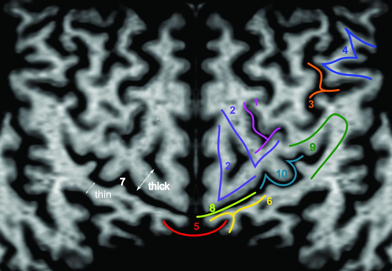
  • Fig 4.
    • Download figure
    • Open in new tab
    • Download powerpoint
    Fig 4.

    Examples of all 10 landmarks in 1 single section: 1 indicates the upper T sign; 2, the L sign (duplicated as a variant); 3, the lower T sign; 4, the M sign; 5, the bracket sign; 6, the bifid post cg sign; 7, the thin post cg sign (fenestrated post cg as a variant); 8, the midline sulcus sign; 9, the subcentral gyrus sign; 10, the handknob. Only 1 hemisphere is depicted and was mirrored for better visualization.

  • Fig 5.
    • Download figure
    • Open in new tab
    • Download powerpoint
    Fig 5.

    Barplots representing the frequency of landmark identification as a percentage of all (blue), right (purple), and left (yellow) hemispheres.

Tables

  • Figures
  • Frequency of all landmarks

    All Hemispheres (n = 178)Right (n = 88)Left (n = 90)
    Upper T sign109 (61.2%)51 (58%)58 (64.4%)
    L sign107 (60.1%)55 (62.5%)52 (57.8%)
    Lower T sign138 (77.5%)69 (78.4%)69 (76.7%)
    M sign157 (88.2%)77 (87.5%)80 (88.9%)
    Bifid pc sign157 (88.2%)76 (86.4%)81 (90%)
    Midline sulcus sign159 (87.5%)77 (87.5%)82 (91.1%)
    Bracket sign82% (of 78 patients)
    Thin post cg sign170 (95.5%)85 (96.6%)85 (94.4%)
    Handknob sign97 (54.5%)49 (55.7%)48 (53.3%)
    Subcentral gyrus sign176 (98.9%)84 (95.5%)88 (97.8%)
    • Note:—Bifid pc sign indicates the bifid post cg sign.

PreviousNext
Back to top

In this issue

American Journal of Neuroradiology: 34 (2)
American Journal of Neuroradiology
Vol. 34, Issue 2
1 Feb 2013
  • Table of Contents
  • Index by author
Advertisement
Print
Download PDF
Email Article

Thank you for your interest in spreading the word on American Journal of Neuroradiology.

NOTE: We only request your email address so that the person you are recommending the page to knows that you wanted them to see it, and that it is not junk mail. We do not capture any email address.

Enter multiple addresses on separate lines or separate them with commas.
The U Sign: Tenth Landmark to the Central Region on Brain Surface Reformatted MR Imaging
(Your Name) has sent you a message from American Journal of Neuroradiology
(Your Name) thought you would like to see the American Journal of Neuroradiology web site.
CAPTCHA
This question is for testing whether or not you are a human visitor and to prevent automated spam submissions.
Cite this article
M. Wagner, A. Jurcoane, E. Hattingen
The U Sign: Tenth Landmark to the Central Region on Brain Surface Reformatted MR Imaging
American Journal of Neuroradiology Feb 2013, 34 (2) 323-326; DOI: 10.3174/ajnr.A3205

Citation Manager Formats

  • BibTeX
  • Bookends
  • EasyBib
  • EndNote (tagged)
  • EndNote 8 (xml)
  • Medlars
  • Mendeley
  • Papers
  • RefWorks Tagged
  • Ref Manager
  • RIS
  • Zotero
0 Responses
Respond to this article
Share
Bookmark this article
The U Sign: Tenth Landmark to the Central Region on Brain Surface Reformatted MR Imaging
M. Wagner, A. Jurcoane, E. Hattingen
American Journal of Neuroradiology Feb 2013, 34 (2) 323-326; DOI: 10.3174/ajnr.A3205
del.icio.us logo Twitter logo Facebook logo Mendeley logo
  • Tweet Widget
  • Facebook Like
  • Google Plus One
Purchase

Jump to section

  • Article
    • Abstract
    • ABBREVIATIONS:
    • Materials and Methods
    • Results
    • Discussion
    • Conclusions
    • References
  • Figures & Data
  • Info & Metrics
  • Responses
  • References
  • PDF

Related Articles

  • No related articles found.
  • PubMed
  • Google Scholar

Cited By...

  • The "White Gray Sign" Identifies the Central Sulcus on 3T High-Resolution T1-Weighted Images
  • Crossref
  • Google Scholar

This article has not yet been cited by articles in journals that are participating in Crossref Cited-by Linking.

More in this TOC Section

  • White Matter Alterations in the Brains of Patients with Active, Remitted, and Cured Cushing Syndrome: A DTI Study
  • Qualitative and Quantitative Analysis of MR Imaging Findings in Patients with Middle Cerebral Artery Stroke Implanted with Mesenchymal Stem Cells
  • Fast Contrast-Enhanced 4D MRA and 4D Flow MRI Using Constrained Reconstruction (HYPRFlow): Potential Applications for Brain Arteriovenous Malformations
Show more Brain

Similar Articles

Advertisement

Indexed Content

  • Current Issue
  • Accepted Manuscripts
  • Article Preview
  • Past Issues
  • Editorials
  • Editor's Choice
  • Fellows' Journal Club
  • Letters to the Editor
  • Video Articles

Cases

  • Case Collection
  • Archive - Case of the Week
  • Archive - Case of the Month
  • Archive - Classic Case

More from AJNR

  • Trainee Corner
  • Imaging Protocols
  • MRI Safety Corner
  • Book Reviews

Multimedia

  • AJNR Podcasts
  • AJNR Scantastics

Resources

  • Turnaround Time
  • Submit a Manuscript
  • Submit a Video Article
  • Submit an eLetter to the Editor/Response
  • Manuscript Submission Guidelines
  • Statistical Tips
  • Fast Publishing of Accepted Manuscripts
  • Graphical Abstract Preparation
  • Imaging Protocol Submission
  • Evidence-Based Medicine Level Guide
  • Publishing Checklists
  • Author Policies
  • Become a Reviewer/Academy of Reviewers
  • News and Updates

About Us

  • About AJNR
  • Editorial Board
  • Editorial Board Alumni
  • Alerts
  • Permissions
  • Not an AJNR Subscriber? Join Now
  • Advertise with Us
  • Librarian Resources
  • Feedback
  • Terms and Conditions
  • AJNR Editorial Board Alumni

American Society of Neuroradiology

  • Not an ASNR Member? Join Now

© 2025 by the American Society of Neuroradiology All rights, including for text and data mining, AI training, and similar technologies, are reserved.
Print ISSN: 0195-6108 Online ISSN: 1936-959X

Powered by HighWire