
Article Figures & Data
Figures
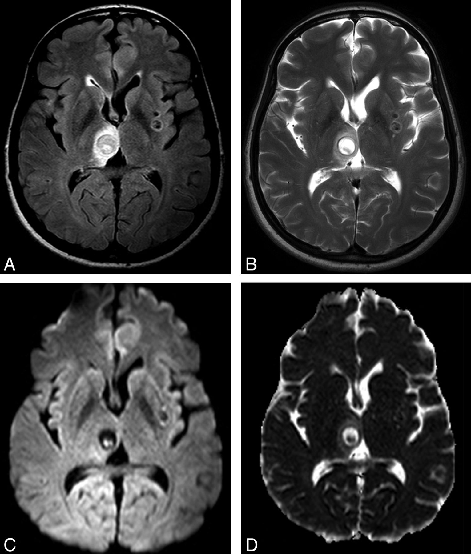
- Fig 1.
Axial FLAIR (A) and axial DWI (B) images show 3 lesions in both frontoparietal areas (arrow and arrowheads in A); there is 1 right parietal lesion (arrow on A), compatible with NCC in the vesicular state. In this lesion, the scolex is clearly seen on DWI as a hyperintense eccentric dot, but it can also be appreciated in the other 2 images. ADC map (C) discloses the scolices as iso-/hypointense dots.
- Fig 2.
Axial FLAIR (A) and axial T2 fast spin-echo (B) images show lesions in the right thalamus and the left lentiform nucleus; the thalamic lesion is compatible with NCC in the colloidal stage. In this lesion, the curvilinear scolex is clearly seen in a transverse section, on DWI (C), as 2 hyperintense contiguous dots. ADC map (D) discloses the scolex as iso-/hypointense dots.
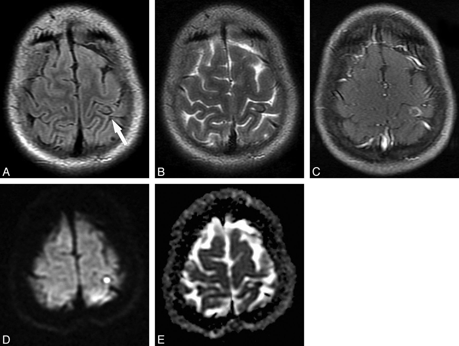
- Fig 3.
NCC lesion in the granular nodular stage (patient E) is appreciated in axial FLAIR (A) and T2-weighted (B) images in the left postcentral gyrus (arrow in A), presenting proteinaceous content (distinct from CSF) with a peripheral rim of low signal presumably due to the beginning of the calcification process, and no perilesional edema. Corresponding axial spin-echo postcontrast T1-weighted (C) image demonstrates residual postcontrast enhancement. Axial DWI image (D) and the corresponding ADC map (E) show homogeneous reduced diffusion of the internal portion of this lesion, possibly related to a more viscous inflammatory content (rADC = 0.54).
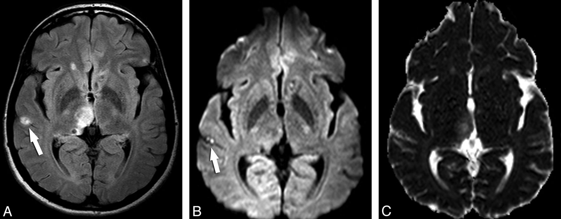
- Fig 4.
A small subarachnoid NCC lesion whose scolex is depicted only in DWI. Axial FLAIR (A) image demonstrates a hyperintense lesion in the right lateral temporal region (arrow). The DWI image (B) shows the curvilinear scolex in a transverse section as 2 hyperintense contiguous dots (arrow). Other lesions can be appreciated in the right thalamus. ADC map (C) discloses the scolex as iso-/hypointense dots.
- Fig 5.
Axial FLAIR sequence (A) in patient F demonstrates many subarachnoid cystic lesions in the right Sylvian cistern, compatible with the racemose form of NCC. Note specifically 1 lesion (arrows) that has signal intensity distinct from CSF. Axial DWI (B) image demonstrates homogeneous reduced diffusion in this small lesion (rADC = 0.79).
- Fig 6.
The intraventricular DWI hyperintense scolex is appreciated inside a lesion in the left lateral ventricle. The lesion and its scolex are well seen on the corresponding FLAIR image (A), but the scolex (arrow) is identified as a hyperintense dot in the DWI sequence (B). Additionally, other intraparenchymal NCC lesions are identified bilaterally.


Tables
Location No. of Lesions DWI-Hyperintense Lesions Eccentric (Punctate or Comma-Shaped) Total/Subtotal Intraparenchymal Vesicular 139 41 0 Colloidal vesicular 93 18 5 Granular nodular 31 0 1 Subarachnoid 65 14 2 Intraventricular 14 2 0 Total 342 75 8 Patient Lesion Stage/Location ADC-Lesion (× 10−3 mm2/s) ADC-WM (× 10−3 mm2/s) rADC A Colloidal 0.94 ± 0.25 0.96 ± 0.19 0.98 B Colloidal 1.41 ± 0.38 1.47 ± 0.13 0.95 C (1) Colloidal 0.72 ± 0.11 0.84 ± 0.12 0.85 C (2) Colloidal 0.52 ± 0.13 0.75 ± 0.12 0.68 D Colloidal 0.54 ± 0.16 0.75 ± 0.12 0.72 E Granular nodular 0.51 ± 0.24 0.94 ± 0.28 0.54 F Subarachnoid (racemose) 0.80 ± 0.38 1.01 ± 0.13 0.79 G Subarachnoid 0.68 ± 0.83 1.18 ± 0.15 0.57


