Advertisement

Main menu

  • Home
  • Content
    • Current Issue
    • Accepted Manuscripts
    • Article Preview
    • Past Issue Archive
    • Video Articles
    • AJNR Case Collection
    • Case of the Week Archive
    • Case of the Month Archive
    • Classic Case Archive
  • Special Collections
    • AJNR Awards
    • Low-Field MRI
    • Alzheimer Disease
    • ASNR Foundation Special Collection
    • Photon-Counting CT
    • View All
  • Multimedia
    • AJNR Podcasts
    • AJNR SCANtastic
    • Trainee Corner
    • MRI Safety Corner
    • Imaging Protocols
  • For Authors
    • Submit a Manuscript
    • Submit a Video Article
    • Submit an eLetter to the Editor/Response
    • Manuscript Submission Guidelines
    • Statistical Tips
    • Fast Publishing of Accepted Manuscripts
    • Graphical Abstract Preparation
    • Imaging Protocol Submission
    • Author Policies
  • About Us
    • About AJNR
    • Editorial Board
    • Editorial Board Alumni
  • More
    • Become a Reviewer/Academy of Reviewers
    • Subscribers
    • Permissions
    • Alerts
    • Feedback
    • Advertisers
    • ASNR Home

User menu

  • Alerts
  • Log in

Search

  • Advanced search
American Journal of Neuroradiology

American Journal of Neuroradiology

ASHNR American Society of Functional Neuroradiology ASHNR American Society of Pediatric Neuroradiology ASSR
  • Alerts
  • Log in

Advanced Search

  • Home
  • Content
    • Current Issue
    • Accepted Manuscripts
    • Article Preview
    • Past Issue Archive
    • Video Articles
    • AJNR Case Collection
    • Case of the Week Archive
    • Case of the Month Archive
    • Classic Case Archive
  • Special Collections
    • AJNR Awards
    • Low-Field MRI
    • Alzheimer Disease
    • ASNR Foundation Special Collection
    • Photon-Counting CT
    • View All
  • Multimedia
    • AJNR Podcasts
    • AJNR SCANtastic
    • Trainee Corner
    • MRI Safety Corner
    • Imaging Protocols
  • For Authors
    • Submit a Manuscript
    • Submit a Video Article
    • Submit an eLetter to the Editor/Response
    • Manuscript Submission Guidelines
    • Statistical Tips
    • Fast Publishing of Accepted Manuscripts
    • Graphical Abstract Preparation
    • Imaging Protocol Submission
    • Author Policies
  • About Us
    • About AJNR
    • Editorial Board
    • Editorial Board Alumni
  • More
    • Become a Reviewer/Academy of Reviewers
    • Subscribers
    • Permissions
    • Alerts
    • Feedback
    • Advertisers
    • ASNR Home
  • Follow AJNR on Twitter
  • Visit AJNR on Facebook
  • Follow AJNR on Instagram
  • Join AJNR on LinkedIn
  • RSS Feeds

AJNR Awards, New Junior Editors, and more. Read the latest AJNR updates

Research ArticleBrain

Intracranial Atherosclerotic Plaque Enhancement in Patients with Ischemic Stroke

M. Skarpathiotakis, D.M. Mandell, R.H. Swartz, G. Tomlinson and D.J. Mikulis
American Journal of Neuroradiology February 2013, 34 (2) 299-304; DOI: https://doi.org/10.3174/ajnr.A3209
M. Skarpathiotakis
aFrom the Department of Medical Imaging, Toronto Western Hospital, Toronto, Ontario, Canada.
  • Find this author on Google Scholar
  • Find this author on PubMed
  • Search for this author on this site
D.M. Mandell
aFrom the Department of Medical Imaging, Toronto Western Hospital, Toronto, Ontario, Canada.
  • Find this author on Google Scholar
  • Find this author on PubMed
  • Search for this author on this site
R.H. Swartz
aFrom the Department of Medical Imaging, Toronto Western Hospital, Toronto, Ontario, Canada.
  • Find this author on Google Scholar
  • Find this author on PubMed
  • Search for this author on this site
G. Tomlinson
aFrom the Department of Medical Imaging, Toronto Western Hospital, Toronto, Ontario, Canada.
  • Find this author on Google Scholar
  • Find this author on PubMed
  • Search for this author on this site
D.J. Mikulis
aFrom the Department of Medical Imaging, Toronto Western Hospital, Toronto, Ontario, Canada.
  • Find this author on Google Scholar
  • Find this author on PubMed
  • Search for this author on this site
  • Article
  • Figures & Data
  • Info & Metrics
  • Responses
  • References
  • PDF
Loading

Article Figures & Data

Figures

  • Tables
  • Fig 1.
    • Download figure
    • Open in new tab
    • Download powerpoint
    Fig 1.

    A, 3D reformatted MRA image demonstrating bilateral MCA stenoses (arrows). B, Diffusion-weighted image of the brain demonstrating areas of restricted diffusion/infarction in the left MCA territory. C, Coronal noncontrast T1-weighted image through the level of the MCAs. Atherosclerotic plaque is present in both the right MCA and left MCA (arrows) at the site of stenoses seen in A. D, Coronal postcontrast T1-weighted image at the same level as in C, with enhancement of the left MCA atherosclerotic plaque but not of the plaques on the right.

  • Fig 2.
    • Download figure
    • Open in new tab
    • Download powerpoint
    Fig 2.

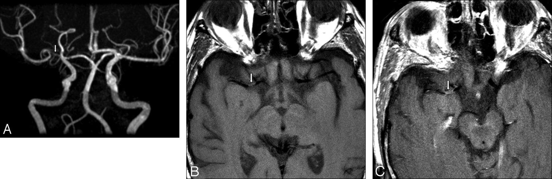
    A, 3D reformatted MRA image demonstrating right MCA stenoses (arrow). B, Axial noncontrast T1-weighted image through the level of the MCAs. Atherosclerotic plaque is present in the right MCA (arrow) at the site of stenosis seen in A. C, Axial postcontrast T1-weighted image at the same level as in B, with enhancement of the right MCA atherosclerotic plaque.

  • Fig 3.
    • Download figure
    • Open in new tab
    • Download powerpoint
    Fig 3.

    A, 3D reformatted MRA image demonstrating midbasilar artery stenosis (arrow). B, Diffusion-weighted image of the brain demonstrating areas of restricted diffusion/infarction in the basilar artery territory. C, Coronal noncontrast T1-weighted image through the level of the basilar artery. Atherosclerotic plaque is present (arrow) at the site of stenosis seen in A. D, Coronal postcontrast T1-weighted image at the same level as in C, with enhancement of midbasilar atherosclerotic plaque.

  • Fig 4.
    • Download figure
    • Open in new tab
    • Download powerpoint
    Fig 4.

    Strength and presence of enhancement versus time elapsed between imaging and initial stroke presentation on a logarithmic scale. Acute, subacute, and chronic timeframes are highlighted.

  • Fig 5.
    • Download figure
    • Open in new tab
    • Download powerpoint
    Fig 5.

    Strength and presence of enhancement versus time elapsed between imaging and initial stroke presentation for 6 patients in whom imaging was performed at 2 separate time points.

Tables

  • Figures
    • View popup
    Table 1:

    Sequence parameters for vessel wall imaging protocol

    TR (msec)TE (msec)TI (msec)Echo-Train LengthSection ThicknessParallel FactorFOVResolution
    T1 FLAIR pre- and postgadolinium (axial and coronal or sagittal)21081286062 mm (initially 3 mm, changed to 2 mm)216 × 22 cm384 × 384
    T2 FRFSE (axial and coronal)345092-172 mm (initially 3 mm, changed to 2 mm)216 × 22 cm512 × 512
    • Note:—FRFSE indicates fast-recovery fast spin-echo.

    • View popup
    Table 2:

    Imaging performed in the acute phase

    Plaques in a Single Vascular Territory in 13 PatientsPlaques in Multiple Vascular Territories in 3 Patients
    Solitary plaque in 10/13Plaque in stroke territory
        All plaques enhanced    Solitary plaque in 3/3
    Multiple plaques in 3/13    All enhanced
        All plaques enhancedPlaque in nonstroke territory
        Solitary plaque in 2/3
            1 plaque enhanced
        Multiple plaques in 1/3
            All plaques enhanced
    • View popup
    Table 3:

    Time since infarct by enhancement

    Time Since InfarctEnhancement
    NoneMildStrongTotal
    Acute phase (0–4 weeks)0 (0%)0 (0%)16 (100%)16 (55.2%)
    Subacute phase (4–12 weeks)1 (20%)3 (60%)1 (20%)5 (17.2%)
    Chronic phase (>12 weeks)4 (50%)3 (37.5%)1 (12.5%)8 (27.6%)
    Total5 (17.2%)6 (20.7%)18 (62.1%)29 (100%)
    • Note:—Fisher exact test for count data, P value = 2.8 × 10⁻⁶. Spearman correlation = −0.84 (95% CIs −0.92, −0.70).

PreviousNext
Back to top

In this issue

American Journal of Neuroradiology: 34 (2)
American Journal of Neuroradiology
Vol. 34, Issue 2
1 Feb 2013
  • Table of Contents
  • Index by author
Advertisement
Print
Download PDF
Email Article

Thank you for your interest in spreading the word on American Journal of Neuroradiology.

NOTE: We only request your email address so that the person you are recommending the page to knows that you wanted them to see it, and that it is not junk mail. We do not capture any email address.

Enter multiple addresses on separate lines or separate them with commas.
Intracranial Atherosclerotic Plaque Enhancement in Patients with Ischemic Stroke
(Your Name) has sent you a message from American Journal of Neuroradiology
(Your Name) thought you would like to see the American Journal of Neuroradiology web site.
CAPTCHA
This question is for testing whether or not you are a human visitor and to prevent automated spam submissions.
Cite this article
M. Skarpathiotakis, D.M. Mandell, R.H. Swartz, G. Tomlinson, D.J. Mikulis
Intracranial Atherosclerotic Plaque Enhancement in Patients with Ischemic Stroke
American Journal of Neuroradiology Feb 2013, 34 (2) 299-304; DOI: 10.3174/ajnr.A3209

Citation Manager Formats

  • BibTeX
  • Bookends
  • EasyBib
  • EndNote (tagged)
  • EndNote 8 (xml)
  • Medlars
  • Mendeley
  • Papers
  • RefWorks Tagged
  • Ref Manager
  • RIS
  • Zotero
0 Responses
Respond to this article
Share
Bookmark this article
Intracranial Atherosclerotic Plaque Enhancement in Patients with Ischemic Stroke
M. Skarpathiotakis, D.M. Mandell, R.H. Swartz, G. Tomlinson, D.J. Mikulis
American Journal of Neuroradiology Feb 2013, 34 (2) 299-304; DOI: 10.3174/ajnr.A3209
del.icio.us logo Twitter logo Facebook logo Mendeley logo
  • Tweet Widget
  • Facebook Like
  • Google Plus One
Purchase

Jump to section

  • Article
    • Abstract
    • Materials and Methods
    • Results
    • Discussion
    • Conclusions
    • Footnotes
    • References
  • Figures & Data
  • Info & Metrics
  • Responses
  • References
  • PDF

Related Articles

  • No related articles found.
  • PubMed
  • Google Scholar

Cited By...

  • Impact of Previous Glycemic Control on High-Resolution MRI Plaque Characteristics and Stroke Mechanisms in Patients with Middle Cerebral Artery Atherosclerosis
  • Vessel wall MRI evaluation for the safety of endovascular recanalization of non-acute intracranial anterior circulation artery occlusions
  • Delayed Enhancement of Intracranial Atherosclerotic Plaque Can Better Differentiate Culprit Lesions: A Multiphase Contrast-Enhanced Vessel Wall MRI Study
  • Impacts of Glycemic Control on Intracranial Plaque in Patients with Type 2 Diabetes Mellitus: A Vessel Wall MRI Study
  • Effect of Time Elapsed since Gadolinium Administration on Atherosclerotic Plaque Enhancement in Clinical Vessel Wall MR Imaging Studies
  • Diagnostic Impact of Intracranial Vessel Wall MRI in 205 Patients with Ischemic Stroke or TIA
  • Differential Features of Culprit Intracranial Atherosclerotic Lesions: A Whole-Brain Vessel Wall Imaging Study in Patients With Acute Ischemic Stroke
  • Identification and Quantitative Assessment of Different Components of Intracranial Atherosclerotic Plaque by Ex Vivo 3T High-Resolution Multicontrast MRI
  • Role of MRI in early detection of stroke secondary to neurosyphilis in an elderly patient coinfected with HIV
  • Quantifying Intracranial Plaque Permeability with Dynamic Contrast-Enhanced MRI: A Pilot Study
  • Intracranial Vessel Wall MRI: Principles and Expert Consensus Recommendations of the American Society of Neuroradiology
  • Vessel wall imaging for intracranial vascular disease evaluation
  • Vessel wall MRI of an inflamed aneurysm with atherosclerosis in a patient with ischemic stroke
  • Gadolinium Enhancement in Intracranial Atherosclerotic Plaque and Ischemic Stroke: A Systematic Review and Meta-Analysis
  • Magnetic Resonance Imaging of Plaque Morphology, Burden, and Distribution in Patients With Symptomatic Middle Cerebral Artery Stenosis
  • Previous Statin Use and High-Resolution Magnetic Resonance Imaging Characteristics of Intracranial Atherosclerotic Plaque: The Intensive Statin Treatment in Acute Ischemic Stroke Patients With Intracranial Atherosclerosis Study
  • High-resolution intracranial vessel wall imaging: imaging beyond the lumen
  • Differential Vascular Pathophysiologic Types of Intracranial Atherosclerotic Stroke: A High-Resolution Wall Magnetic Resonance Imaging Study
  • Imaging the Intracranial Atherosclerotic Vessel Wall Using 7T MRI: Initial Comparison with Histopathology
  • Gadolinium Enhancement of Atherosclerotic Plaque in the Middle Cerebral Artery: Relation to Symptoms and Degree of Stenosis
  • Vessel Wall Magnetic Resonance Imaging in Acute Ischemic Stroke: Effects of Embolism and Mechanical Thrombectomy on the Arterial Wall
  • Imaging Intracranial Vessel Wall Pathology With Magnetic Resonance Imaging: Current Prospects and Future Directions
  • T1 Gadolinium Enhancement of Intracranial Atherosclerotic Plaques Associated with Symptomatic Ischemic Presentations
  • Crossref
  • Google Scholar

This article has not yet been cited by articles in journals that are participating in Crossref Cited-by Linking.

More in this TOC Section

  • Multimodal CT Provides Improved Performance for Lacunar Infarct Detection
  • Optimal MRI Sequence for Identifying Occlusion Location in Acute Stroke: Which Value of Time-Resolved Contrast-Enhanced MRA?
  • Evaluating the Effects of White Matter Multiple Sclerosis Lesions on the Volume Estimation of 6 Brain Tissue Segmentation Methods
Show more Brain

Similar Articles

Advertisement

Indexed Content

  • Current Issue
  • Accepted Manuscripts
  • Article Preview
  • Past Issues
  • Editorials
  • Editor's Choice
  • Fellows' Journal Club
  • Letters to the Editor
  • Video Articles

Cases

  • Case Collection
  • Archive - Case of the Week
  • Archive - Case of the Month
  • Archive - Classic Case

More from AJNR

  • Trainee Corner
  • Imaging Protocols
  • MRI Safety Corner
  • Book Reviews

Multimedia

  • AJNR Podcasts
  • AJNR Scantastics

Resources

  • Turnaround Time
  • Submit a Manuscript
  • Submit a Video Article
  • Submit an eLetter to the Editor/Response
  • Manuscript Submission Guidelines
  • Statistical Tips
  • Fast Publishing of Accepted Manuscripts
  • Graphical Abstract Preparation
  • Imaging Protocol Submission
  • Evidence-Based Medicine Level Guide
  • Publishing Checklists
  • Author Policies
  • Become a Reviewer/Academy of Reviewers
  • News and Updates

About Us

  • About AJNR
  • Editorial Board
  • Editorial Board Alumni
  • Alerts
  • Permissions
  • Not an AJNR Subscriber? Join Now
  • Advertise with Us
  • Librarian Resources
  • Feedback
  • Terms and Conditions
  • AJNR Editorial Board Alumni

American Society of Neuroradiology

  • Not an ASNR Member? Join Now

© 2025 by the American Society of Neuroradiology All rights, including for text and data mining, AI training, and similar technologies, are reserved.
Print ISSN: 0195-6108 Online ISSN: 1936-959X

Powered by HighWire