Advertisement

Main menu

  • Home
  • Content
    • Current Issue
    • Accepted Manuscripts
    • Article Preview
    • Past Issue Archive
    • Video Articles
    • AJNR Case Collection
    • Case of the Week Archive
    • Case of the Month Archive
    • Classic Case Archive
  • Special Collections
    • AJNR Awards
    • Low-Field MRI
    • Alzheimer Disease
    • ASNR Foundation Special Collection
    • Photon-Counting CT
    • View All
  • Multimedia
    • AJNR Podcasts
    • AJNR SCANtastic
    • Trainee Corner
    • MRI Safety Corner
    • Imaging Protocols
  • For Authors
    • Submit a Manuscript
    • Submit a Video Article
    • Submit an eLetter to the Editor/Response
    • Manuscript Submission Guidelines
    • Statistical Tips
    • Fast Publishing of Accepted Manuscripts
    • Graphical Abstract Preparation
    • Imaging Protocol Submission
    • Author Policies
  • About Us
    • About AJNR
    • Editorial Board
    • Editorial Board Alumni
  • More
    • Become a Reviewer/Academy of Reviewers
    • Subscribers
    • Permissions
    • Alerts
    • Feedback
    • Advertisers
    • ASNR Home

User menu

  • Alerts
  • Log in

Search

  • Advanced search
American Journal of Neuroradiology

American Journal of Neuroradiology

ASHNR American Society of Functional Neuroradiology ASHNR American Society of Pediatric Neuroradiology ASSR
  • Alerts
  • Log in

Advanced Search

  • Home
  • Content
    • Current Issue
    • Accepted Manuscripts
    • Article Preview
    • Past Issue Archive
    • Video Articles
    • AJNR Case Collection
    • Case of the Week Archive
    • Case of the Month Archive
    • Classic Case Archive
  • Special Collections
    • AJNR Awards
    • Low-Field MRI
    • Alzheimer Disease
    • ASNR Foundation Special Collection
    • Photon-Counting CT
    • View All
  • Multimedia
    • AJNR Podcasts
    • AJNR SCANtastic
    • Trainee Corner
    • MRI Safety Corner
    • Imaging Protocols
  • For Authors
    • Submit a Manuscript
    • Submit a Video Article
    • Submit an eLetter to the Editor/Response
    • Manuscript Submission Guidelines
    • Statistical Tips
    • Fast Publishing of Accepted Manuscripts
    • Graphical Abstract Preparation
    • Imaging Protocol Submission
    • Author Policies
  • About Us
    • About AJNR
    • Editorial Board
    • Editorial Board Alumni
  • More
    • Become a Reviewer/Academy of Reviewers
    • Subscribers
    • Permissions
    • Alerts
    • Feedback
    • Advertisers
    • ASNR Home
  • Follow AJNR on Twitter
  • Visit AJNR on Facebook
  • Follow AJNR on Instagram
  • Join AJNR on LinkedIn
  • RSS Feeds

AJNR Awards, New Junior Editors, and more. Read the latest AJNR updates

Research ArticleNeurointervention

Spontaneous Delayed Migration/Shortening of the Pipeline Embolization Device: Report of 5 Cases

N. Chalouhi, S.I. Tjoumakaris, L.F. Gonzalez, D. Hasan, P.J. Pema, G. Gould, R.H. Rosenwasser and P.M. Jabbour
American Journal of Neuroradiology December 2013, 34 (12) 2326-2330; DOI: https://doi.org/10.3174/ajnr.A3632
N. Chalouhi
aFrom the Department of Neurosurgery (N.C., S.I.T., L.F.G., G.G., R.H.R., P.M.J.), Thomas Jefferson University and Jefferson Hospital for Neuroscience, Philadelphia, Pennsylvania
  • Find this author on Google Scholar
  • Find this author on PubMed
  • Search for this author on this site
S.I. Tjoumakaris
aFrom the Department of Neurosurgery (N.C., S.I.T., L.F.G., G.G., R.H.R., P.M.J.), Thomas Jefferson University and Jefferson Hospital for Neuroscience, Philadelphia, Pennsylvania
  • Find this author on Google Scholar
  • Find this author on PubMed
  • Search for this author on this site
L.F. Gonzalez
aFrom the Department of Neurosurgery (N.C., S.I.T., L.F.G., G.G., R.H.R., P.M.J.), Thomas Jefferson University and Jefferson Hospital for Neuroscience, Philadelphia, Pennsylvania
  • Find this author on Google Scholar
  • Find this author on PubMed
  • Search for this author on this site
D. Hasan
bDepartment of Neurosurgery (D.H.), University of Iowa, Iowa City, Iowa
  • Find this author on Google Scholar
  • Find this author on PubMed
  • Search for this author on this site
P.J. Pema
cDepartment of Radiology (P.J.P.), Riverside Methodist Hospital, Columbus, Ohio.
  • Find this author on Google Scholar
  • Find this author on PubMed
  • Search for this author on this site
G. Gould
aFrom the Department of Neurosurgery (N.C., S.I.T., L.F.G., G.G., R.H.R., P.M.J.), Thomas Jefferson University and Jefferson Hospital for Neuroscience, Philadelphia, Pennsylvania
  • Find this author on Google Scholar
  • Find this author on PubMed
  • Search for this author on this site
R.H. Rosenwasser
aFrom the Department of Neurosurgery (N.C., S.I.T., L.F.G., G.G., R.H.R., P.M.J.), Thomas Jefferson University and Jefferson Hospital for Neuroscience, Philadelphia, Pennsylvania
  • Find this author on Google Scholar
  • Find this author on PubMed
  • Search for this author on this site
P.M. Jabbour
aFrom the Department of Neurosurgery (N.C., S.I.T., L.F.G., G.G., R.H.R., P.M.J.), Thomas Jefferson University and Jefferson Hospital for Neuroscience, Philadelphia, Pennsylvania
  • Find this author on Google Scholar
  • Find this author on PubMed
  • Search for this author on this site
  • Article
  • Figures & Data
  • Info & Metrics
  • Responses
  • References
  • PDF
Loading

Article Figures & Data

Figures

  • Tables
  • Fig 1.
    • Download figure
    • Open in new tab
    • Download powerpoint
    Fig 1.

    A, Angiogram after deployment of a PED extending from the proximal superior division of the M2 segment to the mid-M1 segment. B, Angiogram showing complete MCA occlusion 5 months later with distal PED migration.

  • Fig 2.
    • Download figure
    • Open in new tab
    • Download powerpoint
    Fig 2.

    A, Angiogram showing a PED that was deployed from the mid-M1 segment to the proximal cavernous ICA. B, DSA showing proximal PED migration. Note the new rupture site on the aneurysmal dome facing the displaced PED. C, Noncontrast head CT scan on postoperative day 3 showing an extensive subarachnoid hemorrhage, with displacement of the distal limb of the device now located centrally within the aneurysmal sac.

  • Fig 3.
    • Download figure
    • Open in new tab
    • Download powerpoint
    Fig 3.

    A, A PED is deployed across 2 aneurysms extending from the petrous segment to the paraclinoid segment of the ICA. B, Follow-up angiography 4 months later showing that the distal portion of the PED had migrated slightly proximally, with the neck of the SHA aneurysm no longer covered by the device.
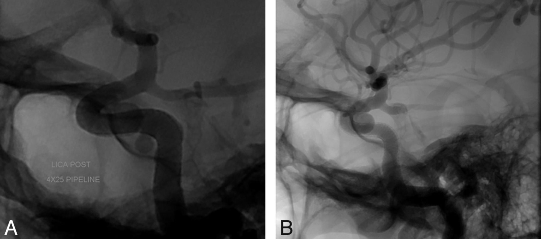
  • Fig 4.
    • Download figure
    • Open in new tab
    • Download powerpoint
    Fig 4.

    A, Angiogram showing the initial position of the PED; the distal end is at the clinoid segment. B, Angiogram showing proximal migration of the PED; the distal end of the device is now within the aneurysmal sac.

Tables

  • Figures
  • PED migration characteristics

    Patient No.Length of PED Extending to Aneurysm (mm)Migration Distance (mm)Migration of Proximal or Distal End of PED?
    Proximal to AneurysmDistal to Aneurysm
    1769Proximal
    2101520Distal
    31535Distal
    4107>10Distal and proximal
    5133.57.5Distal
PreviousNext
Back to top

In this issue

American Journal of Neuroradiology: 34 (12)
American Journal of Neuroradiology
Vol. 34, Issue 12
1 Dec 2013
  • Table of Contents
  • Index by author
  • Complete Issue (PDF)
Advertisement
Print
Download PDF
Email Article

Thank you for your interest in spreading the word on American Journal of Neuroradiology.

NOTE: We only request your email address so that the person you are recommending the page to knows that you wanted them to see it, and that it is not junk mail. We do not capture any email address.

Enter multiple addresses on separate lines or separate them with commas.
Spontaneous Delayed Migration/Shortening of the Pipeline Embolization Device: Report of 5 Cases
(Your Name) has sent you a message from American Journal of Neuroradiology
(Your Name) thought you would like to see the American Journal of Neuroradiology web site.
CAPTCHA
This question is for testing whether or not you are a human visitor and to prevent automated spam submissions.
Cite this article
N. Chalouhi, S.I. Tjoumakaris, L.F. Gonzalez, D. Hasan, P.J. Pema, G. Gould, R.H. Rosenwasser, P.M. Jabbour
Spontaneous Delayed Migration/Shortening of the Pipeline Embolization Device: Report of 5 Cases
American Journal of Neuroradiology Dec 2013, 34 (12) 2326-2330; DOI: 10.3174/ajnr.A3632

Citation Manager Formats

  • BibTeX
  • Bookends
  • EasyBib
  • EndNote (tagged)
  • EndNote 8 (xml)
  • Medlars
  • Mendeley
  • Papers
  • RefWorks Tagged
  • Ref Manager
  • RIS
  • Zotero
0 Responses
Respond to this article
Share
Bookmark this article
Spontaneous Delayed Migration/Shortening of the Pipeline Embolization Device: Report of 5 Cases
N. Chalouhi, S.I. Tjoumakaris, L.F. Gonzalez, D. Hasan, P.J. Pema, G. Gould, R.H. Rosenwasser, P.M. Jabbour
American Journal of Neuroradiology Dec 2013, 34 (12) 2326-2330; DOI: 10.3174/ajnr.A3632
del.icio.us logo Twitter logo Facebook logo Mendeley logo
  • Tweet Widget
  • Facebook Like
  • Google Plus One
Purchase

Jump to section

  • Article
    • Abstract
    • ABBREVIATIONS:
    • Materials and Methods
    • Discussion
    • Conclusions
    • Footnotes
    • REFERENCES
  • Figures & Data
  • Info & Metrics
  • Responses
  • References
  • PDF

Related Articles

  • No related articles found.
  • PubMed
  • Google Scholar

Cited By...

  • Flow diverter braid deformation following treatment of cerebral aneurysms: incidence, clinical relevance, and potential risk factors
  • Spontaneous delayed migration or shortening after pipeline embolization device treatment of intracranial aneurysm: incidence, management, and risk factors
  • Efficacy and safety of the dual-layer flow-diverting stent (FRED) for the treatment of intracranial aneurysms
  • Ophthalmic artery occlusion after Pipeline Embolization Device placement with reconstitution of flow via an endoleak: a report of two cases
  • Republished: Novel balloon application for rescue and realignment of a proximal end migrated pipeline flex embolization device into the aneurysmal sac: complication management
  • Republished: Spontaneous regression of intracranial aneurysm following remote ruptured aneurysm treatment with pipeline stent assisted coiling
  • Endovascular treatment of intracranial aneurysms using the Pipeline Flex embolization device: a case series of 30 consecutive patients
  • Novel balloon application for rescue and realignment of a proximal end migrated pipeline flex embolization device into the aneurysmal sac: complication management
  • Flow Diversion versus Standard Endovascular Techniques for the Treatment of Unruptured Carotid-Ophthalmic Aneurysms
  • Initial Experience with p64: A Novel Mechanically Detachable Flow Diverter for the Treatment of Intracranial Saccular Sidewall Aneurysms
  • Utilization of Pipeline embolization device for treatment of ruptured intracranial aneurysms: US multicenter experience
  • Focal, transient mechanical narrowing of a pipeline embolization device following treatment of an internal carotid artery aneurysm
  • Preliminary experience with the Pipeline Flex Embolization Device: technical note
  • Spontaneous regression of intracranial aneurysm following remote ruptured aneurysm treatment with pipeline stent assisted coiling
  • Focal, transient mechanical narrowing of a pipeline embolization device following treatment of an internal carotid artery aneurysm
  • Effect of Structural Remodeling (Retraction and Recoil) of the Pipeline Embolization Device on Aneurysm Occlusion Rate
  • A Single Pipeline Embolization Device is Sufficient for Treatment of Intracranial Aneurysms
  • Crossref
  • Google Scholar

This article has not yet been cited by articles in journals that are participating in Crossref Cited-by Linking.

More in this TOC Section

  • Rescue Reentry in Carotid Near-Occlusion
  • Contour Neurovascular System: Five Year Follow Up
  • Effect of SARS-CoV2 on Endovascular Thrombectomy
Show more Neurointervention

Similar Articles

Advertisement

Indexed Content

  • Current Issue
  • Accepted Manuscripts
  • Article Preview
  • Past Issues
  • Editorials
  • Editor's Choice
  • Fellows' Journal Club
  • Letters to the Editor
  • Video Articles

Cases

  • Case Collection
  • Archive - Case of the Week
  • Archive - Case of the Month
  • Archive - Classic Case

More from AJNR

  • Trainee Corner
  • Imaging Protocols
  • MRI Safety Corner
  • Book Reviews

Multimedia

  • AJNR Podcasts
  • AJNR Scantastics

Resources

  • Turnaround Time
  • Submit a Manuscript
  • Submit a Video Article
  • Submit an eLetter to the Editor/Response
  • Manuscript Submission Guidelines
  • Statistical Tips
  • Fast Publishing of Accepted Manuscripts
  • Graphical Abstract Preparation
  • Imaging Protocol Submission
  • Evidence-Based Medicine Level Guide
  • Publishing Checklists
  • Author Policies
  • Become a Reviewer/Academy of Reviewers
  • News and Updates

About Us

  • About AJNR
  • Editorial Board
  • Editorial Board Alumni
  • Alerts
  • Permissions
  • Not an AJNR Subscriber? Join Now
  • Advertise with Us
  • Librarian Resources
  • Feedback
  • Terms and Conditions
  • AJNR Editorial Board Alumni

American Society of Neuroradiology

  • Not an ASNR Member? Join Now

© 2025 by the American Society of Neuroradiology All rights, including for text and data mining, AI training, and similar technologies, are reserved.
Print ISSN: 0195-6108 Online ISSN: 1936-959X

Powered by HighWire