Advertisement

Main menu

  • Home
  • Content
    • Current Issue
    • Accepted Manuscripts
    • Article Preview
    • Past Issue Archive
    • Video Articles
    • AJNR Case Collection
    • Case of the Week Archive
    • Case of the Month Archive
    • Classic Case Archive
  • Special Collections
    • AJNR Awards
    • Low-Field MRI
    • Alzheimer Disease
    • ASNR Foundation Special Collection
    • Photon-Counting CT
    • View All
  • Multimedia
    • AJNR Podcasts
    • AJNR SCANtastic
    • Trainee Corner
    • MRI Safety Corner
    • Imaging Protocols
  • For Authors
    • Submit a Manuscript
    • Submit a Video Article
    • Submit an eLetter to the Editor/Response
    • Manuscript Submission Guidelines
    • Statistical Tips
    • Fast Publishing of Accepted Manuscripts
    • Graphical Abstract Preparation
    • Imaging Protocol Submission
    • Author Policies
  • About Us
    • About AJNR
    • Editorial Board
    • Editorial Board Alumni
  • More
    • Become a Reviewer/Academy of Reviewers
    • Subscribers
    • Permissions
    • Alerts
    • Feedback
    • Advertisers
    • ASNR Home

User menu

  • Alerts
  • Log in

Search

  • Advanced search
American Journal of Neuroradiology

American Journal of Neuroradiology

ASHNR American Society of Functional Neuroradiology ASHNR American Society of Pediatric Neuroradiology ASSR
  • Alerts
  • Log in

Advanced Search

  • Home
  • Content
    • Current Issue
    • Accepted Manuscripts
    • Article Preview
    • Past Issue Archive
    • Video Articles
    • AJNR Case Collection
    • Case of the Week Archive
    • Case of the Month Archive
    • Classic Case Archive
  • Special Collections
    • AJNR Awards
    • Low-Field MRI
    • Alzheimer Disease
    • ASNR Foundation Special Collection
    • Photon-Counting CT
    • View All
  • Multimedia
    • AJNR Podcasts
    • AJNR SCANtastic
    • Trainee Corner
    • MRI Safety Corner
    • Imaging Protocols
  • For Authors
    • Submit a Manuscript
    • Submit a Video Article
    • Submit an eLetter to the Editor/Response
    • Manuscript Submission Guidelines
    • Statistical Tips
    • Fast Publishing of Accepted Manuscripts
    • Graphical Abstract Preparation
    • Imaging Protocol Submission
    • Author Policies
  • About Us
    • About AJNR
    • Editorial Board
    • Editorial Board Alumni
  • More
    • Become a Reviewer/Academy of Reviewers
    • Subscribers
    • Permissions
    • Alerts
    • Feedback
    • Advertisers
    • ASNR Home
  • Follow AJNR on Twitter
  • Visit AJNR on Facebook
  • Follow AJNR on Instagram
  • Join AJNR on LinkedIn
  • RSS Feeds

AJNR Awards, New Junior Editors, and more. Read the latest AJNR updates

Research ArticleBrain

Regional Analysis of the Magnetization Transfer Ratio of the Brain in Mild Alzheimer Disease and Amnestic Mild Cognitive Impairment

M. Mascalchi, A. Ginestroni, V. Bessi, N. Toschi, S. Padiglioni, S. Ciulli, C. Tessa, M. Giannelli, L. Bracco and S. Diciotti
American Journal of Neuroradiology November 2013, 34 (11) 2098-2104; DOI: https://doi.org/10.3174/ajnr.A3568
M. Mascalchi
aFrom the Quantitative and Functional Neuroradiology Research Unit, Department of Experimental and Clinical Biomedical Sciences (M.M., A.G., S.C., S.D.)
  • Find this author on Google Scholar
  • Find this author on PubMed
  • Search for this author on this site
A. Ginestroni
aFrom the Quantitative and Functional Neuroradiology Research Unit, Department of Experimental and Clinical Biomedical Sciences (M.M., A.G., S.C., S.D.)
  • Find this author on Google Scholar
  • Find this author on PubMed
  • Search for this author on this site
V. Bessi
bDepartment of Neurological and Psychiatric Sciences (V.B., S.P., L.B.), University of Florence, Florence, Italy
  • Find this author on Google Scholar
  • Find this author on PubMed
  • Search for this author on this site
N. Toschi
cMedical Physics Section (N.T.), Faculty of Medicine, University of Rome “Tor Vergata,” Rome, Italy
  • Find this author on Google Scholar
  • Find this author on PubMed
  • Search for this author on this site
S. Padiglioni
bDepartment of Neurological and Psychiatric Sciences (V.B., S.P., L.B.), University of Florence, Florence, Italy
  • Find this author on Google Scholar
  • Find this author on PubMed
  • Search for this author on this site
S. Ciulli
aFrom the Quantitative and Functional Neuroradiology Research Unit, Department of Experimental and Clinical Biomedical Sciences (M.M., A.G., S.C., S.D.)
  • Find this author on Google Scholar
  • Find this author on PubMed
  • Search for this author on this site
C. Tessa
dRadiology Department (C.T.), Versilia Hospital, Lido di Camaiore (Lucca), Italy
  • Find this author on Google Scholar
  • Find this author on PubMed
  • Search for this author on this site
M. Giannelli
eUnit of Medical Physics (M.G.), Pisa University Hospital, “Azienda Ospedaliero-Universitaria Pisana,” Pisa, Italy.
  • Find this author on Google Scholar
  • Find this author on PubMed
  • Search for this author on this site
L. Bracco
bDepartment of Neurological and Psychiatric Sciences (V.B., S.P., L.B.), University of Florence, Florence, Italy
  • Find this author on Google Scholar
  • Find this author on PubMed
  • Search for this author on this site
S. Diciotti
aFrom the Quantitative and Functional Neuroradiology Research Unit, Department of Experimental and Clinical Biomedical Sciences (M.M., A.G., S.C., S.D.)
  • Find this author on Google Scholar
  • Find this author on PubMed
  • Search for this author on this site
  • Article
  • Figures & Data
  • Supplemental
  • Info & Metrics
  • Responses
  • References
  • PDF
Loading

Article Figures & Data

Figures

  • Fig 1.
    • Download figure
    • Open in new tab
    • Download powerpoint
    Fig 1.

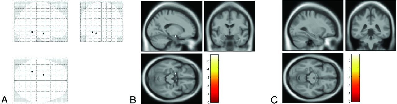
    SPM8 “glass brain” representation (A) of voxelwise analysis between control subjects and patients with Alzheimer disease showing significant clusters of decreased magnetization transfer ratio in the left hippocampus and amygdala and in the posterior mesial temporal cortex (fusiform gyrus) (P < .05, with family-wise error rate correction). Superimposition onto T1 template of the cluster in the hippocampus and amygdala are demonstrated in B and of the cluster in the fusiform gyrus in C.

  • Fig 2.
    • Download figure
    • Open in new tab
    • Download powerpoint
    Fig 2.

    Example of the FreeSurfer automatic VOI segmentation of the hippocampus (A) and amygdala (B) eroded with a 3D structural element with 1-mm radius.

  • Fig 3.
    • Download figure
    • Open in new tab
    • Download powerpoint
    Fig 3.

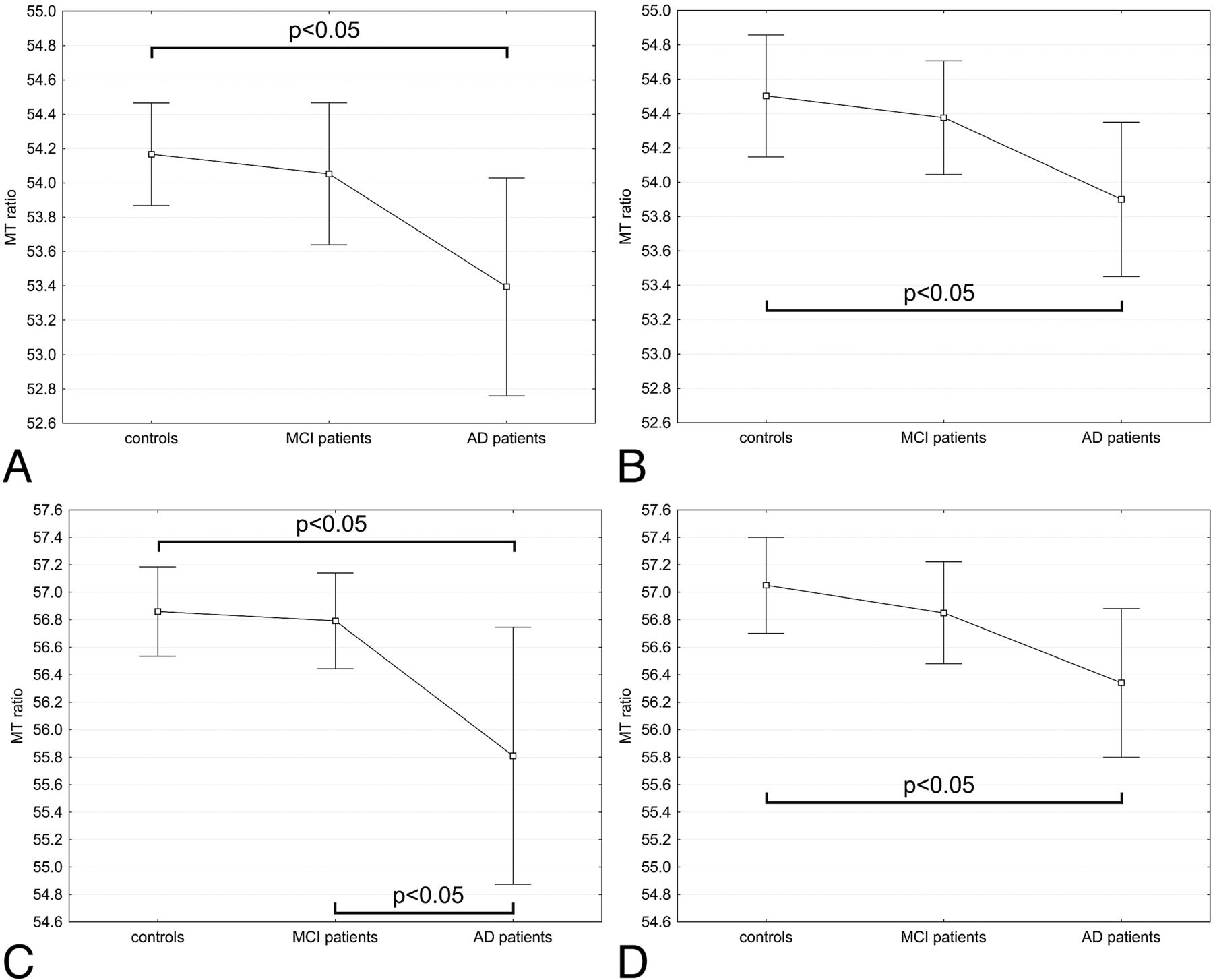
    Magnetization transfer ratio of in the hippocampi [left (A) and right (B)] and amygdalae [left (C) and right (D)] in the 3 groups. Mean and 95% confidence intervals of mean MT ratio are reported. The MT ratio in the bilateral hippocampus and amygdala was significantly lower in patients with Alzheimer disease when compared with control subjects (ANOVA with post hoc Bonferroni correction, with P < .05). The values of the mean MT ratio in the hippocampus and amygdala in the patients with amnestic mild cognitive impairment were between those of healthy control subjects and those of patients with mild AD, and the differences were not significant with the exception of the MT ratio in the left amygdala, which was significantly lower in AD than in amnestic MCI.

PreviousNext
Back to top

In this issue

American Journal of Neuroradiology: 34 (11)
American Journal of Neuroradiology
Vol. 34, Issue 11
1 Nov 2013
  • Table of Contents
  • Index by author
  • Complete Issue (PDF)
Advertisement
Print
Download PDF
Email Article

Thank you for your interest in spreading the word on American Journal of Neuroradiology.

NOTE: We only request your email address so that the person you are recommending the page to knows that you wanted them to see it, and that it is not junk mail. We do not capture any email address.

Enter multiple addresses on separate lines or separate them with commas.
Regional Analysis of the Magnetization Transfer Ratio of the Brain in Mild Alzheimer Disease and Amnestic Mild Cognitive Impairment
(Your Name) has sent you a message from American Journal of Neuroradiology
(Your Name) thought you would like to see the American Journal of Neuroradiology web site.
CAPTCHA
This question is for testing whether or not you are a human visitor and to prevent automated spam submissions.
Cite this article
M. Mascalchi, A. Ginestroni, V. Bessi, N. Toschi, S. Padiglioni, S. Ciulli, C. Tessa, M. Giannelli, L. Bracco, S. Diciotti
Regional Analysis of the Magnetization Transfer Ratio of the Brain in Mild Alzheimer Disease and Amnestic Mild Cognitive Impairment
American Journal of Neuroradiology Nov 2013, 34 (11) 2098-2104; DOI: 10.3174/ajnr.A3568

Citation Manager Formats

  • BibTeX
  • Bookends
  • EasyBib
  • EndNote (tagged)
  • EndNote 8 (xml)
  • Medlars
  • Mendeley
  • Papers
  • RefWorks Tagged
  • Ref Manager
  • RIS
  • Zotero
0 Responses
Respond to this article
Share
Bookmark this article
Regional Analysis of the Magnetization Transfer Ratio of the Brain in Mild Alzheimer Disease and Amnestic Mild Cognitive Impairment
M. Mascalchi, A. Ginestroni, V. Bessi, N. Toschi, S. Padiglioni, S. Ciulli, C. Tessa, M. Giannelli, L. Bracco, S. Diciotti
American Journal of Neuroradiology Nov 2013, 34 (11) 2098-2104; DOI: 10.3174/ajnr.A3568
del.icio.us logo Twitter logo Facebook logo Mendeley logo
  • Tweet Widget
  • Facebook Like
  • Google Plus One
Purchase

Jump to section

  • Article
    • Abstract
    • ABBREVIATIONS:
    • Materials and Methods
    • Results
    • Discussion
    • Conclusions
    • REFERENCES
  • Figures & Data
  • Supplemental
  • Info & Metrics
  • Responses
  • References
  • PDF

Related Articles

  • PubMed
  • Google Scholar

Cited By...

  • No citing articles found.
  • Crossref
  • Google Scholar

This article has not yet been cited by articles in journals that are participating in Crossref Cited-by Linking.

More in this TOC Section

  • Multimodal CT Provides Improved Performance for Lacunar Infarct Detection
  • Optimal MRI Sequence for Identifying Occlusion Location in Acute Stroke: Which Value of Time-Resolved Contrast-Enhanced MRA?
  • Evaluating the Effects of White Matter Multiple Sclerosis Lesions on the Volume Estimation of 6 Brain Tissue Segmentation Methods
Show more Brain

Similar Articles

Advertisement

Indexed Content

  • Current Issue
  • Accepted Manuscripts
  • Article Preview
  • Past Issues
  • Editorials
  • Editor's Choice
  • Fellows' Journal Club
  • Letters to the Editor
  • Video Articles

Cases

  • Case Collection
  • Archive - Case of the Week
  • Archive - Case of the Month
  • Archive - Classic Case

More from AJNR

  • Trainee Corner
  • Imaging Protocols
  • MRI Safety Corner
  • Book Reviews

Multimedia

  • AJNR Podcasts
  • AJNR Scantastics

Resources

  • Turnaround Time
  • Submit a Manuscript
  • Submit a Video Article
  • Submit an eLetter to the Editor/Response
  • Manuscript Submission Guidelines
  • Statistical Tips
  • Fast Publishing of Accepted Manuscripts
  • Graphical Abstract Preparation
  • Imaging Protocol Submission
  • Evidence-Based Medicine Level Guide
  • Publishing Checklists
  • Author Policies
  • Become a Reviewer/Academy of Reviewers
  • News and Updates

About Us

  • About AJNR
  • Editorial Board
  • Editorial Board Alumni
  • Alerts
  • Permissions
  • Not an AJNR Subscriber? Join Now
  • Advertise with Us
  • Librarian Resources
  • Feedback
  • Terms and Conditions
  • AJNR Editorial Board Alumni

American Society of Neuroradiology

  • Not an ASNR Member? Join Now

© 2025 by the American Society of Neuroradiology All rights, including for text and data mining, AI training, and similar technologies, are reserved.
Print ISSN: 0195-6108 Online ISSN: 1936-959X

Powered by HighWire