Advertisement

Main menu

  • Home
  • Content
    • Current Issue
    • Accepted Manuscripts
    • Article Preview
    • Past Issue Archive
    • Video Articles
    • AJNR Case Collection
    • Case of the Week Archive
    • Case of the Month Archive
    • Classic Case Archive
  • Special Collections
    • AJNR Awards
    • Low-Field MRI
    • Alzheimer Disease
    • ASNR Foundation Special Collection
    • Photon-Counting CT
    • View All
  • Multimedia
    • AJNR Podcasts
    • AJNR SCANtastic
    • Trainee Corner
    • MRI Safety Corner
    • Imaging Protocols
  • For Authors
    • Submit a Manuscript
    • Submit a Video Article
    • Submit an eLetter to the Editor/Response
    • Manuscript Submission Guidelines
    • Statistical Tips
    • Fast Publishing of Accepted Manuscripts
    • Graphical Abstract Preparation
    • Imaging Protocol Submission
    • Author Policies
  • About Us
    • About AJNR
    • Editorial Board
    • Editorial Board Alumni
  • More
    • Become a Reviewer/Academy of Reviewers
    • Subscribers
    • Permissions
    • Alerts
    • Feedback
    • Advertisers
    • ASNR Home

User menu

  • Alerts
  • Log in

Search

  • Advanced search
American Journal of Neuroradiology

American Journal of Neuroradiology

ASHNR American Society of Functional Neuroradiology ASHNR American Society of Pediatric Neuroradiology ASSR
  • Alerts
  • Log in

Advanced Search

  • Home
  • Content
    • Current Issue
    • Accepted Manuscripts
    • Article Preview
    • Past Issue Archive
    • Video Articles
    • AJNR Case Collection
    • Case of the Week Archive
    • Case of the Month Archive
    • Classic Case Archive
  • Special Collections
    • AJNR Awards
    • Low-Field MRI
    • Alzheimer Disease
    • ASNR Foundation Special Collection
    • Photon-Counting CT
    • View All
  • Multimedia
    • AJNR Podcasts
    • AJNR SCANtastic
    • Trainee Corner
    • MRI Safety Corner
    • Imaging Protocols
  • For Authors
    • Submit a Manuscript
    • Submit a Video Article
    • Submit an eLetter to the Editor/Response
    • Manuscript Submission Guidelines
    • Statistical Tips
    • Fast Publishing of Accepted Manuscripts
    • Graphical Abstract Preparation
    • Imaging Protocol Submission
    • Author Policies
  • About Us
    • About AJNR
    • Editorial Board
    • Editorial Board Alumni
  • More
    • Become a Reviewer/Academy of Reviewers
    • Subscribers
    • Permissions
    • Alerts
    • Feedback
    • Advertisers
    • ASNR Home
  • Follow AJNR on Twitter
  • Visit AJNR on Facebook
  • Follow AJNR on Instagram
  • Join AJNR on LinkedIn
  • RSS Feeds

AJNR Awards, New Junior Editors, and more. Read the latest AJNR updates

Getting new auth cookie, if you see this message a lot, tell someone!
Research ArticlePatient Safety
Open Access

Endovascular Catheter for Magnetic Navigation under MR Imaging Guidance: Evaluation of Safety In Vivo at 1.5T

S.W. Hetts, M. Saeed, A.J. Martin, L. Evans, A.F. Bernhardt, V. Malba, F. Settecase, L. Do, E.J. Yee, A. Losey, R. Sincic, P. Lillaney, S. Roy, R.L. Arenson and M.W. Wilson
American Journal of Neuroradiology November 2013, 34 (11) 2083-2091; DOI: https://doi.org/10.3174/ajnr.A3530
S.W. Hetts
aFrom the Departments of Radiology and Biomedical Imaging (S.W.H., M.S., A.J.M., F.S., L.D., E.J.Y., R.S., P.L., R.L.A., M.W.W.)
  • Find this author on Google Scholar
  • Find this author on PubMed
  • Search for this author on this site
M. Saeed
aFrom the Departments of Radiology and Biomedical Imaging (S.W.H., M.S., A.J.M., F.S., L.D., E.J.Y., R.S., P.L., R.L.A., M.W.W.)
  • Find this author on Google Scholar
  • Find this author on PubMed
  • Search for this author on this site
A.J. Martin
aFrom the Departments of Radiology and Biomedical Imaging (S.W.H., M.S., A.J.M., F.S., L.D., E.J.Y., R.S., P.L., R.L.A., M.W.W.)
  • Find this author on Google Scholar
  • Find this author on PubMed
  • Search for this author on this site
L. Evans
dLawrence Livermore National Laboratory (L.E., A.F.B., V.M.), Livermore, California.
  • Find this author on Google Scholar
  • Find this author on PubMed
  • Search for this author on this site
A.F. Bernhardt
dLawrence Livermore National Laboratory (L.E., A.F.B., V.M.), Livermore, California.
  • Find this author on Google Scholar
  • Find this author on PubMed
  • Search for this author on this site
V. Malba
dLawrence Livermore National Laboratory (L.E., A.F.B., V.M.), Livermore, California.
  • Find this author on Google Scholar
  • Find this author on PubMed
  • Search for this author on this site
F. Settecase
aFrom the Departments of Radiology and Biomedical Imaging (S.W.H., M.S., A.J.M., F.S., L.D., E.J.Y., R.S., P.L., R.L.A., M.W.W.)
  • Find this author on Google Scholar
  • Find this author on PubMed
  • Search for this author on this site
L. Do
aFrom the Departments of Radiology and Biomedical Imaging (S.W.H., M.S., A.J.M., F.S., L.D., E.J.Y., R.S., P.L., R.L.A., M.W.W.)
  • Find this author on Google Scholar
  • Find this author on PubMed
  • Search for this author on this site
E.J. Yee
aFrom the Departments of Radiology and Biomedical Imaging (S.W.H., M.S., A.J.M., F.S., L.D., E.J.Y., R.S., P.L., R.L.A., M.W.W.)
  • Find this author on Google Scholar
  • Find this author on PubMed
  • Search for this author on this site
A. Losey
cSchool of Medicine (A.L.), University of California, San Francisco
  • Find this author on Google Scholar
  • Find this author on PubMed
  • Search for this author on this site
R. Sincic
aFrom the Departments of Radiology and Biomedical Imaging (S.W.H., M.S., A.J.M., F.S., L.D., E.J.Y., R.S., P.L., R.L.A., M.W.W.)
  • Find this author on Google Scholar
  • Find this author on PubMed
  • Search for this author on this site
P. Lillaney
aFrom the Departments of Radiology and Biomedical Imaging (S.W.H., M.S., A.J.M., F.S., L.D., E.J.Y., R.S., P.L., R.L.A., M.W.W.)
  • Find this author on Google Scholar
  • Find this author on PubMed
  • Search for this author on this site
S. Roy
bBioengineering (S.R.)
  • Find this author on Google Scholar
  • Find this author on PubMed
  • Search for this author on this site
R.L. Arenson
aFrom the Departments of Radiology and Biomedical Imaging (S.W.H., M.S., A.J.M., F.S., L.D., E.J.Y., R.S., P.L., R.L.A., M.W.W.)
  • Find this author on Google Scholar
  • Find this author on PubMed
  • Search for this author on this site
M.W. Wilson
aFrom the Departments of Radiology and Biomedical Imaging (S.W.H., M.S., A.J.M., F.S., L.D., E.J.Y., R.S., P.L., R.L.A., M.W.W.)
  • Find this author on Google Scholar
  • Find this author on PubMed
  • Search for this author on this site
  • Article
  • Figures & Data
  • Supplemental
  • Info & Metrics
  • Responses
  • References
  • PDF
Loading

Article Figures & Data

Figures

  • Tables
  • Fig 1.
    • Download figure
    • Open in new tab
    • Download powerpoint
    Fig 1.

    MARC catheter system. A, Hand-wound copper coil solenoid on an alumina substrate tube. B, Catheter diagram with thermocouple. A. Catheter length. B. Catheter tip. C. Coil feedwire. D. Power lead E. Power phone jack lead AA. Catheter tip. C, Magnetic microcatheter III with thermocouple. A light blue Rapid Transit microcatheter (Cordis, Miami Lakes, Florida) with a 2.3F distal tip has been used as a substrate. A 30-turn copper solenoid coil mounted on an alumina tube is attached to the distal tip of the microcatheter with brown shrink-wrap. A copper-constantan thermocouple terminates adjacent to the coils within the shrink-wrap. Current-carrying copper wires to the solenoid coil run down the catheter lumen and are attached to a phone jack adaptor that, itself, can be plugged into a power source for activation experiments. The dark blue thermocouple plug at the catheter hub can be attached to a data logger for temperature measurements. D, Distal tip of magnetic microcatheter III. A 30-turn copper solenoid coil mounted on an alumina tube is attached to the distal tip of the microcatheter. Brown shrink-wrap attaches the alumina tube–copper coil assembly to the distal tip of the microcatheter. The final outer diameter is approximately 2 mm.
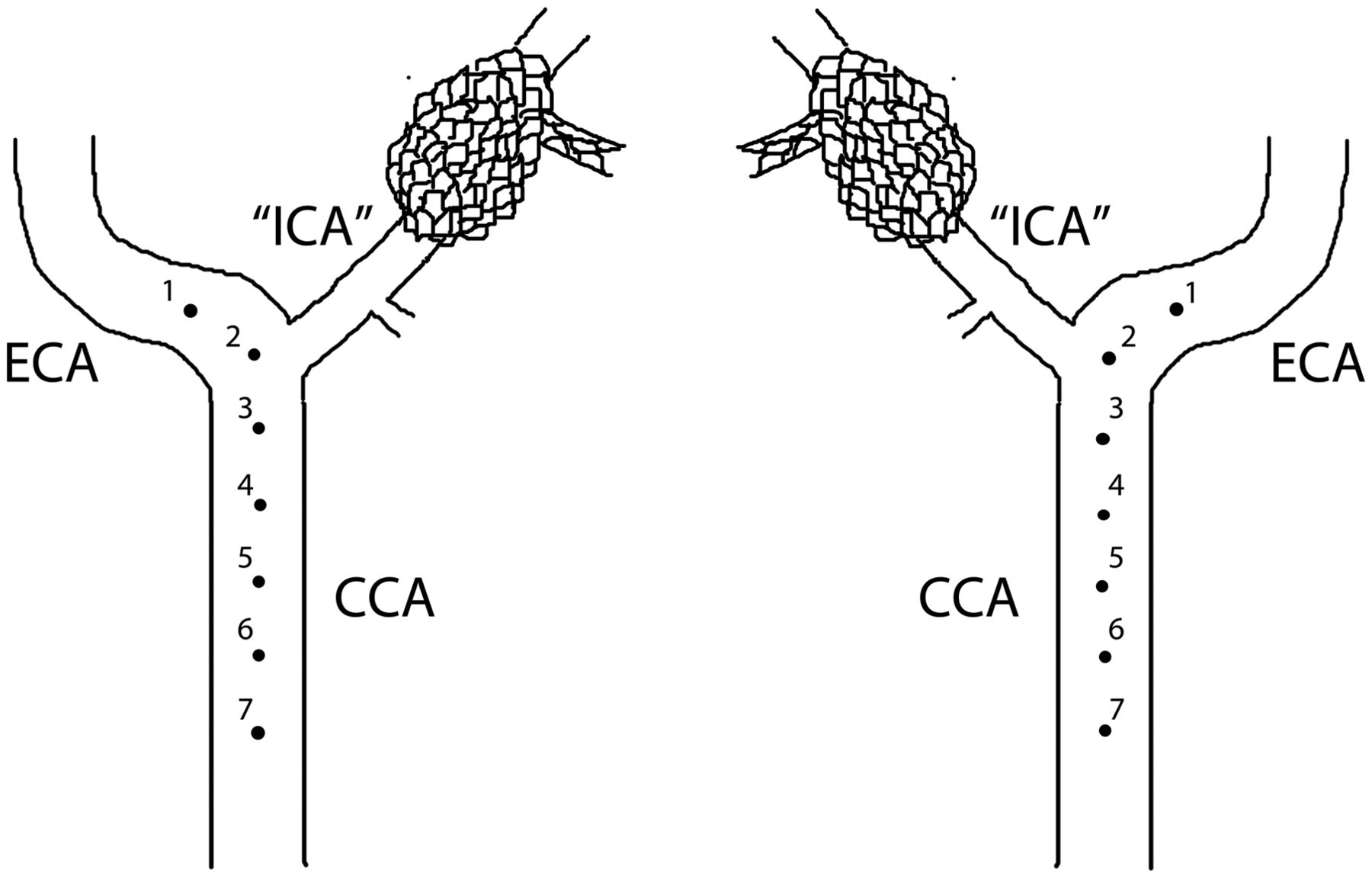
  • Fig 2.
    • Download figure
    • Open in new tab
    • Download powerpoint
    Fig 2.

    Schematic diagram depicting coil-tipped microcatheter heating activation points in vivo (swine right and left common carotid arteries, respectively). The first activation point is 1 cm distal to the origin of the internal carotid artery (ascending pharyngeal artery). Each subsequent activation point is separated by a 1-cm manual pull-back of the catheter, confirmed by imaging. This ensures adequate spacing between points in case of potential thermal damage to the arterial wall at any given point.
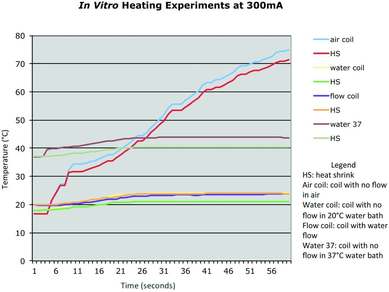
  • Fig 3.
    • Download figure
    • Open in new tab
    • Download powerpoint
    Fig 3.

    In vitro heating data for alumina tube construct tested at 300 mA in air, in a 25°C water bath, and in a 37°C water bath.
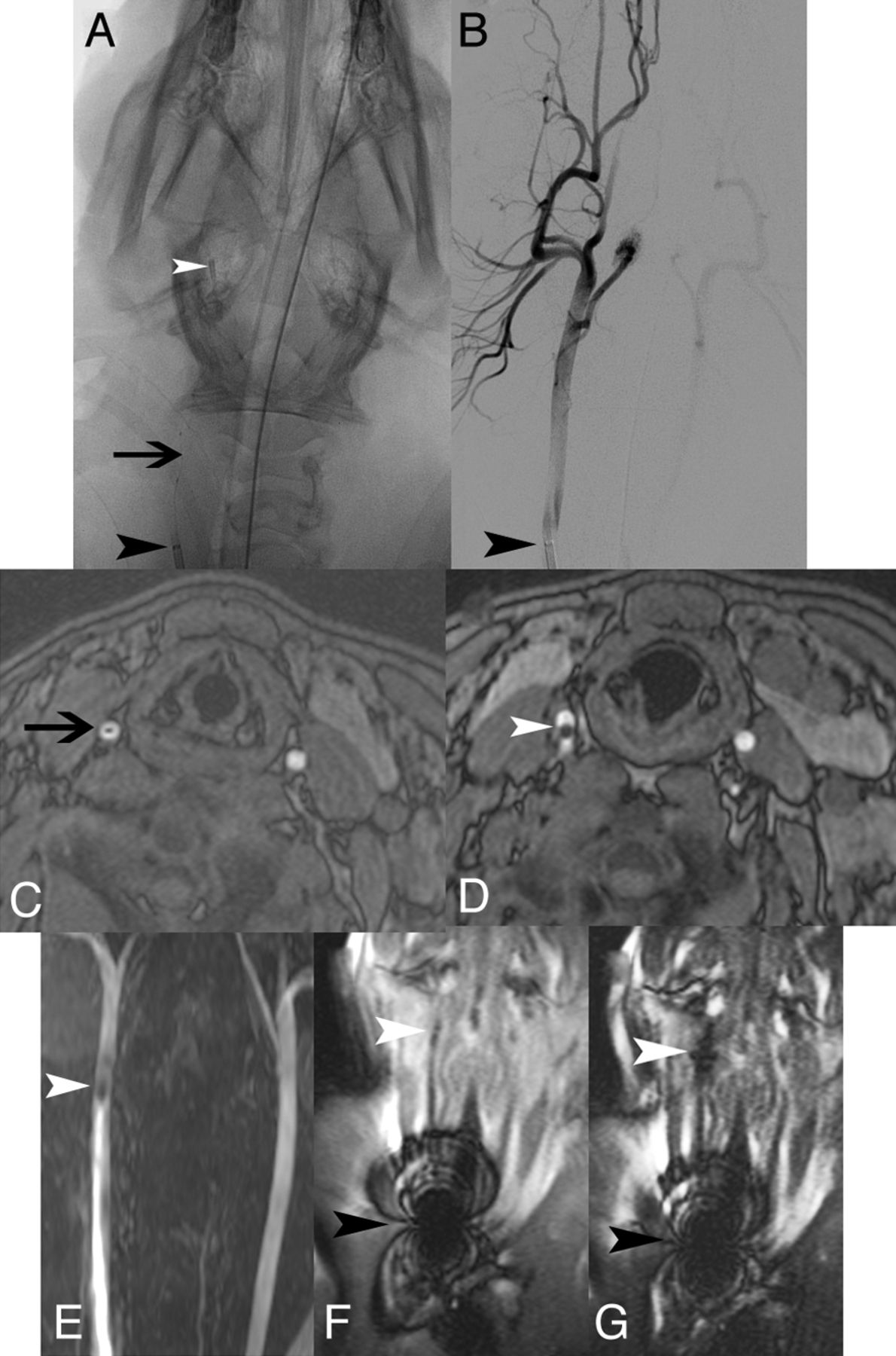
  • Fig 4.
    • Download figure
    • Open in new tab
    • Download powerpoint
    Fig 4.

    In vivo x-ray, DSA, axial MRA, coronal MRA MIP, and coronal SSFP. Unsubtracted x-ray image (A) demonstrates MARC catheter tip coils (white arrowhead), microcatheter shaft with lead wires (black arrow), and guiding catheter (black arrowhead) in the right CCA. Only the guiding catheter tip marker is readily evident on the equivalent DSA image (B). Susceptibility from the catheter shaft lead wires (black arrow) and catheter tip (white arrowhead) is seen on axial MRA (C and D), coronal MRA MIP (E), and coronal SSFP (F). With a 300-mA current applied (G), the catheter tip coils are more apparent (white arrowhead). Guide catheter tip artifacts resulting from a metallic marker band are very prominent on the SSFP sequence (F and G).

  • Fig 5.
    • Download figure
    • Open in new tab
    • Download powerpoint
    Fig 5.

    Porcine carotid artery wall histologic appearance after use of endovascular catheter tip coils at a 300-mA tip current for 1 minute at normal flow (A-D) or a 600-mA tip current for 2 minutes at zero flow (E and F). There is no evidence of vessel wall damage on hematoxylin-eosin (A and B) or Masson trichrome (C and D) at 300 mA. At 600 mA, however, luminal thrombus (E, black arrowhead), extensive medial vacuolization (F, black arrow), and medial hemorrhage (G, white arrow) all indicate thermal damage to the arterial wall.
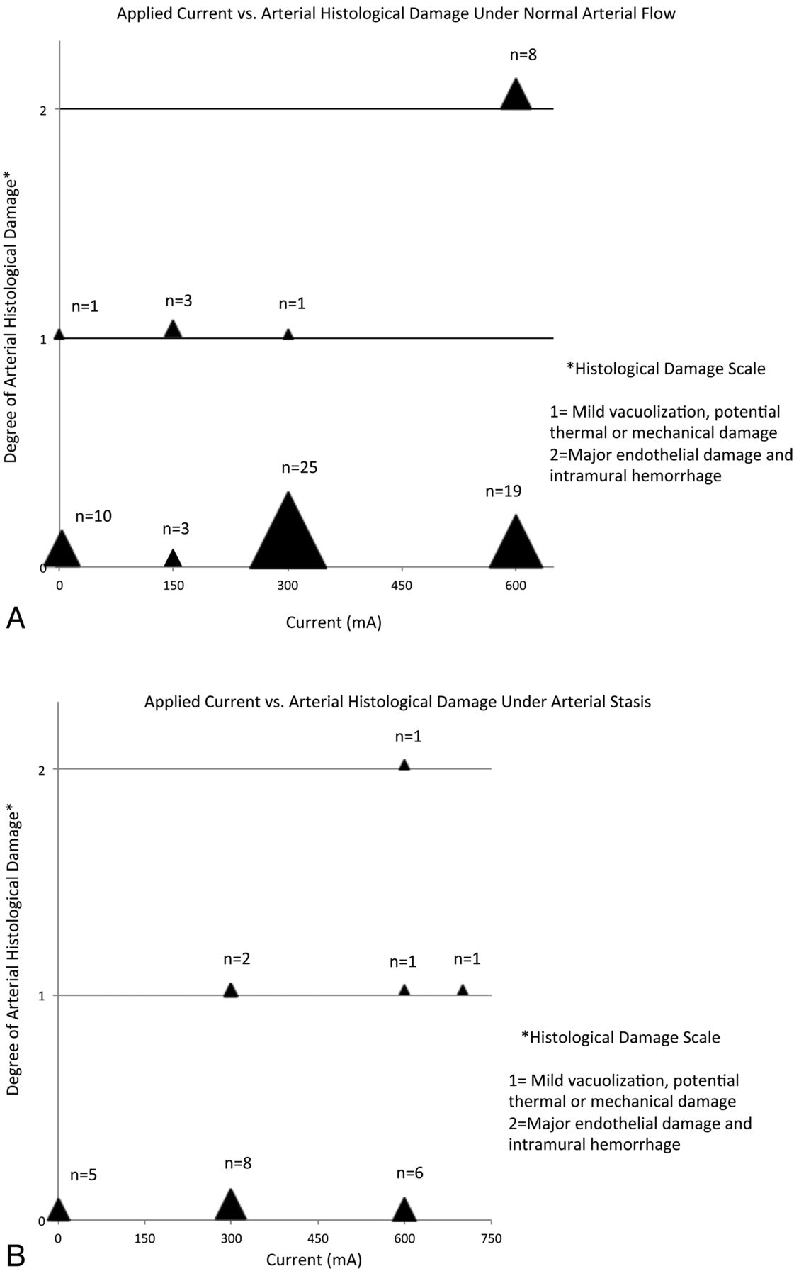
  • Fig 6.
    • Download figure
    • Open in new tab
    • Download powerpoint
    Fig 6.

    Plot of applied current vs degree of histologic damage for catheter activation experiments in vivo (n=94) in porcine carotid arteries. A, Normal arterial flow. B, Arterial stasis.

Tables

  • Figures
    • View popup
    Table 1:

    Microcatheter constructs

    Experimental Catheter No.Substrate MicrocatheterCatheter Length (cm)Solenoid Coil (no. of turns)Resistance (ohms)Thermocouple
    I2.3F RapidTransit170302None
    II2.7F Tracker-18150755.5None
    III2.3F RapidTransit170302.5Copper/constantan type T
    IV2.3F RapidTransit170302.5Copper/constantan type T
    • View popup
    Table 2:

    Definite vs no or questionable histologic damage to carotid arteries under various conditions of catheter testing in vivo

    Condition TestedDefinite Damage (%)No/Questionable Damage (%)Odds Ratio95% CIP Value*
    Arterial stasis (n = 24)4963.00.36–137.44
    Normal flow (n = 70)1189
    Current ≤300 mA (n = 58)010000–0.21.0001
    Current > 300 mA (n = 36)2575
    Activations ≤1 min (n = 52)6940.370.056–1.9.29
    Activations > 1 min (n = 42)1486
    Work ≤100 J (n = 68)3970.0820.008–0.5.0015
    Work >100 J (n = 26)2773
    Saline drip ≤2 mL/min (n = 39)15853.20.61–21.16
    Saline drip > 2 mL/min (n = 55)595
    ≤5°C catheter tip coil temperature rise (n = 36)010000–0.36.0022
    > 5°C temperature rise (n = 29)2476
    MR and x-ray guidance (n = 45)11881.90.35–13.47
    X-ray guidance only (n = 49)694
    • CI indicates confidence interval; J, Joules; mA, milliamperes.

    • Note:—Definite damage denotes a histologic score of 2; questionable damage, histologic score of 1; and no damage, histologic score of 0.

    • ↵* P value = 2-tailed Fisher exact test.

    • View popup
    Table 3:

    Definite or questionable vs no histologic damage to carotid arteries under various conditions of catheter testing in vivo

    Condition TestedDefinite/Questionable Damage (%)No Damage (%)Odds Ratio95% CIP Value*
    Arterial stasis (n = 24)21790.870.25–3.5.77
    Normal flow (n = 57)2377
    Current ≤300 mA (n = 51)14860.310.092–1.020.034
    Current > 300 mA (n = 36)3169
    Activations ≤1 minute (n = 52)8920.170.037–0.61.003
    Activations > 1 min (n = 42)3367
    Work ≤100 J (n = 68)13870.290.018–1.037
    Work > 100 J (n = 26)3565
    Saline drip ≤2 mL/min (n = 39)21791.20.35–3.7.80
    Saline drip >2 mL/min (n = 55)1882
    ≤5°C catheter tip coil temperature rise (n = 36)8920.170.028–0.80.013
    > 5°C temperature rise (n = 29)3468
    MR and x-ray guidance (n = 45)16840.640.19–2.0.44
    X-ray guidance only (n = 49)2278
    • ↵* P value = 2-tailed Fisher exact test.

PreviousNext
Back to top

In this issue

American Journal of Neuroradiology: 34 (11)
American Journal of Neuroradiology
Vol. 34, Issue 11
1 Nov 2013
  • Table of Contents
  • Index by author
  • Complete Issue (PDF)
Advertisement
Print
Download PDF
Email Article

Thank you for your interest in spreading the word on American Journal of Neuroradiology.

NOTE: We only request your email address so that the person you are recommending the page to knows that you wanted them to see it, and that it is not junk mail. We do not capture any email address.

Enter multiple addresses on separate lines or separate them with commas.
Endovascular Catheter for Magnetic Navigation under MR Imaging Guidance: Evaluation of Safety In Vivo at 1.5T
(Your Name) has sent you a message from American Journal of Neuroradiology
(Your Name) thought you would like to see the American Journal of Neuroradiology web site.
CAPTCHA
This question is for testing whether or not you are a human visitor and to prevent automated spam submissions.
Cite this article
S.W. Hetts, M. Saeed, A.J. Martin, L. Evans, A.F. Bernhardt, V. Malba, F. Settecase, L. Do, E.J. Yee, A. Losey, R. Sincic, P. Lillaney, S. Roy, R.L. Arenson, M.W. Wilson
Endovascular Catheter for Magnetic Navigation under MR Imaging Guidance: Evaluation of Safety In Vivo at 1.5T
American Journal of Neuroradiology Nov 2013, 34 (11) 2083-2091; DOI: 10.3174/ajnr.A3530

Citation Manager Formats

  • BibTeX
  • Bookends
  • EasyBib
  • EndNote (tagged)
  • EndNote 8 (xml)
  • Medlars
  • Mendeley
  • Papers
  • RefWorks Tagged
  • Ref Manager
  • RIS
  • Zotero
0 Responses
Respond to this article
Share
Bookmark this article
Endovascular Catheter for Magnetic Navigation under MR Imaging Guidance: Evaluation of Safety In Vivo at 1.5T
S.W. Hetts, M. Saeed, A.J. Martin, L. Evans, A.F. Bernhardt, V. Malba, F. Settecase, L. Do, E.J. Yee, A. Losey, R. Sincic, P. Lillaney, S. Roy, R.L. Arenson, M.W. Wilson
American Journal of Neuroradiology Nov 2013, 34 (11) 2083-2091; DOI: 10.3174/ajnr.A3530
del.icio.us logo Twitter logo Facebook logo Mendeley logo
  • Tweet Widget
  • Facebook Like
  • Google Plus One
Purchase

Jump to section

  • Article
    • Abstract
    • ABBREVIATIONS:
    • Materials and Methods
    • Results
    • Discussion
    • Conclusions
    • Acknowledgments
    • Footnotes
    • REFERENCES
  • Figures & Data
  • Supplemental
  • Info & Metrics
  • Responses
  • References
  • PDF

Related Articles

  • PubMed
  • Google Scholar

Cited By...

  • No citing articles found.
  • Crossref
  • Google Scholar

This article has not yet been cited by articles in journals that are participating in Crossref Cited-by Linking.

More in this TOC Section

  • Safety of Intrathecal Gadobutrol in Various Doses
  • Impact of Kidney Function on CNS Gadolinium Deposition in Patients Receiving Repeated Doses of Gadobutrol
  • Contrast-Induced Acute Kidney Injury in Radiologic Management of Acute Ischemic Stroke in the Emergency Setting
Show more Patient Safety

Similar Articles

Advertisement

Indexed Content

  • Current Issue
  • Accepted Manuscripts
  • Article Preview
  • Past Issues
  • Editorials
  • Editor's Choice
  • Fellows' Journal Club
  • Letters to the Editor
  • Video Articles

Cases

  • Case Collection
  • Archive - Case of the Week
  • Archive - Case of the Month
  • Archive - Classic Case

More from AJNR

  • Trainee Corner
  • Imaging Protocols
  • MRI Safety Corner
  • Book Reviews

Multimedia

  • AJNR Podcasts
  • AJNR Scantastics

Resources

  • Turnaround Time
  • Submit a Manuscript
  • Submit a Video Article
  • Submit an eLetter to the Editor/Response
  • Manuscript Submission Guidelines
  • Statistical Tips
  • Fast Publishing of Accepted Manuscripts
  • Graphical Abstract Preparation
  • Imaging Protocol Submission
  • Evidence-Based Medicine Level Guide
  • Publishing Checklists
  • Author Policies
  • Become a Reviewer/Academy of Reviewers
  • News and Updates

About Us

  • About AJNR
  • Editorial Board
  • Editorial Board Alumni
  • Alerts
  • Permissions
  • Not an AJNR Subscriber? Join Now
  • Advertise with Us
  • Librarian Resources
  • Feedback
  • Terms and Conditions
  • AJNR Editorial Board Alumni

American Society of Neuroradiology

  • Not an ASNR Member? Join Now

© 2025 by the American Society of Neuroradiology All rights, including for text and data mining, AI training, and similar technologies, are reserved.
Print ISSN: 0195-6108 Online ISSN: 1936-959X

Powered by HighWire