
Article Figures & Data
Figures
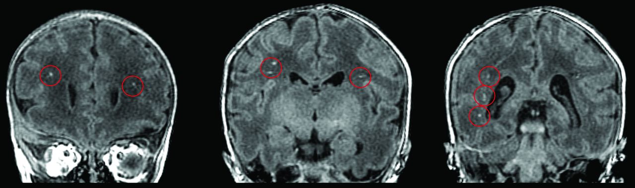
- Fig 1.
Infant with hypoplastic left heart syndrome born at 31 weeks postconceptional age and imaged at 35.5 weeks postconceptional age. Three coronal cuts from the 3D T1-weighted spoiled gradient recalled echo sequence demonstrating punctate T1-hyperintense lesions (circled in red) in the periventricular white matter and corona radiata just rostral to the genu of the corpus callosum (left), at the level of the pre- and post-central gyri (middle), and posterior to the trigone of the lateral ventricle (right), consistent with bilateral pWMLs or periventricular leukomalacia.
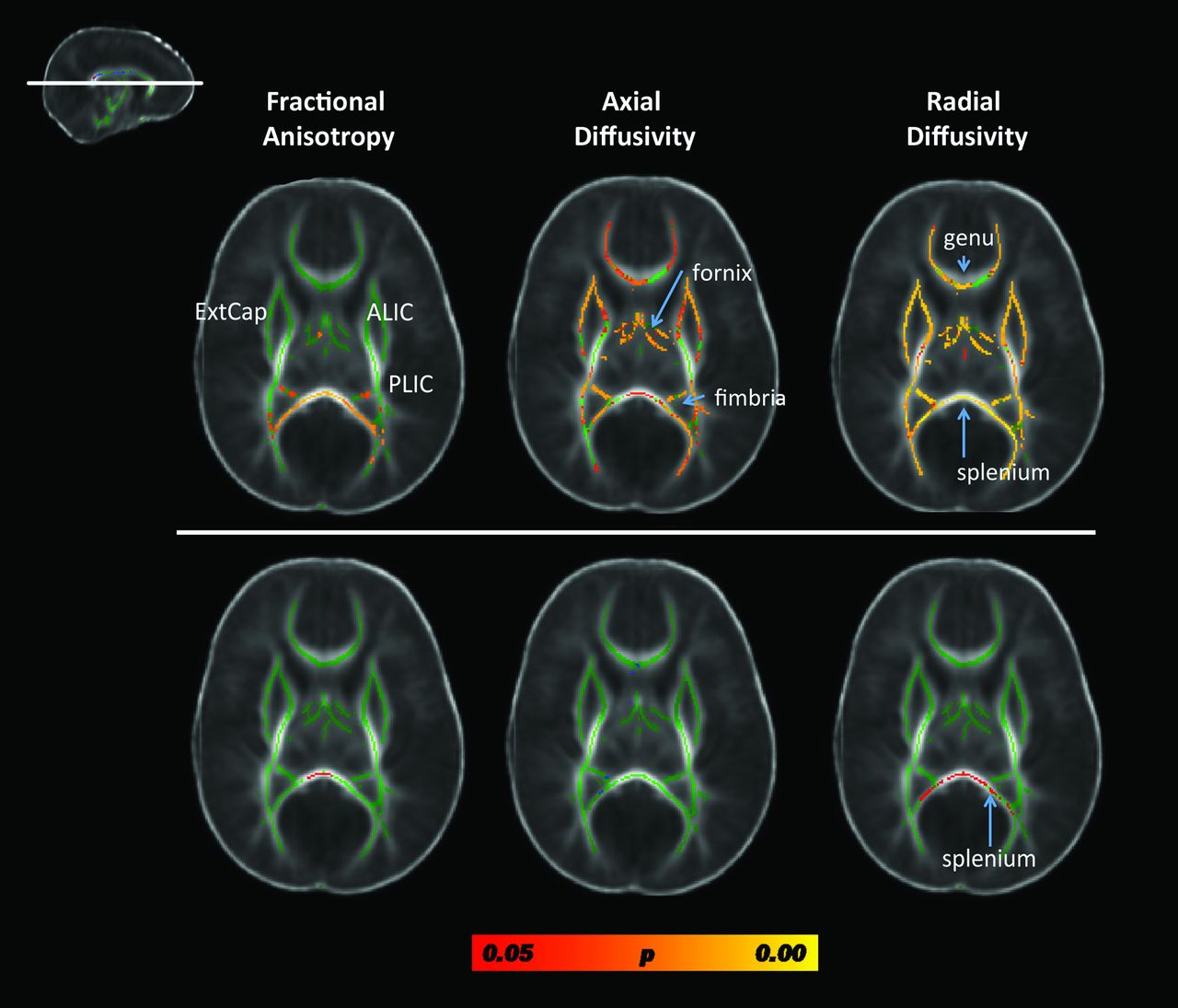
- Fig 2.
Results from the whole-brain, voxelwise TBSS analysis contrasting fractional anisotropy, axial diffusivity, and radial diffusivity metrics between preterm patients with CHD and term neonates without CHD, controlling for postconceptional age. Results from a single cut at the level of the genu and splenium of the corpus callosum are displayed (see inlay in the upper left). The top row includes data from comparing all preterm CHD cases to the term neonates without CHD. Diffuse microstructural abnormalities are seen in nearly all white matter regions. The bottom row includes data from comparing only the preterm CHD cases without pWMLs with the term neonates without CHD. The only structure showing micotructural abnormality is the splenium. Voxels showing a significant reduction in FA and axial diffusivity and a significant increase in radial diffusivity are shown in red-yellow, with the color bar denoting statistical significance, corrected for multiple comparisons. Note that most of the group differences are due to the cases with pWMLs or periventricular leukomalacia. ExtCap indicates external capsule; ALIC = anterior limb of the internal capsule; PLIC = posterior limb of the internal capsule.
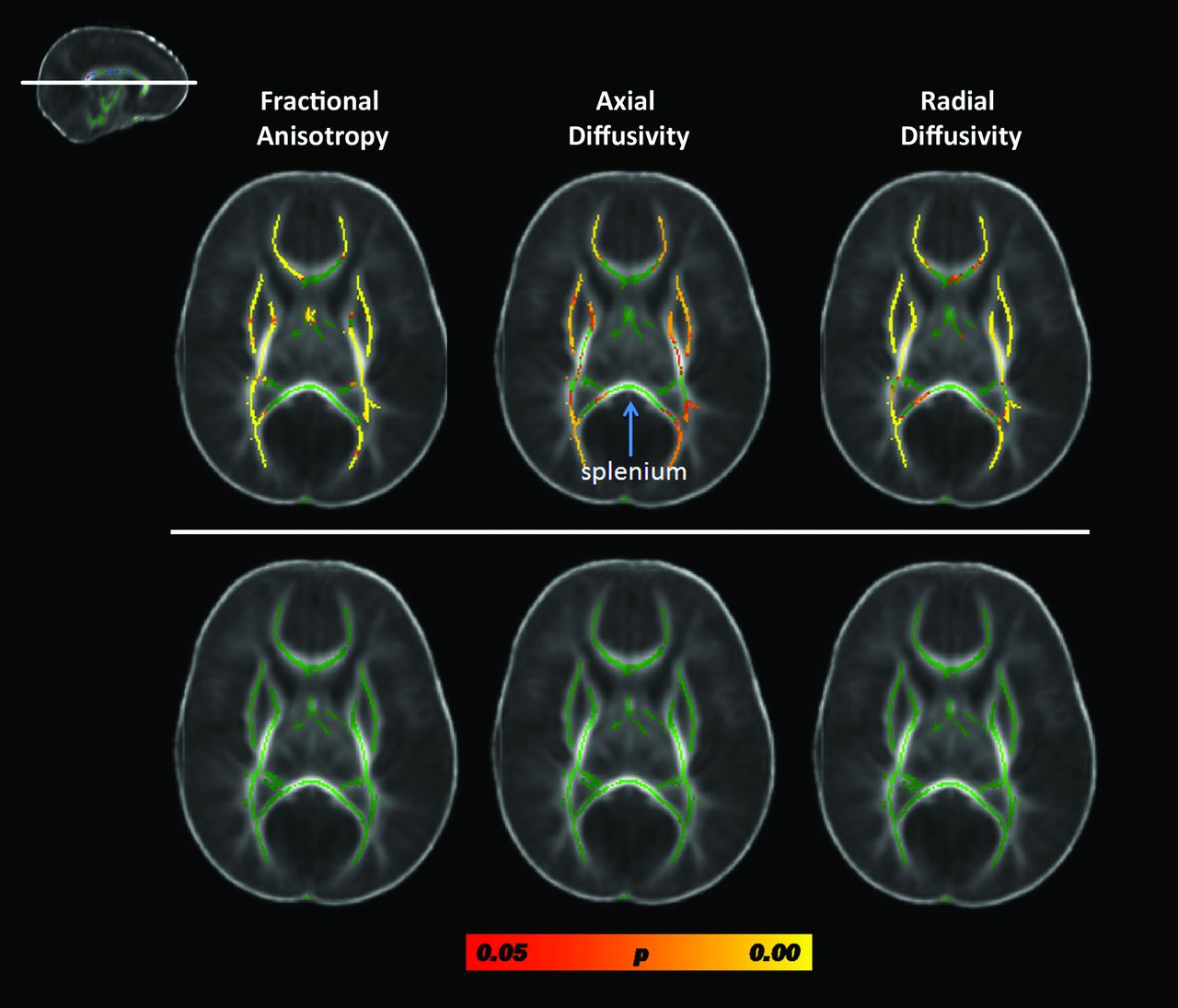
- Fig 3.
Results from the TBSS analysis contrasting FA, axial diffusivity (AD), and radial diffusivity (RD) in preterm patients with CHD with complex CHD lesions compared with preterm patients with CHD other heart lesions. Results from a single cut at the level of the genu and splenium of the corpus callosum are displayed (see inlay). The top row depicts the results of the contrasts between the preterm neonates with complex heart lesions relative to the preterm neonates with other heart lesions; the bottom row depicts the results from the contrasts of the same cases after correction for number of pWMLs. Both analyses are corrected for postconceptional age. Voxels showing a significant reduction in FA and AD and a significant increase in RD are shown in red-yellow, with the color bar denoting statistical significance, corrected for multiple comparisons. Of note, there is no difference in DTI metrics values in the splenium of the corpus callosum (arrow) between heart types.


Tables
Heart Defects n Hypoplastic left heart syndrome 5 Ebstein anomaly 2 Coarctation of the aorta 3 Transposition of the great arteries 2 Atrial septal defect, ventricular septal defect, patent ductus arteriosus requiring surgery 8 Double-outlet right ventricle 1 - Table 2:
Comparison of clinical variables between preterm neonates with CHD with pWMLs and preterm neonates without CHD
CHD Preterm with pWMLs (n = 8) Control Preterm (n = 27) P Valuea PCA, weeks, mean (SD) 38.63 (3.36) 42.61 (6.51) .13a GA, weeks, mean (SD) 33.29 (1.89) 30.67 (4.62) .156a PNA MRI, weeks, mean (SD) 5.35 (4.38) 11.095 (8.24) .087a PCA MRI, weeks, mean (SD) 38.63 (3.36) 42.61 (6.51) .13a Apgar 1 minute, median (n) 6 (6) 6 (21) .860c Apgar 5 minutes, median (n) 8.5 (6) 8 (21) .878c Apgar 10 minutes, median (n) 6 (2) 7 (3) .747c Size for GA 33 8 .216b Small, % 67 84 Appropriate, % 0 8 Large, % 6 25 ISAM, % (n) 0 (5) 8.3 (24) .763d Postnatal sepsis, % (n) 42.9 (7) 60.0 (25) .706d Hydrocortisone for BP, % (n) 50 (6) 18.5 (27) .271d Days on hydrocortisone, mean (SD) 0.5 (0.55) 1.741 (6.06) .62a Inotropes, % (n) 100 (6) 46.2 (26) .02a Days on dopamine, mean (SD) 4.33 (4.23) 3.81 (5.98) .84a - Table 3:
Preterm CHD group: comparison of perioperative variables between pWML versus non-pWML groups
Surgical Variables CHD Preterm Group with pWML CHD Preterm without pWML P Value* Mean (SD) n Mean (SD) n Birth weight 1963.5 (591.6) 8 1676.2 (566.6) 11 .341 Age at 1st surgery, days 3.5 (2.8) 8 12.0 (12.7) 12 .05 PCA at 1st surgery, weeks 37 (1.7) 8 43.1 (10.9) 12 .20 1st ABG pH 7.24 (0.08) 5 7.29 (1.10) 5 .36 1st ABG pO2 81.75 (74.82) 4 39 (−) 1 .64 Pre-op ABG pH 7.33 (0.1) 8 7.36 (0.05) 12 .41 Pre-op ABG pO2 62.2 (21.2) 8 69.75 (34.6) 8 .67 Post-op ABG pH 7.30 (0.07) 8 7.32 (0.05) 12 .38 Post-op ABG pO2 67.83 (48.3) 8 101 (83.52) 12 .37 Pre-op epi, no. of days 0 (0) 8 0.08 (0.3) 12 .49 Pre-op dobutamine, no. of days 0 (0) 8 0.08 (0.3) 12 .49 Pre-op dopamine, no. of days 1.167 (0.41) 8 0.417 (1.2) 12 .62 Pre-op hydrocortisone, no. of days 0.167 (0.41) 8 0.08 (0.3) 12 .61 Pre-op milirinone, no. of days 0 (0) 8 0.08 (0.3) 12 .49 Post-op epi, no. of days 1.67 (2.7) 8 1.5 (3) 12 .90 Post-op dobutamine, no. of days 0 (0) 8 0.25 (0.5) 12 .20 Post-op dopamine, no. of days 4.17 (4.4) 8 3.17 (4.3) 12 .64 Post-op hydrocortisone, no. of days 0.33 (0.5) 8 1.67 (5.5) 12 .50 Post-op milirinone, no. of days 3.33 (4.13) 8 3.5 (4.0) 12 .93 Post-op nitroprusside, no. of days 0.0 (0) 8 0.33 (0.9) 12 .37 Note:—PCA indicates postconceptional age; ABG, arterial blood gas; epi, epinephrine; Pre-op, preoperative; Post-op, postoperative.


Jump to section
Related Articles
Cited By...
- Development and Validation of a Paralimbic Related Subcortical Brain Dysmaturation MRI Score in Infants with Congenital Heart Disease
- Postnatal Brain Magnetic Resonance Imaging Trajectories and Maternal Intelligence Predict Neurodevelopmental Outcomes in Complex Congenital Heart Disease
- Single Ventricle Reconstruction III: Brain Connectome and Neurodevelopmental Outcomes: Design, Recruitment, and Technical Challenges of a Multicenter, Observational Neuroimaging Study
- Cerebral Autoregulation in Neonates With and Without Congenital Heart Disease
- Neurodevelopmental Abnormalities and Congenital Heart Disease: Insights Into Altered Brain Maturation