Advertisement

Main menu

  • Home
  • Content
    • Current Issue
    • Accepted Manuscripts
    • Article Preview
    • Past Issue Archive
    • Video Articles
    • AJNR Case Collection
    • Case of the Week Archive
    • Case of the Month Archive
    • Classic Case Archive
  • Special Collections
    • AJNR Awards
    • Low-Field MRI
    • Alzheimer Disease
    • ASNR Foundation Special Collection
    • Photon-Counting CT
    • View All
  • Multimedia
    • AJNR Podcasts
    • AJNR SCANtastic
    • Trainee Corner
    • MRI Safety Corner
    • Imaging Protocols
  • For Authors
    • Submit a Manuscript
    • Submit a Video Article
    • Submit an eLetter to the Editor/Response
    • Manuscript Submission Guidelines
    • Statistical Tips
    • Fast Publishing of Accepted Manuscripts
    • Graphical Abstract Preparation
    • Imaging Protocol Submission
    • Author Policies
  • About Us
    • About AJNR
    • Editorial Board
    • Editorial Board Alumni
  • More
    • Become a Reviewer/Academy of Reviewers
    • Subscribers
    • Permissions
    • Alerts
    • Feedback
    • Advertisers
    • ASNR Home

User menu

  • Alerts
  • Log in

Search

  • Advanced search
American Journal of Neuroradiology

American Journal of Neuroradiology

ASHNR American Society of Functional Neuroradiology ASHNR American Society of Pediatric Neuroradiology ASSR
  • Alerts
  • Log in

Advanced Search

  • Home
  • Content
    • Current Issue
    • Accepted Manuscripts
    • Article Preview
    • Past Issue Archive
    • Video Articles
    • AJNR Case Collection
    • Case of the Week Archive
    • Case of the Month Archive
    • Classic Case Archive
  • Special Collections
    • AJNR Awards
    • Low-Field MRI
    • Alzheimer Disease
    • ASNR Foundation Special Collection
    • Photon-Counting CT
    • View All
  • Multimedia
    • AJNR Podcasts
    • AJNR SCANtastic
    • Trainee Corner
    • MRI Safety Corner
    • Imaging Protocols
  • For Authors
    • Submit a Manuscript
    • Submit a Video Article
    • Submit an eLetter to the Editor/Response
    • Manuscript Submission Guidelines
    • Statistical Tips
    • Fast Publishing of Accepted Manuscripts
    • Graphical Abstract Preparation
    • Imaging Protocol Submission
    • Author Policies
  • About Us
    • About AJNR
    • Editorial Board
    • Editorial Board Alumni
  • More
    • Become a Reviewer/Academy of Reviewers
    • Subscribers
    • Permissions
    • Alerts
    • Feedback
    • Advertisers
    • ASNR Home
  • Follow AJNR on Twitter
  • Visit AJNR on Facebook
  • Follow AJNR on Instagram
  • Join AJNR on LinkedIn
  • RSS Feeds

AJNR Awards, New Junior Editors, and more. Read the latest AJNR updates

Research ArticleBrain

Parenchymal Hypointense Foci Associated with Developmental Venous Anomalies: Evaluation by Phase-Sensitive MR Imaging at 3T

M. Takasugi, S. Fujii, Y. Shinohara, T. Kaminou, T. Watanabe and T. Ogawa
American Journal of Neuroradiology October 2013, 34 (10) 1940-1944; DOI: https://doi.org/10.3174/ajnr.A3495
M. Takasugi
aFrom the Division of Radiology (M.T., S.F., Y.S., T.K., T.O.), Department of Pathophysiological and Therapeutic Science
  • Find this author on Google Scholar
  • Find this author on PubMed
  • Search for this author on this site
S. Fujii
aFrom the Division of Radiology (M.T., S.F., Y.S., T.K., T.O.), Department of Pathophysiological and Therapeutic Science
  • Find this author on Google Scholar
  • Find this author on PubMed
  • Search for this author on this site
Y. Shinohara
aFrom the Division of Radiology (M.T., S.F., Y.S., T.K., T.O.), Department of Pathophysiological and Therapeutic Science
  • Find this author on Google Scholar
  • Find this author on PubMed
  • Search for this author on this site
T. Kaminou
aFrom the Division of Radiology (M.T., S.F., Y.S., T.K., T.O.), Department of Pathophysiological and Therapeutic Science
  • Find this author on Google Scholar
  • Find this author on PubMed
  • Search for this author on this site
T. Watanabe
bDivision of Neurosurgery (T.W.), Department of Brain and Neurosciences, Faculty of Medicine, Tottori University, Yonago, Tottori, Japan.
  • Find this author on Google Scholar
  • Find this author on PubMed
  • Search for this author on this site
T. Ogawa
aFrom the Division of Radiology (M.T., S.F., Y.S., T.K., T.O.), Department of Pathophysiological and Therapeutic Science
  • Find this author on Google Scholar
  • Find this author on PubMed
  • Search for this author on this site
  • Article
  • Figures & Data
  • Info & Metrics
  • Responses
  • References
  • PDF
Loading

Article Figures & Data

Figures

  • Tables
  • Fig 1.
    • Download figure
    • Open in new tab
    • Download powerpoint
    Fig 1.

    A 64-year-old man with DVA in the right frontal lobe. A, PSI shows DVA in the right frontal lobe (arrow) and hypointense foci around the DVA (arrowhead). B, T2-weighted image shows WMH around the DVA (arrow). C, DVA is enhanced by contrast agent administration, whereas hypointense foci are not enhanced on T1-weighted image.
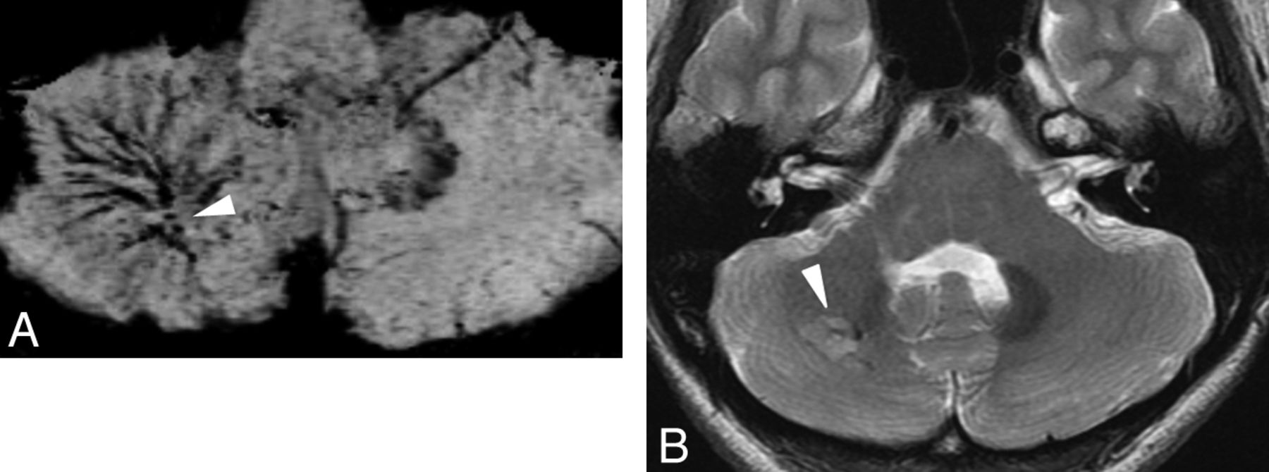
  • Fig 2.
    • Download figure
    • Open in new tab
    • Download powerpoint
    Fig 2.

    A 25-year-old woman with DVA in the right cerebellum. A, PSI shows DVA in the right cerebellum and hypointense foci around the medullary veins (arrowhead). B, T2-weighted image shows WMH around the DVA (arrowhead).

  • Fig 3.
    • Download figure
    • Open in new tab
    • Download powerpoint
    Fig 3.

    A 75-year-old man with DVA in the right parieto-temporal lobe. A, PSI shows DVA in the right parieto-temporal lobe (arrow) and minute hypointense foci around the DVA (arrowhead). B, T2-weighted image shows WMH around the DVA (arrow).

Tables

  • Figures
    • View popup
    Table 1:

    Location of cavernous malformation

    n
    Peripheral18
    Central11
    Both6
    • Note:—Total n = 35. Three patients were excluded because the hypointense foci were too large to assess.

    • View popup
    Table 2:

    Associations between hypointense foci and other factors

    Hypointense Foci (+) (n = 38)Hypointense Foci (−) (n = 23)
    n%n%
    WMHYes2668.4730.4
    No1231.61669.6
    LocationBasal ganglia/thalamus615.800
    Lobe1950.01565.2
    Cerebellum1026.3521.7
    Pons37.9313.0
    DepthPeriventricular1642.11356.5
    Juxtacortical821.1521.7
    Subcortical1436.8521.7
    Draining veinLong1539.51669.6
    Medium1436.8521.7
    Short923.728.7
    DirectionDeep1128.9417.4
    Superficial2771.11982.6
    • Note:—Total n = 61.

    • View popup
    Table 3:

    Patient symptoms and indications for examinations in the 2 groups based on hypointense foci

    Hypointense Foci (+) (n = 38)Hypointense Foci (−) (n = 21)n
    n%n%
    Symptoms or clinical indications for examinationsNo/follow-up of primary disease1152.41047.621
    Seizures583.3116.76
    Transient ischemic attack/stroke583.3116.76
    Dizziness, vertigo1253754
    Headache4100004
    Sensory disturbance266.7133.33
    Double vision2100002
    Dysarthria2100002
    Nerve palsy1501502
    SDH1501502
    Consciousness disturbance1100001
    ICH1100001
    Visual field disturbance1100001
    SAH0011001
    Syncope0011001
    Trauma0011001
    Unknown101
    • Note:—SDH indicates subdural hematoma.

PreviousNext
Back to top

In this issue

American Journal of Neuroradiology: 34 (10)
American Journal of Neuroradiology
Vol. 34, Issue 10
1 Oct 2013
  • Table of Contents
  • Index by author
  • Complete Issue (PDF)
Advertisement
Print
Download PDF
Email Article

Thank you for your interest in spreading the word on American Journal of Neuroradiology.

NOTE: We only request your email address so that the person you are recommending the page to knows that you wanted them to see it, and that it is not junk mail. We do not capture any email address.

Enter multiple addresses on separate lines or separate them with commas.
Parenchymal Hypointense Foci Associated with Developmental Venous Anomalies: Evaluation by Phase-Sensitive MR Imaging at 3T
(Your Name) has sent you a message from American Journal of Neuroradiology
(Your Name) thought you would like to see the American Journal of Neuroradiology web site.
CAPTCHA
This question is for testing whether or not you are a human visitor and to prevent automated spam submissions.
Cite this article
M. Takasugi, S. Fujii, Y. Shinohara, T. Kaminou, T. Watanabe, T. Ogawa
Parenchymal Hypointense Foci Associated with Developmental Venous Anomalies: Evaluation by Phase-Sensitive MR Imaging at 3T
American Journal of Neuroradiology Oct 2013, 34 (10) 1940-1944; DOI: 10.3174/ajnr.A3495

Citation Manager Formats

  • BibTeX
  • Bookends
  • EasyBib
  • EndNote (tagged)
  • EndNote 8 (xml)
  • Medlars
  • Mendeley
  • Papers
  • RefWorks Tagged
  • Ref Manager
  • RIS
  • Zotero
0 Responses
Respond to this article
Share
Bookmark this article
Parenchymal Hypointense Foci Associated with Developmental Venous Anomalies: Evaluation by Phase-Sensitive MR Imaging at 3T
M. Takasugi, S. Fujii, Y. Shinohara, T. Kaminou, T. Watanabe, T. Ogawa
American Journal of Neuroradiology Oct 2013, 34 (10) 1940-1944; DOI: 10.3174/ajnr.A3495
del.icio.us logo Twitter logo Facebook logo Mendeley logo
  • Tweet Widget
  • Facebook Like
  • Google Plus One
Purchase

Jump to section

  • Article
    • Abstract
    • ABBREVIATIONS:
    • Materials and Methods
    • Results
    • Discussion
    • Conclusions
    • ACKNOWLEDGMENTS
    • REFERENCES
  • Figures & Data
  • Info & Metrics
  • Responses
  • References
  • PDF

Related Articles

  • No related articles found.
  • PubMed
  • Google Scholar

Cited By...

  • Symptomatic Developmental Venous Anomaly: State-of-the-Art Review on Genetics, Pathophysiology, and Imaging Approach to Diagnosis
  • Neonatal Developmental Venous Anomalies: Clinicoradiologic Characterization and Follow-Up
  • Increased Prevalence of Developmental Venous Anomalies in Children with Intracranial Neoplasms
  • Brain Parenchymal Signal Abnormalities Associated with Developmental Venous Anomalies in Children and Young Adults
  • Diffusion and Perfusion MRI Findings of the Signal-Intensity Abnormalities of Brain Associated with Developmental Venous Anomaly
  • Crossref
  • Google Scholar

This article has not yet been cited by articles in journals that are participating in Crossref Cited-by Linking.

More in this TOC Section

  • Multimodal CT Provides Improved Performance for Lacunar Infarct Detection
  • Optimal MRI Sequence for Identifying Occlusion Location in Acute Stroke: Which Value of Time-Resolved Contrast-Enhanced MRA?
  • Evaluating the Effects of White Matter Multiple Sclerosis Lesions on the Volume Estimation of 6 Brain Tissue Segmentation Methods
Show more Brain

Similar Articles

Advertisement

Indexed Content

  • Current Issue
  • Accepted Manuscripts
  • Article Preview
  • Past Issues
  • Editorials
  • Editor's Choice
  • Fellows' Journal Club
  • Letters to the Editor
  • Video Articles

Cases

  • Case Collection
  • Archive - Case of the Week
  • Archive - Case of the Month
  • Archive - Classic Case

More from AJNR

  • Trainee Corner
  • Imaging Protocols
  • MRI Safety Corner
  • Book Reviews

Multimedia

  • AJNR Podcasts
  • AJNR Scantastics

Resources

  • Turnaround Time
  • Submit a Manuscript
  • Submit a Video Article
  • Submit an eLetter to the Editor/Response
  • Manuscript Submission Guidelines
  • Statistical Tips
  • Fast Publishing of Accepted Manuscripts
  • Graphical Abstract Preparation
  • Imaging Protocol Submission
  • Evidence-Based Medicine Level Guide
  • Publishing Checklists
  • Author Policies
  • Become a Reviewer/Academy of Reviewers
  • News and Updates

About Us

  • About AJNR
  • Editorial Board
  • Editorial Board Alumni
  • Alerts
  • Permissions
  • Not an AJNR Subscriber? Join Now
  • Advertise with Us
  • Librarian Resources
  • Feedback
  • Terms and Conditions
  • AJNR Editorial Board Alumni

American Society of Neuroradiology

  • Not an ASNR Member? Join Now

© 2025 by the American Society of Neuroradiology All rights, including for text and data mining, AI training, and similar technologies, are reserved.
Print ISSN: 0195-6108 Online ISSN: 1936-959X

Powered by HighWire