
Article Figures & Data
Figures
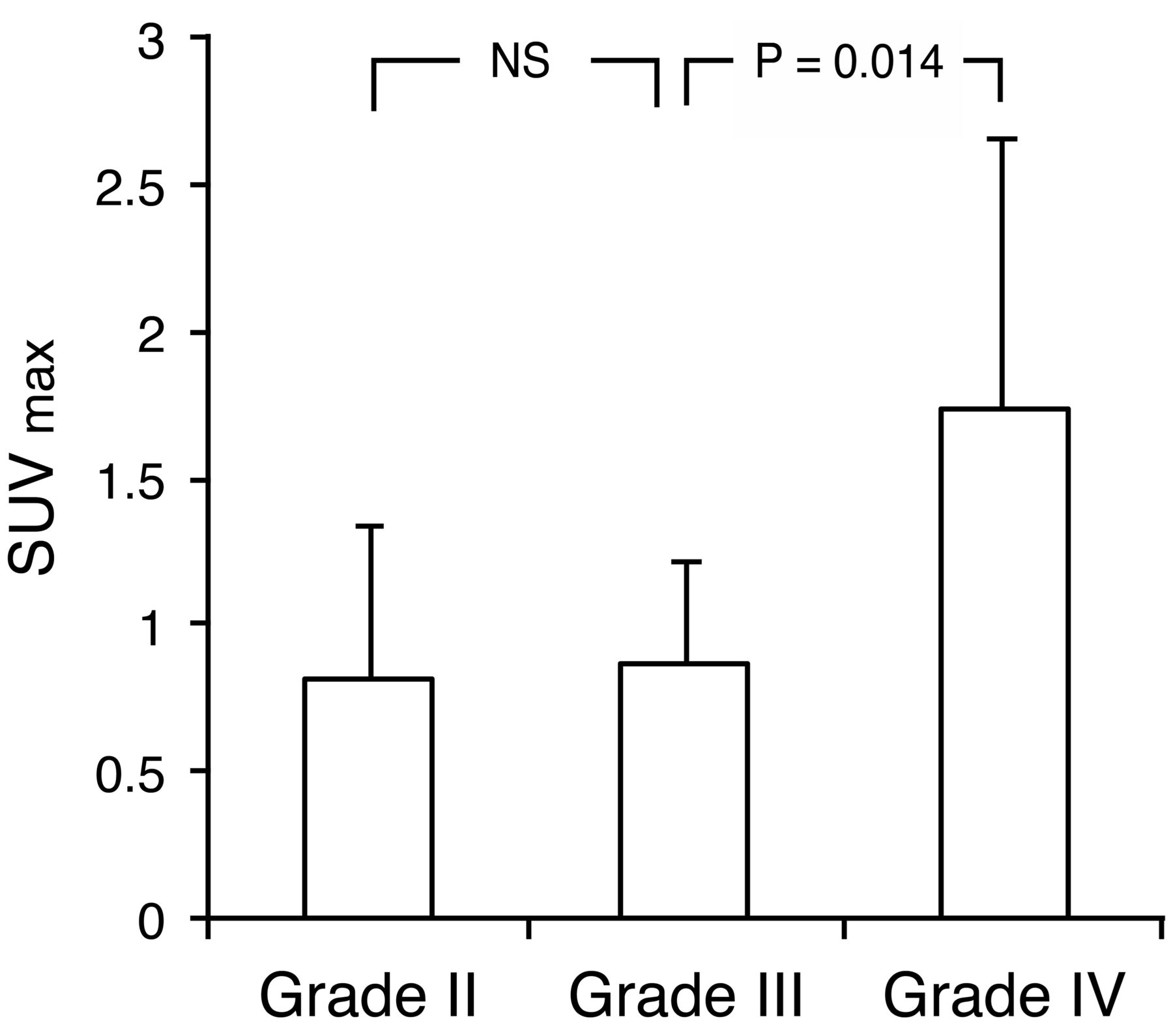
- Fig 1.
62Cu-ATSM SUVmax in gliomas of different grades (2007 WHO grading) showing a significant difference in uptake between grades IV and III tumors but not between grades III and II tumors (P = .88, Steel-Dwass test). NS = not significant.
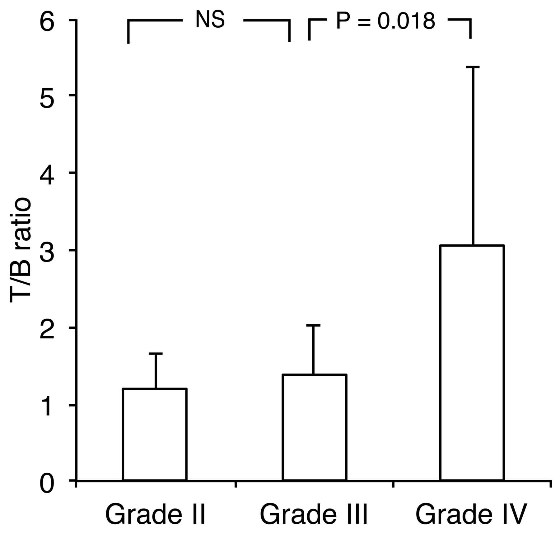
- Fig 2.
62Cu-ATSM T/B ratio in gliomas of different grades (2007 WHO grading) showing a significant difference between grades IV and III tumors but not between grades III and II tumors (P = .92, Steel-Dwass test).
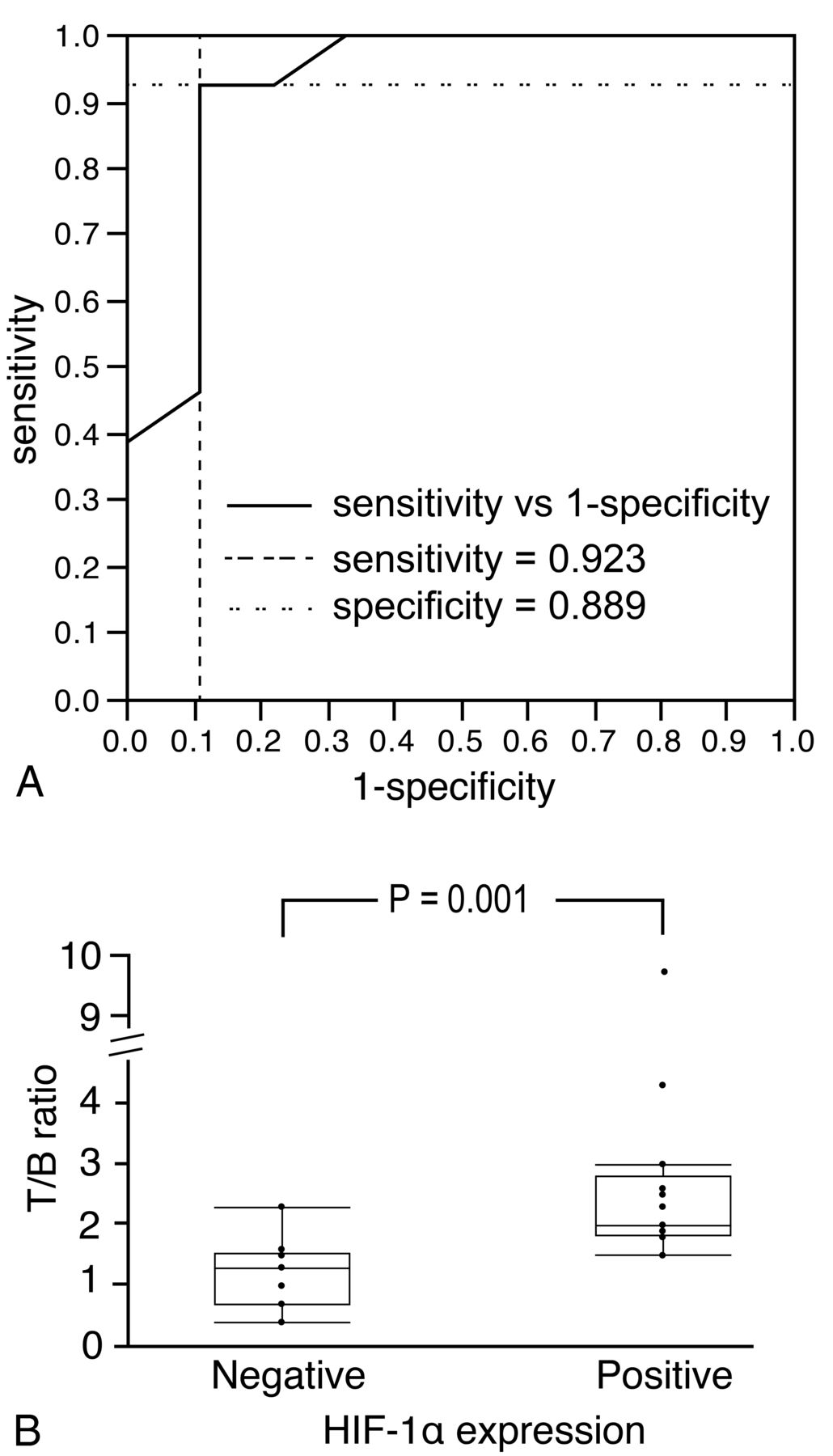
- Fig 3.
A, ROC analysis indicating that 62Cu-ATSM uptake is predictive of HIF-1α positivity, with a sensitivity of 92.3% and a specificity of 88.9% for a T/B ratio cutoff threshold of 1.8 (area under the curve = 0.92). B, The 62Cu-ATSM T/B ratio is significantly higher in HIF-1α-positive than in HIF-1α-negative gliomas (P = .001, Wilcoxon signed rank test). Circles above bars represent outliers (1.5×, the interquartile range).
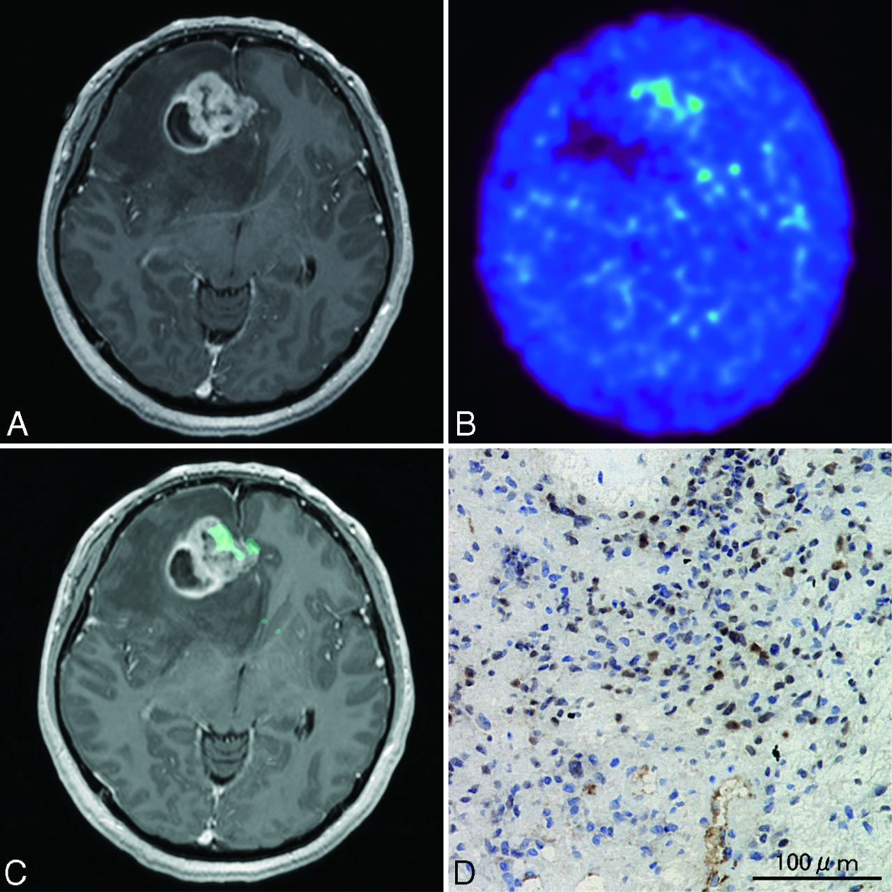
- Fig 4.
Case 2. A 61-year-old woman with glioblastoma. A, Axial T1-weighted gadolinium-enhanced MR imaging demonstrating an enhanced lesion with necrosis in the right temporal lobe. B, 62Cu-ATSM PET showing high uptake in the lesion. C, PET/MR imaging fusion image showing 62Cu-ATSM uptake (T/B cutoff threshold of ≥1.8) within contrast-enhanced lesion. D, Photomicrographs of tissue with the highest 62Cu-ATSM uptake stained with anti-HIF-1α showing intense HIF-1α immunoreactivity. Original magnification × 200.
- Fig 5.
Case 8. A 28-year-old woman with glioblastoma. A, Axial T1-weighted gadolinium-enhanced MR imaging demonstrating an enhanced lesion with necrosis in the right frontal lobe. B, 62Cu-ATSM PET showing mild uptake in the tumor. C, PET/MR imaging fusion image demonstrating 62Cu-ATSM accumulation (T/B cutoff threshold of ≥1.8) within contrast-enhanced lesion. D, Photomicrograph of a tissue sample with 62Cu-ATSM accumulation showing high HIF-1α expression. Original magnification × 200.
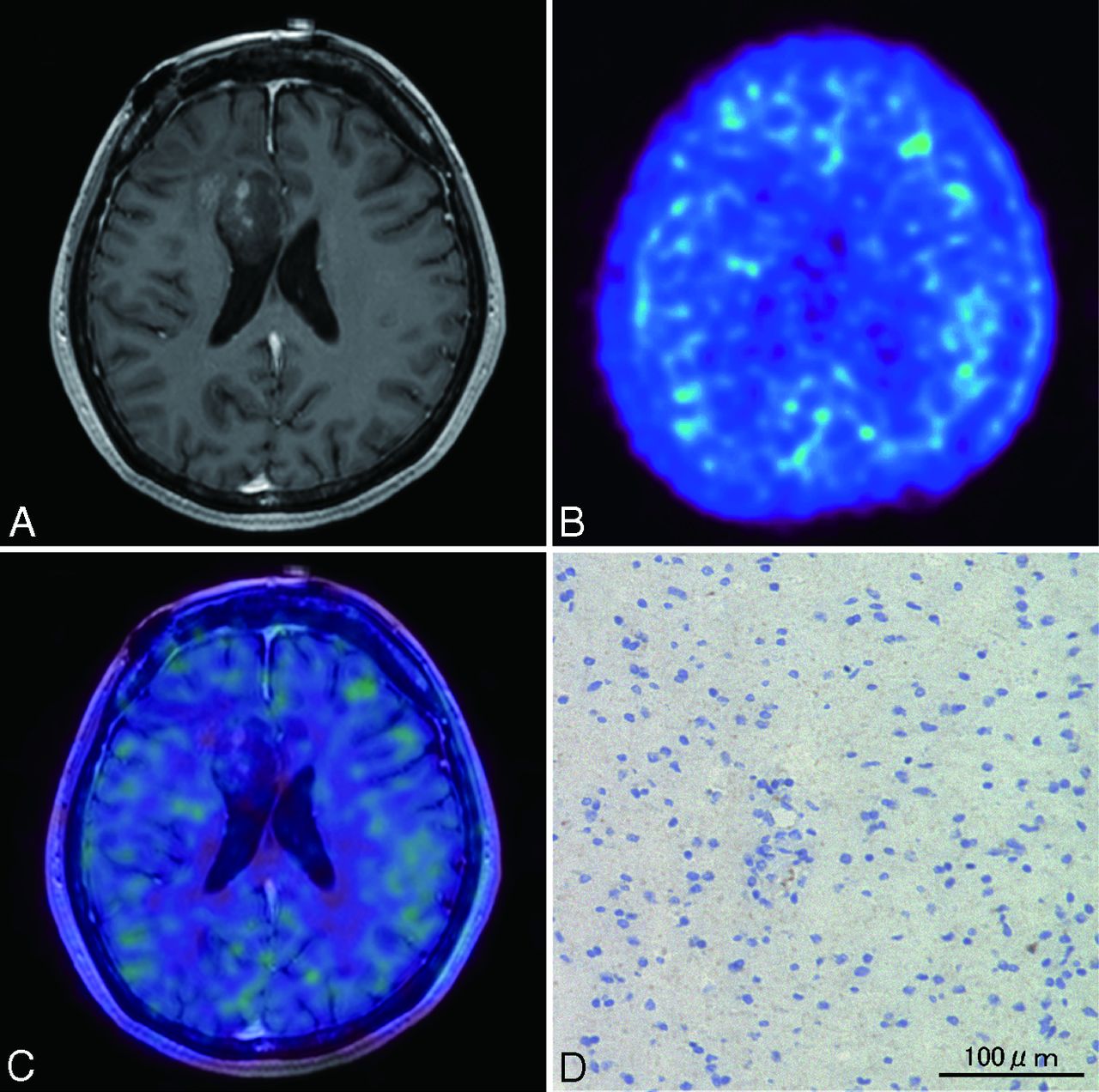
- Fig 6.
Case 13. A 42-year-old woman with oligoastrocytoma. A, Axial T1-weighted gadolinium-enhanced MR imaging demonstrating a mildly enhanced lesion in the right frontal lobe. 62Cu-ATSM PET image (B) and PET/MR imaging fusion image (C), showing absent 62Cu-ATSM uptake in the tumor. D, Photomicrograph of a lesion tissue sample showing no HIF-1α immunoreactivity. Original magnification × 200.


Tables
Case No. Age (years), Sex Histologic Diagnosisa Tumor Diagnosis Previous RT Location 1 30, F AOA, grade III New Rt frontal 2 61, F GB, grade IV New Rt temporal 3 53, M DA, grade II Rec No Lt parietal 4 50, M AA, grade III Rec Yes Lt thalamus 5 66, F AOD, grade III New Lt thalamus 6 69, F GB, grade IV New Lt parietal 7 66, F AOD, grade III New Lt frontal 8 28, F GB, grade IV New Rt frontal 9 23, F OA, grade II New Rt frontal 10 60, M AOD, grade III Rec No Rt parietal 11 63, F GB, grade IV Rec Yes Rt temporal 12 75, F GB, grade IV New Rt occipital 13 42, F OA, grade II Rec No Rt frontal 14 79, F GB, grade IV Rec Yes Rt frontal 15 59, M GB, grade IV New Rt frontal 16 69, F DA, grade II New Lt frontal 17 41, M AOA, grade III Rec Yes Lt parietal 18 75, M GB, grade IV New Lt temporal 19 37, F AOA, grade III New Lt frontal 20 71, F GB, grade IV Rec Yes Lt parietal 21 59, F GB, grade IV New Lt temporal 22 65, F GB, grade IV Rec Yes Lt frontal Note:—AA indicates anaplastic astrocytoma; AOA, anaplastic oligoastrocytoma; AOD, anaplastic oligodendroglioma; DA, diffuse astrocytoma; F, female; GB, glioblastoma; Lt, left; M, male; New, new lesion; OA, oligoastrocytoma; Rec, recurrent; Rt, right; RT, radiation therapy.
↵a Histologic diagnosis and grading according to the 2007 WHO classification.
- Table 2:
Summary of tumor grades, 62Cu-ATSM uptake values, MR findings, and HIF-1α expression
Case No. WHO Grade 62Cu-ATSM Enhancement on MRI HIF-1α Expressiona SUVmax SUVmean ± SD T/B Ratio 1 III 0.51 0.38 ± 0.07 0.7 E Negative 2 IV 4.33 1.79 ± 0.58 9.7 E+N Positive 3 II 0.59 0.43 ± 0.10 0.7 None Negative 4 III 0.92 0.59 ± 0.14 1.8 E Positive 5 III 0.73 0.51 ± 0.09 2.3 E+N Negative 6 IV 1.84 1.30 ± 0.21 2.5 E+N Positive 7 III 1.02 0.73 ± 0.24 1.6 E Negative 8 IV 1.15 0.76 ± 0.14 2.0 E+N Positive 9 II 0.44 0.32 ± 0.07 1.0 E Negative 10 III 1.01 0.58 ± 0.16 1.5 E Negative 11 IV 2.04 1.29 ± 0.17 1.9 E+N Positive 12 IV 1.17 0.84 ± 0.10 3.0 E+N Positive 13 II 0.67 0.33 ± 0.09 1.3 E Negative 14 IV 1.32 0.96 ± 0.09 4.3 E+N Positive 15 IV 1.35 0.84 ± 0.19 1.9 E+N Positive 16 II 1.59 0.80 ± 0.14 1.8 E Positive 17 III 0.46 0.30 ± 0.05 0.4 E Negative 18 IV 1.62 0.69 ± 0.17 1.5 E+N Positive 19 III 1.46 0.80 ± 0.13 1.3 E Negative 20 IV 1.54 0.77 ± 0.09 1.9 E+N Positive 21 IV 1.82 0.91 ± 0.15 2.6 E+N Positive 22 IV 0.94 0.68 ± 0.17 2.3 E+N Positive Note:—E indicates contrast enhancement without necrosis; E+N, contrast enhancement with necrosis; None, no enhancement; SUVmax, maximum standard uptake value; SUVmean, mean standard uptake value.
↵a Negative indicates <5% HIF-1α-positive cells; positive indicates ≥5% HIF-1α-positive cells.
Necrosis on MRI No. Tumors with 62Cu-ATSM Uptake Uptake Non-Uptake Yes 11 1 No 2 7 Note:—62Cu-ATSM uptake was defined as T/B cutoff threshold of ≥1.8. No indicates contrast-enhanced lesion without necrotic component; Yes indicates contrast-enhanced lesion with necrotic component;
In this issue


Jump to section
Related Articles
Cited By...
- 62Cu-Diacetyl-Bis (N4-Methylthiosemicarbazone) PET in Human Gliomas: Comparative Study with [18F]Fluorodeoxyglucose and L-Methyl-[11C]Methionine PET
- Interrogating Tumor Metabolism and Tumor Microenvironments Using Molecular Positron Emission Tomography Imaging. Theranostic Approaches to Improve Therapeutics