Advertisement

Main menu

  • Home
  • Content
    • Current Issue
    • Accepted Manuscripts
    • Article Preview
    • Past Issue Archive
    • Video Articles
    • AJNR Case Collection
    • Case of the Week Archive
    • Case of the Month Archive
    • Classic Case Archive
  • Special Collections
    • AJNR Awards
    • Low-Field MRI
    • Alzheimer Disease
    • ASNR Foundation Special Collection
    • Photon-Counting CT
    • View All
  • Multimedia
    • AJNR Podcasts
    • AJNR SCANtastic
    • Trainee Corner
    • MRI Safety Corner
    • Imaging Protocols
  • For Authors
    • Submit a Manuscript
    • Submit a Video Article
    • Submit an eLetter to the Editor/Response
    • Manuscript Submission Guidelines
    • Statistical Tips
    • Fast Publishing of Accepted Manuscripts
    • Graphical Abstract Preparation
    • Imaging Protocol Submission
    • Author Policies
  • About Us
    • About AJNR
    • Editorial Board
    • Editorial Board Alumni
  • More
    • Become a Reviewer/Academy of Reviewers
    • Subscribers
    • Permissions
    • Alerts
    • Feedback
    • Advertisers
    • ASNR Home

User menu

  • Alerts
  • Log in

Search

  • Advanced search
American Journal of Neuroradiology

American Journal of Neuroradiology

ASHNR American Society of Functional Neuroradiology ASHNR American Society of Pediatric Neuroradiology ASSR
  • Alerts
  • Log in

Advanced Search

  • Home
  • Content
    • Current Issue
    • Accepted Manuscripts
    • Article Preview
    • Past Issue Archive
    • Video Articles
    • AJNR Case Collection
    • Case of the Week Archive
    • Case of the Month Archive
    • Classic Case Archive
  • Special Collections
    • AJNR Awards
    • Low-Field MRI
    • Alzheimer Disease
    • ASNR Foundation Special Collection
    • Photon-Counting CT
    • View All
  • Multimedia
    • AJNR Podcasts
    • AJNR SCANtastic
    • Trainee Corner
    • MRI Safety Corner
    • Imaging Protocols
  • For Authors
    • Submit a Manuscript
    • Submit a Video Article
    • Submit an eLetter to the Editor/Response
    • Manuscript Submission Guidelines
    • Statistical Tips
    • Fast Publishing of Accepted Manuscripts
    • Graphical Abstract Preparation
    • Imaging Protocol Submission
    • Author Policies
  • About Us
    • About AJNR
    • Editorial Board
    • Editorial Board Alumni
  • More
    • Become a Reviewer/Academy of Reviewers
    • Subscribers
    • Permissions
    • Alerts
    • Feedback
    • Advertisers
    • ASNR Home
  • Follow AJNR on Twitter
  • Visit AJNR on Facebook
  • Follow AJNR on Instagram
  • Join AJNR on LinkedIn
  • RSS Feeds

AJNR Awards, New Junior Editors, and more. Read the latest AJNR updates

Research ArticleSpine Imaging and Spine Image-Guided Interventions

Diagnostic Yield of Fluoroscopy-Guided Biopsy for Infectious Spondylitis

B.J. Kim, J.W. Lee, S.J. Kim, G.Y. Lee and H.S. Kang
American Journal of Neuroradiology January 2013, 34 (1) 233-238; DOI: https://doi.org/10.3174/ajnr.A3120
B.J. Kim
aFrom the Department of Radiology (B.J.K., J.W.L., G.Y.L., H.S.K.), Seoul National University Bundang Hospital, Gyeonggi-do, Korea
  • Find this author on Google Scholar
  • Find this author on PubMed
  • Search for this author on this site
J.W. Lee
aFrom the Department of Radiology (B.J.K., J.W.L., G.Y.L., H.S.K.), Seoul National University Bundang Hospital, Gyeonggi-do, Korea
  • Find this author on Google Scholar
  • Find this author on PubMed
  • Search for this author on this site
S.J. Kim
bDepartment of Radiology (S.J.K.), Boramae Medical Center, Seoul, Korea.
  • Find this author on Google Scholar
  • Find this author on PubMed
  • Search for this author on this site
G.Y. Lee
aFrom the Department of Radiology (B.J.K., J.W.L., G.Y.L., H.S.K.), Seoul National University Bundang Hospital, Gyeonggi-do, Korea
  • Find this author on Google Scholar
  • Find this author on PubMed
  • Search for this author on this site
H.S. Kang
aFrom the Department of Radiology (B.J.K., J.W.L., G.Y.L., H.S.K.), Seoul National University Bundang Hospital, Gyeonggi-do, Korea
  • Find this author on Google Scholar
  • Find this author on PubMed
  • Search for this author on this site
  • Article
  • Figures & Data
  • Info & Metrics
  • Responses
  • References
  • PDF
Loading

Article Figures & Data

Figures

  • Tables
  • Fig 1.
    • Download figure
    • Open in new tab
    • Download powerpoint
    Fig 1.

    A 35-year-old woman with neck pain and a sense of heat in both shoulders for 6 months. A, Sagittal T2-weighted MR image shows low signal change with upper endplate destruction at the C6 upper body. Percutaneous biopsy was performed at the C6 upper body with a right unilateral anterior approach under fluoroscopic guidance, which is seen on anteroposterior (B) and lateral (C) spot radiographs. Biopsy was confirmed as chronic osteomyelitis, but the causative organism was not isolated.

  • Fig 2.
    • Download figure
    • Open in new tab
    • Download powerpoint
    Fig 2.

    A 69-year-old man with fever 15 days after an operation for mechanical obstruction. Sagittal T1-weighted (A) and enhancement (B) MR images show low bone marrow signal change with enhancement at T12 and the L1 body and abscess formation at the anterior epidural space of the T12-L1 level. Percutaneous biopsy was performed at the T12 lower body with a left unilateral transpedicular approach under fluoroscopic guidance, which is demonstrated on anteroposterior (C) and lateral (D) spot radiographs. Pathologic findings were consistent with infectious spondylitis. The isolated causative organism was Klebsiella pneumoniae.
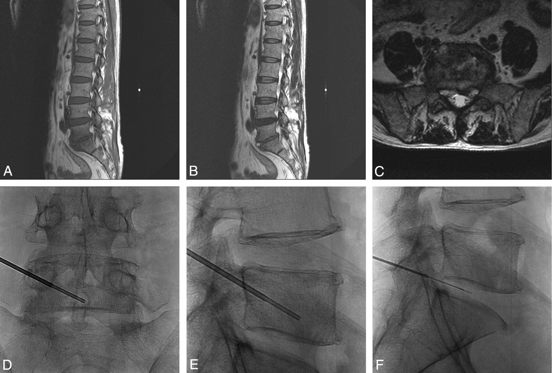
  • Fig 3.
    • Download figure
    • Open in new tab
    • Download powerpoint
    Fig 3.

    A 53-year-old man with low back pain and a chilling sensation for 1 week. Sagittal T1- (A), T2- (B), and axial T2-weighted (C) MR images show T2 high-fluid signal intensity in the L5/S1 disk space with T1 low marrow signal change in the peripheral portion of the L5 lower and S1 upper bodies and paravertebral extension. As seen on anteroposterior (D) and lateral (E and F) spot radiographs, percutaneous biopsy and disk aspiration were performed at the left inferior lower body of the L5 vertebra with a left unilateral transpedicular approach under fluoroscopic guidance. Biopsy confirmed infectious spondylitis, but the causative organism was not isolated. After empiric antibiotic treatment, clinical symptoms were improved.

Tables

  • Figures
    • View popup
    Table 1:

    Level in bone biopsy

    LevelNo.
    Cervical spine1
    Thoracic spine22
    Lumbar spine147
    Total170
    • View popup
    Table 2:

    The classification of the case by pathologic reporta

    No.
    Histology(+)115
        Culture(+)51
        Culture(−)64
    Histology(−)55
        Infection32
        Noninfection13
        Follow-up loss1
        Inadequate specimen5
        Etc4
    Total170
    • Note:—Etc. indicates the case that infectious spondylitis was suspected on MRI image, but clinical symptoms were improved without empirical antibiotic treatment.

    • ↵a For Histology(+), the pathology report was recorded as follows: “consistent with infectious spondylitis,” “active or acute inflammation,” “suggestive of osteomyelitis,” “chronic granulomatous inflammation.” Histology(−) is the rest except the histology(+). Culture(+) indicates cases of identification of the causative organism. Culture(−) is cases of no identification of the causative organism.

    • View popup
    Table 3:

    Clinical outcome in histology(+) and culture(−) and histology(−) casesa

    Histology(+), Culture(−)Culture(−)
    Infection5330
        Confirmed by operation134
        Isolation of the causative organism by blood culture125
        Improvement after empiric antibiotic treatment2519
        Aggravation on follow-up MRI31
    Noninfection1120
        Compression fracture67
        Degenerative change (Modic type I)23
        Hemorrhage by previous trauma02
        CML01
        Follow-up loss11
        Etc26
    6450
    • Note:—Etc. indicates the case that infectious spondylitis was suspected on MRI image, but clinical symptoms were improved without empirical antibiotic treatment; CML, chronic myelomonocytic leukemia.

    • ↵a For Histology(+), the pathology report was recorded as “consistent with infectious spondylitis, “active or acute inflammation,” “suggestive of osteomyelitis,” and “chronic granulomatous inflammation.” Histology(−) indicates the rest except the histology(+). Culture(+) is identification of the causative organism. Culture(−) is no identification of the causative organism.

PreviousNext
Back to top

In this issue

American Journal of Neuroradiology: 34 (1)
American Journal of Neuroradiology
Vol. 34, Issue 1
1 Jan 2013
  • Table of Contents
  • Index by author
Advertisement
Print
Download PDF
Email Article

Thank you for your interest in spreading the word on American Journal of Neuroradiology.

NOTE: We only request your email address so that the person you are recommending the page to knows that you wanted them to see it, and that it is not junk mail. We do not capture any email address.

Enter multiple addresses on separate lines or separate them with commas.
Diagnostic Yield of Fluoroscopy-Guided Biopsy for Infectious Spondylitis
(Your Name) has sent you a message from American Journal of Neuroradiology
(Your Name) thought you would like to see the American Journal of Neuroradiology web site.
CAPTCHA
This question is for testing whether or not you are a human visitor and to prevent automated spam submissions.
Cite this article
B.J. Kim, J.W. Lee, S.J. Kim, G.Y. Lee, H.S. Kang
Diagnostic Yield of Fluoroscopy-Guided Biopsy for Infectious Spondylitis
American Journal of Neuroradiology Jan 2013, 34 (1) 233-238; DOI: 10.3174/ajnr.A3120

Citation Manager Formats

  • BibTeX
  • Bookends
  • EasyBib
  • EndNote (tagged)
  • EndNote 8 (xml)
  • Medlars
  • Mendeley
  • Papers
  • RefWorks Tagged
  • Ref Manager
  • RIS
  • Zotero
0 Responses
Respond to this article
Share
Bookmark this article
Diagnostic Yield of Fluoroscopy-Guided Biopsy for Infectious Spondylitis
B.J. Kim, J.W. Lee, S.J. Kim, G.Y. Lee, H.S. Kang
American Journal of Neuroradiology Jan 2013, 34 (1) 233-238; DOI: 10.3174/ajnr.A3120
del.icio.us logo Twitter logo Facebook logo Mendeley logo
  • Tweet Widget
  • Facebook Like
  • Google Plus One
Purchase

Jump to section

  • Article
    • Abstract
    • ABBREVIATION:
    • Materials and Methods
    • Results
    • Discussion
    • References
  • Figures & Data
  • Info & Metrics
  • Responses
  • References
  • PDF

Related Articles

  • No related articles found.
  • PubMed
  • Google Scholar

Cited By...

  • Yield of Image-Guided Needle Biopsy for Infectious Discitis: A Systematic Review and Meta-Analysis
  • Imaging Psoas Sign in Lumbar Spinal Infections: Evaluation of Diagnostic Accuracy and Comparison with Established Imaging Characteristics
  • Crossref
  • Google Scholar

This article has not yet been cited by articles in journals that are participating in Crossref Cited-by Linking.

More in this TOC Section

  • Optimization of Photon Counting CT Myelography
  • Characteristics of SIH Type I Culprit Lesions
  • Management Outcomes For VO Spine Biopsy
Show more Spine Imaging and Spine Image-Guided Interventions

Similar Articles

Advertisement

Indexed Content

  • Current Issue
  • Accepted Manuscripts
  • Article Preview
  • Past Issues
  • Editorials
  • Editor's Choice
  • Fellows' Journal Club
  • Letters to the Editor
  • Video Articles

Cases

  • Case Collection
  • Archive - Case of the Week
  • Archive - Case of the Month
  • Archive - Classic Case

More from AJNR

  • Trainee Corner
  • Imaging Protocols
  • MRI Safety Corner
  • Book Reviews

Multimedia

  • AJNR Podcasts
  • AJNR Scantastics

Resources

  • Turnaround Time
  • Submit a Manuscript
  • Submit a Video Article
  • Submit an eLetter to the Editor/Response
  • Manuscript Submission Guidelines
  • Statistical Tips
  • Fast Publishing of Accepted Manuscripts
  • Graphical Abstract Preparation
  • Imaging Protocol Submission
  • Evidence-Based Medicine Level Guide
  • Publishing Checklists
  • Author Policies
  • Become a Reviewer/Academy of Reviewers
  • News and Updates

About Us

  • About AJNR
  • Editorial Board
  • Editorial Board Alumni
  • Alerts
  • Permissions
  • Not an AJNR Subscriber? Join Now
  • Advertise with Us
  • Librarian Resources
  • Feedback
  • Terms and Conditions
  • AJNR Editorial Board Alumni

American Society of Neuroradiology

  • Not an ASNR Member? Join Now

© 2025 by the American Society of Neuroradiology All rights, including for text and data mining, AI training, and similar technologies, are reserved.
Print ISSN: 0195-6108 Online ISSN: 1936-959X

Powered by HighWire