
Article Figures & Data
Figures
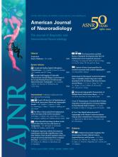
- Fig 1.
Ideal candidate for cervical arthroplasty. A, Lateral radiograph depicting minimal facet arthropathy and degenerative disease. B and C, Extension (B) and flexion (C) radiographs depicting normal segmental motion at the index level and throughout the cervical spine. D, Sagittal T2-weighted MR image depicting single-level degenerative disk disease, endplate changes, relative preservation of disk height, and posterior disk bulge without marked osteophyte formation.
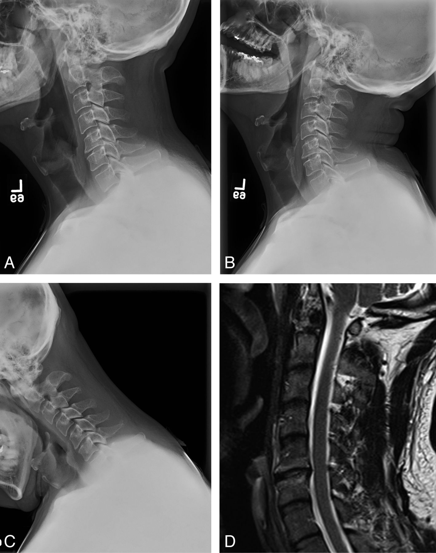
- Fig 2.
Case illustrating the importance of patient positioning for optimized biomechanics. A, A 36-year-old man with radicular symptoms referable to this C6–7 paramedian disk herniation seen on a sagittal T2-weighted MR image. The patient was positioned in mild cervical lordosis during ProDisc-C placement. B and C, As a result, postoperative extension (B) and flexion (C) radiographs obtained at 6 months revealed no movement at the instrumented level. D, Segmental motion was evident only with maximal extension beyond the typical physiologic range of motion.
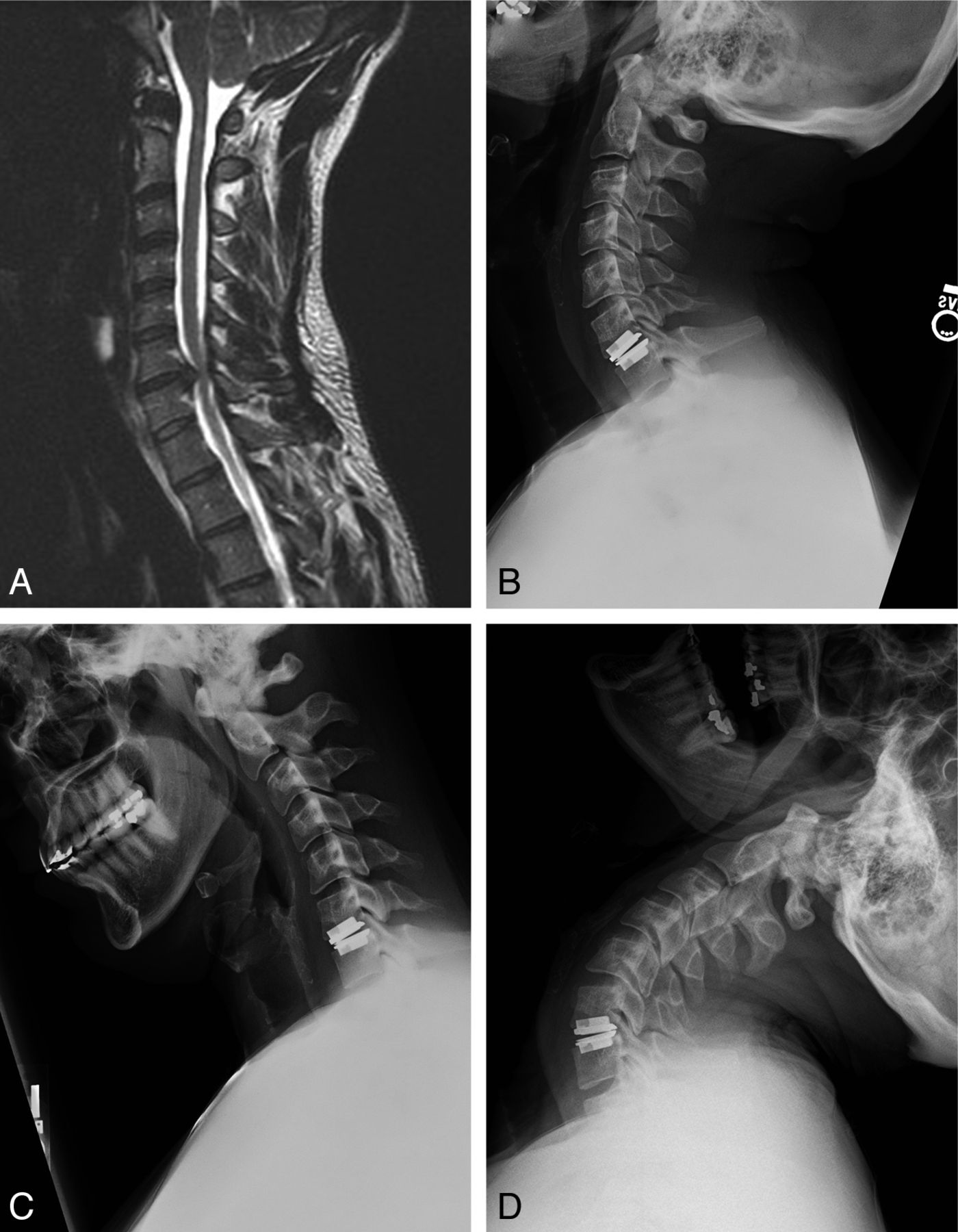
- Fig 3.
Dynamic flexion (A) and extension (B) postoperative lateral cervical radiographs following Prestige-ST arthroplasty in a 56-year-old woman. Segmental range of motion is preserved by the Prestige-ST device. Despite motion preservation, this particular patient ultimately required CT myelography, removal of the arthroplasty device, and 2-level anterior cervical fusion for symptomatic adjacent-level disease 1 year after arthroplasty.
- Fig 4.
Case illustrating the importance of device positioning. A 37-year-old man with prior history of noninstrumented L5-S1 microdiskectomy, now presenting with axial back pain. A, Sagittal T2-weighted MR image depicting degenerative disk disease at the previously operated level. Normal segmental motion without radiographically detectable instability was identified on preoperative flexion/extension dynamic radiographs. B and C, Postoperative lateral (B) and anteroposterior (C) views depicting the off-midline position of the ProDisc-L device. At the time of device placement, visualization of the L5-S1 level was limited due to immobile vascular structures. D, The patient awoke with right S1 radicular pain attributable to foraminal encroachment from the device as seen on this axial CT scan. The patient failed a short course of conservative management and ultimately required a right-sided L5-S1 hemilaminotomy, foraminotomy, and partial facetectomy for relief of symptoms.


Tables
FDA-approved cervical and lumbar arthroplasty devices
Device Application Design Biomaterials Endplate Fixation Kinematics FDA IDE Approval Manufacturer Prestige ST Cervical Uniarticular ball and trough Metal-on-metal articulation, stainless steel Roughened surface Vertebral body screws Unconstrained July 2007 Medtronic Bryan Cervical Biarticular Titanium alloy shells with polyurethane nucleus, saline lubricant Applied porous coating Milled, press-fit Unconstrained May 2009 Medtronic ProDisc-C Cervical Uniarticular ball and socket CCM endplate with UHMWE inlay, metal-on-polyurethane articulation Roughened titanium Central keel Semi-constrained December-2007 Synthes Spine CHARITÉ Lumbar Biarticular ball and socket CC endplates with UHMWPE sliding core Titanium and calcium phosphate plasma spray 6 Fixation teeth at cranial/caudal endplates Unconstrained October-2004 Depuy Spine ProDisc-L Lumbar Uniarticular ball and socket CCM endplates with UHMWPE insert Titanium plasma spray Large central keel, 2 lateral spikes Semi-constrained August 2006 Synthes Spine -
Note:—CCM indicates cobalt-chrome-molybdenum, CC, cobalt-chrome alloy; IDE, Investigational Device Exemption.
-


