
Article Figures & Data
Figures
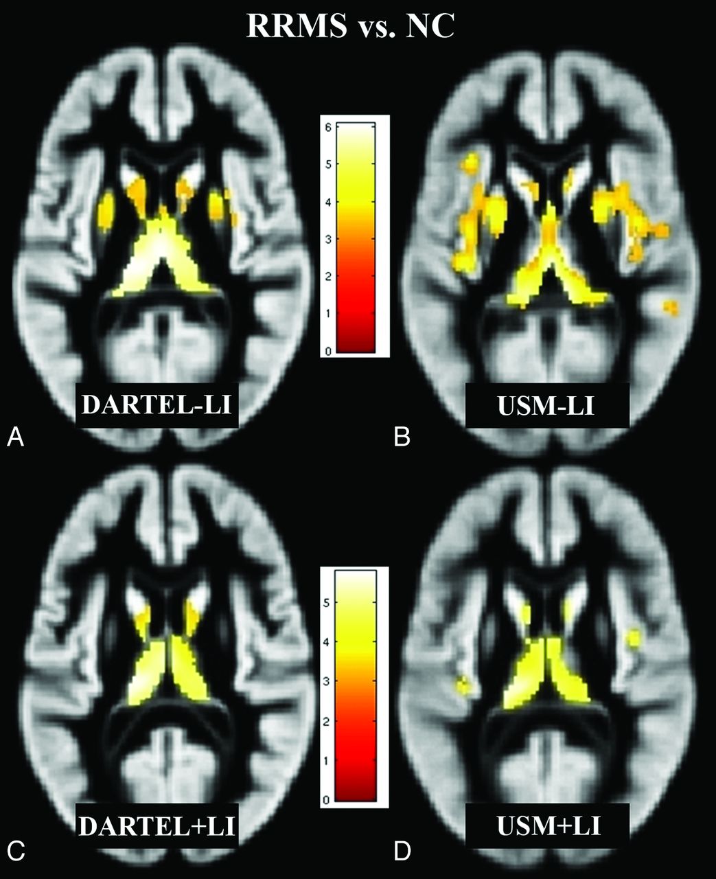
- Fig 1.
Regions of decreased GM volume in patients with RRMS (n = 26) compared with NC (n = 28) (P < .05, corrected for multiple comparisons by using false discovery rate, cluster size >20), overlaid on a GM template (A–D). Comparisons of all 4 methods (DARTEL + LI, DARTEL − LI, USM + LI, and USM − LI) are illustrated. The pattern of GM atrophy in patients with RRMS shown by DARTEL is more focal, while with USM, it is more widespread. DARTEL + LI (C) likely yielded the most accurate results compared with DARTEL − LI (A), USM − LI (B), and USM + LI (D) (see “Discussion”). Bar is color-coded for t values. Images are presented in the neurologic convention (right side of image = right side of brain). See “Materials and Methods” and “Results” sections for more details.
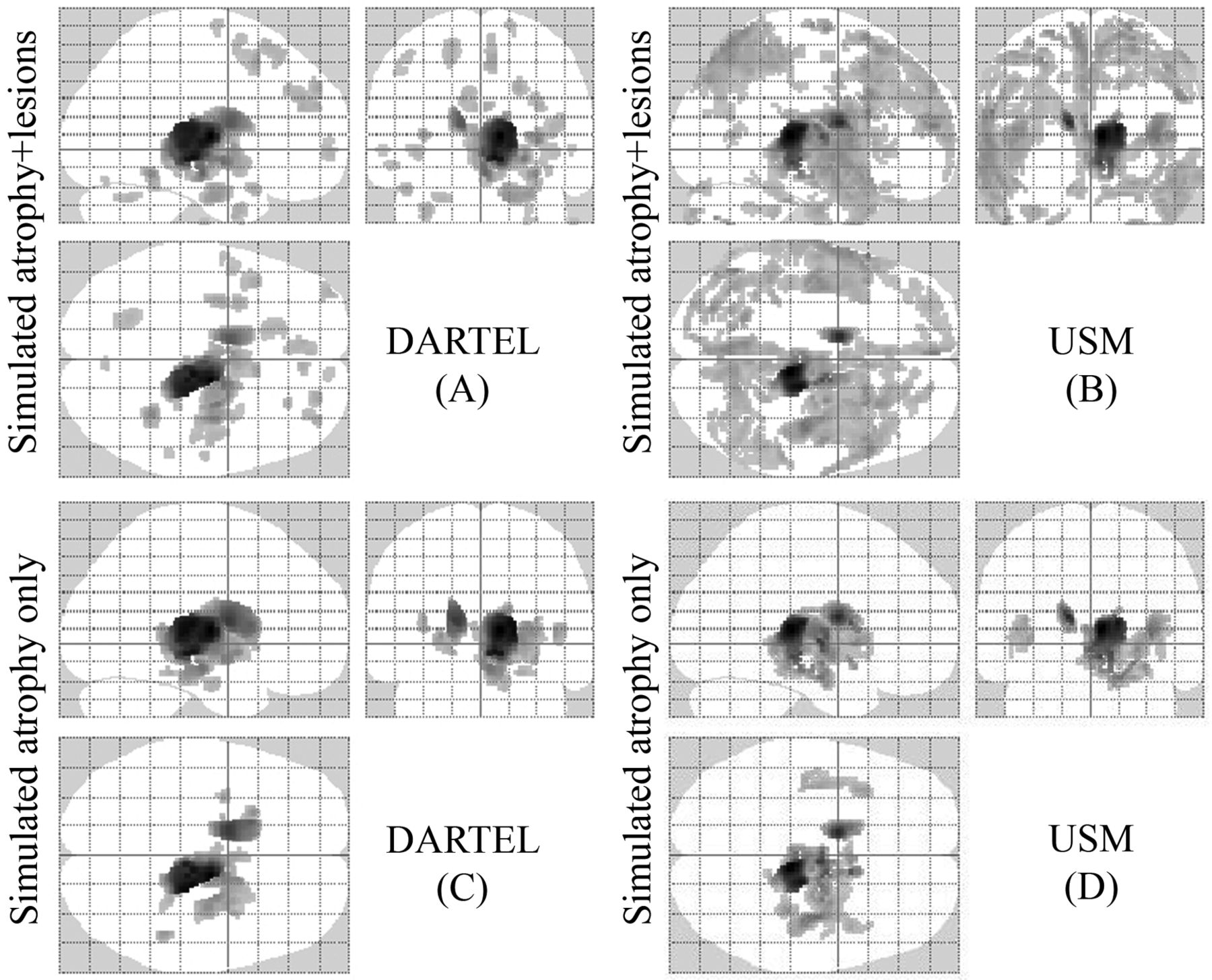
- Fig 2.
Regions of GM atrophy by using simulated ground truth data (A–D). Comparisons based on all methods are illustrated. Sixteen normal brain T1-weighted images were used as a control group and were compared with 2 groups of simulated patients (P < .05, corrected for multiple comparisons by using false discovery rate, cluster size >20). The results are displayed on a 3D glass brain. In 1 group (C and D), we only simulated atrophy of the thalamus and caudate, and in the other (A and B), we simulated atrophy in the same regions and added artificial lesions. DARTEL in the presence of only atrophy (C) yielded the most accurate results, detecting the atrophy under the ground truth and reducing the number of FPs compared with USM (D). The presence of lesions increased the number of FP errors in both DARTEL and USM (A and B). See “Materials and Methods” and “Results” section for more details.


Tables
NC RRMS No. of subjects 28 26 Men/womenb 7:21 6:20 Age (yr)b 43.0 ± 7.1 39.2 ± 9.4 Disease duration (yr) – 8.5 ± 5.9 EDSS score – 1.2 ± 0.9 FLAIR cerebral lesion volume (mL) – 15.0 ± 14.7 T1 hypointense cerebral lesion volume (mL) – 7.7 ± 9.6 Number of T1 hypointense cerebral lesions – 57.0 ± 57.6 Intracranial volume 1380.6 ± 124.1 1414.6 ± 139.6 Similarity DARTEL + LI (n = 54) DARTEL − LI (n = 54) P (DARTEL + LI vs DARTEL − LI) USM + LI (n = 54) USM − LI (n = 54) P (USM + LI vs USM − LI) NMI 1.2630 (0.0064) 1.2602 (0.0063) <.0001 1.1998 (0.0063) 1.1981 (0.0062) <.0001 CC 0.9675 (0.0039) 0.9669 (0.0040) <.0001 0.9090 (0.0106) 0.9084 (0.0107) <.0001 -
↵a Values are mean (SD). Overall, DARTEL showed higher NMI and CC (P < .0001) compared with USM.
-
In this issue


Jump to section
Related Articles
Cited By...
- Distributed and gradual microstructure changes track the emergence of behavioural benefit from memory reactivation
- Cueing motor memory reactivation during NREM sleep engenders learning-related changes in precuneus and sensorimotor structures
- Regional microglial activation in the substantia nigra is linked with fatigue in MS
- Serum lipid antibodies are associated with cerebral tissue damage in multiple sclerosis