Advertisement

Main menu

  • Home
  • Content
    • Current Issue
    • Accepted Manuscripts
    • Article Preview
    • Past Issue Archive
    • Video Articles
    • AJNR Case Collection
    • Case of the Week Archive
    • Case of the Month Archive
    • Classic Case Archive
  • Special Collections
    • AJNR Awards
    • Low-Field MRI
    • Alzheimer Disease
    • ASNR Foundation Special Collection
    • Photon-Counting CT
    • View All
  • Multimedia
    • AJNR Podcasts
    • AJNR SCANtastic
    • Trainee Corner
    • MRI Safety Corner
    • Imaging Protocols
  • For Authors
    • Submit a Manuscript
    • Submit a Video Article
    • Submit an eLetter to the Editor/Response
    • Manuscript Submission Guidelines
    • Statistical Tips
    • Fast Publishing of Accepted Manuscripts
    • Graphical Abstract Preparation
    • Imaging Protocol Submission
    • Author Policies
  • About Us
    • About AJNR
    • Editorial Board
    • Editorial Board Alumni
  • More
    • Become a Reviewer/Academy of Reviewers
    • Subscribers
    • Permissions
    • Alerts
    • Feedback
    • Advertisers
    • ASNR Home

User menu

  • Alerts
  • Log in

Search

  • Advanced search
American Journal of Neuroradiology

American Journal of Neuroradiology

ASHNR American Society of Functional Neuroradiology ASHNR American Society of Pediatric Neuroradiology ASSR
  • Alerts
  • Log in

Advanced Search

  • Home
  • Content
    • Current Issue
    • Accepted Manuscripts
    • Article Preview
    • Past Issue Archive
    • Video Articles
    • AJNR Case Collection
    • Case of the Week Archive
    • Case of the Month Archive
    • Classic Case Archive
  • Special Collections
    • AJNR Awards
    • Low-Field MRI
    • Alzheimer Disease
    • ASNR Foundation Special Collection
    • Photon-Counting CT
    • View All
  • Multimedia
    • AJNR Podcasts
    • AJNR SCANtastic
    • Trainee Corner
    • MRI Safety Corner
    • Imaging Protocols
  • For Authors
    • Submit a Manuscript
    • Submit a Video Article
    • Submit an eLetter to the Editor/Response
    • Manuscript Submission Guidelines
    • Statistical Tips
    • Fast Publishing of Accepted Manuscripts
    • Graphical Abstract Preparation
    • Imaging Protocol Submission
    • Author Policies
  • About Us
    • About AJNR
    • Editorial Board
    • Editorial Board Alumni
  • More
    • Become a Reviewer/Academy of Reviewers
    • Subscribers
    • Permissions
    • Alerts
    • Feedback
    • Advertisers
    • ASNR Home
  • Follow AJNR on Twitter
  • Visit AJNR on Facebook
  • Follow AJNR on Instagram
  • Join AJNR on LinkedIn
  • RSS Feeds

AJNR Awards, New Junior Editors, and more. Read the latest AJNR updates

Getting new auth cookie, if you see this message a lot, tell someone!
Research ArticleBrain

A Preliminary Prediction Model with MR Plaque Imaging to Estimate Risk for New Ischemic Brain Lesions on Diffusion-Weighted Imaging after Endarterectomy or Stenting in Patients with Carotid Stenosis

N. Akutsu, K. Hosoda, A. Fujita and E. Kohmura
American Journal of Neuroradiology September 2012, 33 (8) 1557-1564; DOI: https://doi.org/10.3174/ajnr.A3002
N. Akutsu
aFrom the Department of Neurosurgery, Kobe University Graduate School of Medicine, Kobe, Japan.
  • Find this author on Google Scholar
  • Find this author on PubMed
  • Search for this author on this site
K. Hosoda
aFrom the Department of Neurosurgery, Kobe University Graduate School of Medicine, Kobe, Japan.
  • Find this author on Google Scholar
  • Find this author on PubMed
  • Search for this author on this site
A. Fujita
aFrom the Department of Neurosurgery, Kobe University Graduate School of Medicine, Kobe, Japan.
  • Find this author on Google Scholar
  • Find this author on PubMed
  • Search for this author on this site
E. Kohmura
aFrom the Department of Neurosurgery, Kobe University Graduate School of Medicine, Kobe, Japan.
  • Find this author on Google Scholar
  • Find this author on PubMed
  • Search for this author on this site
  • Article
  • Figures & Data
  • Info & Metrics
  • Responses
  • References
  • PDF
Loading

Article Figures & Data

Figures

  • Tables
  • Fig 1.
    • Download figure
    • Open in new tab
    • Download powerpoint
    Fig 1.

    Coregistered multisequence MR imaging cross-sections of carotid plaque. Left: T1WI-BB. Right: T2WI-BB. The regions of interest are outlined in green. Arrows indicate the lumen of the ICA.

  • Fig 2.
    • Download figure
    • Open in new tab
    • Download powerpoint
    Fig 2.

    Forest plot of ORs of the full penalized logistic regression model for new occurrences of hyperintense lesions on DWI after CEA or CAS. All odds of continuous variables (T1-SIR, T2-SIR, stenosis, and clinical score) are represented as odds per increase, described in parentheses. Squares and horizontal bars represent ORs and 95% CIs, respectively, on a log scale. The size of the squares represents precision.

  • Fig 3.
    • Download figure
    • Open in new tab
    • Download powerpoint
    Fig 3.

    Effects of all of the predictors on the probability of new occurrences of hyperintense lesions on DWI after CEA or CAS, holding other predictors to their medians. See text for details.
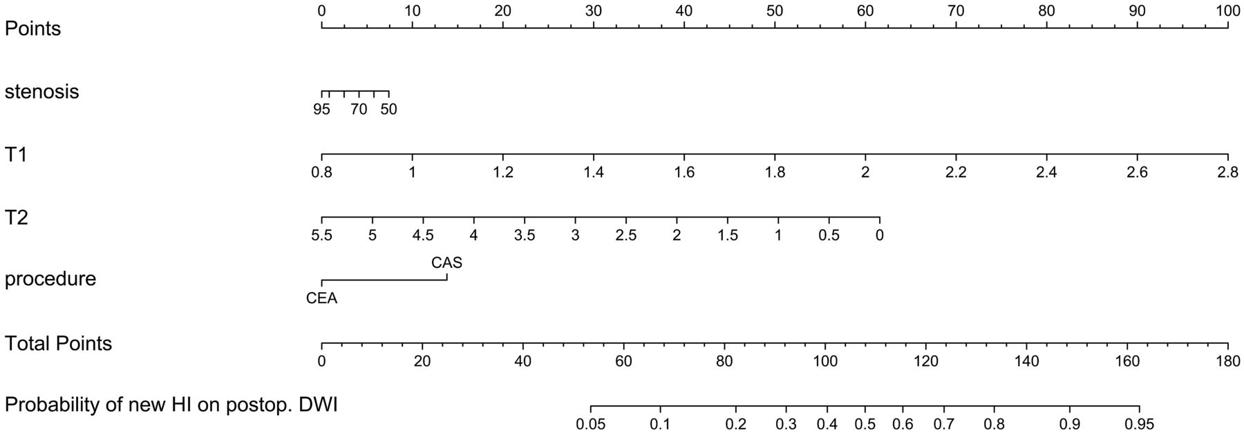
  • Fig 4.
    • Download figure
    • Open in new tab
    • Download powerpoint
    Fig 4.

    Preliminary nomogram for predicting the probability of new occurrences of hyperintense lesions on DWI after treatment (CEA or CAS) from the full penalized logistic model, by using an approximate model fitted by using ordinary least squares (R2 = 0.992 against the predicted logits of the full penalized logistic model). Points are assigned for each of the 4 predictors. The total points correspond to an absolute predicted risk for occurrence of new hyperintense lesions on DWI after treatment. For each predictor, read the points assigned on the 0–100 scale and add these points. Read the results on the Total Points scale and then read the corresponding predictions below it. Postop. indicates postoperative.

  • Fig 5.
    • Download figure
    • Open in new tab
    • Download powerpoint
    Fig 5.

    An example using the preliminary nomogram for predicting the probability of new occurrences of hyperintense lesions on DWI after treatment (CEA or CAS). Left: right carotid angiogram. Right upper: T1WI-BB. Right lower: T2WI-BB. Arrows indicate the lumen of the ICA. See text and Table 2 for details.

Tables

  • Figures
    • View popup
    Table 1:

    Baseline characteristics of patients with or without hyperintense lesions on DWI after CEA or CAS

    PredictorHyperintense Lesions on Postprocedural DWIP Value
    Positive (n = 25)Negative(n = 79)
    Age (yr)72.9 ± 6.271.3 ± 7.2.30
    Male (%)23 (92%)70 (89%)1
    Vascular risk factors (%)
        Hypertension20 (80%)54 (68%).32
        Hyperlipidemia21 (84%)57 (72%).30
        Statin use20 (80%)64 (81%)1
        DM10 (40%)23 (29%).33
        Smoking17 (68%)43 (54%).26
        IHD8 (32%)16 (20%).28
    Symptomatic stenosis (%)15 (60%)38 (48%).36
    Clinical score0.073 ± 0.97−0.023 ± 1.02.67
    Degree of stenosis (%)75.2 ± 11.475.8 ± 9.8.82
    CAS (%)14 (56%)27 (34%).063
    Plaque imaging
        T1-SIR1.69 ± 0.401.36 ± 0.29.00054
        T2-SIR1.49 ± 0.451.75 ± 0.80.045
    • View popup
    Table 2:

    Hypothetic examples with the calculated predicted probability of new HI on DWI after treatment

    PredictorExample
    Risk of CEARisk of CAS
    ValuesPointsValuesPoints
    Stenosis (%)852852
    T1-SIR1.4301.430
    T2-SIR1.1491.149
    CEA or CASCEA0CAS14
    Total points8195
    Probability of new HI on postop. DWI19%34%
    • Note:—postop. indicates postoperative; HI, hyperintensity.

PreviousNext
Back to top

In this issue

American Journal of Neuroradiology: 33 (8)
American Journal of Neuroradiology
Vol. 33, Issue 8
1 Sep 2012
  • Table of Contents
  • Index by author
Advertisement
Print
Download PDF
Email Article

Thank you for your interest in spreading the word on American Journal of Neuroradiology.

NOTE: We only request your email address so that the person you are recommending the page to knows that you wanted them to see it, and that it is not junk mail. We do not capture any email address.

Enter multiple addresses on separate lines or separate them with commas.
A Preliminary Prediction Model with MR Plaque Imaging to Estimate Risk for New Ischemic Brain Lesions on Diffusion-Weighted Imaging after Endarterectomy or Stenting in Patients with Carotid Stenosis
(Your Name) has sent you a message from American Journal of Neuroradiology
(Your Name) thought you would like to see the American Journal of Neuroradiology web site.
CAPTCHA
This question is for testing whether or not you are a human visitor and to prevent automated spam submissions.
Cite this article
N. Akutsu, K. Hosoda, A. Fujita, E. Kohmura
A Preliminary Prediction Model with MR Plaque Imaging to Estimate Risk for New Ischemic Brain Lesions on Diffusion-Weighted Imaging after Endarterectomy or Stenting in Patients with Carotid Stenosis
American Journal of Neuroradiology Sep 2012, 33 (8) 1557-1564; DOI: 10.3174/ajnr.A3002

Citation Manager Formats

  • BibTeX
  • Bookends
  • EasyBib
  • EndNote (tagged)
  • EndNote 8 (xml)
  • Medlars
  • Mendeley
  • Papers
  • RefWorks Tagged
  • Ref Manager
  • RIS
  • Zotero
0 Responses
Respond to this article
Share
Bookmark this article
A Preliminary Prediction Model with MR Plaque Imaging to Estimate Risk for New Ischemic Brain Lesions on Diffusion-Weighted Imaging after Endarterectomy or Stenting in Patients with Carotid Stenosis
N. Akutsu, K. Hosoda, A. Fujita, E. Kohmura
American Journal of Neuroradiology Sep 2012, 33 (8) 1557-1564; DOI: 10.3174/ajnr.A3002
del.icio.us logo Twitter logo Facebook logo Mendeley logo
  • Tweet Widget
  • Facebook Like
  • Google Plus One
Purchase

Jump to section

  • Article
    • Abstract
    • ABBREVIATIONS:
    • Materials and Methods
    • Results
    • Discussion
    • Conclusions
    • Acknowledgments
    • Footnotes
    • References
  • Figures & Data
  • Info & Metrics
  • Responses
  • References
  • PDF

Related Articles

  • No related articles found.
  • PubMed
  • Google Scholar

Cited By...

  • Ischemic cerebral lesions after Carotid Stenting versus Carotid Endarterectomy: A Systematic review and Meta-Analysis
  • Predicting Impaired Cerebrovascular Reactivity and Hyperperfusion Syndrome with BeamSAT MRI in Carotid Artery Stenosis
  • Absence of the Anterior Communicating Artery on Selective MRA is Associated with New Ischemic Lesions on MRI after Carotid Revascularization
  • The association between carotid intraplaque hemorrhage and outcomes of carotid stenting: a systematic review and meta-analysis
  • Reduced cerebrovascular reserve is associated with an increased risk of postoperative ischemic lesions during carotid artery stenting
  • Crossref
  • Google Scholar

This article has not yet been cited by articles in journals that are participating in Crossref Cited-by Linking.

More in this TOC Section

  • Quiet PROPELLER MRI Techniques Match the Quality of Conventional PROPELLER Brain Imaging Techniques
  • Predictors of Reperfusion in Patients with Acute Ischemic Stroke
  • Enhanced Axonal Metabolism during Early Natalizumab Treatment in Relapsing-Remitting Multiple Sclerosis
Show more Brain

Similar Articles

Advertisement

Indexed Content

  • Current Issue
  • Accepted Manuscripts
  • Article Preview
  • Past Issues
  • Editorials
  • Editor's Choice
  • Fellows' Journal Club
  • Letters to the Editor
  • Video Articles

Cases

  • Case Collection
  • Archive - Case of the Week
  • Archive - Case of the Month
  • Archive - Classic Case

More from AJNR

  • Trainee Corner
  • Imaging Protocols
  • MRI Safety Corner
  • Book Reviews

Multimedia

  • AJNR Podcasts
  • AJNR Scantastics

Resources

  • Turnaround Time
  • Submit a Manuscript
  • Submit a Video Article
  • Submit an eLetter to the Editor/Response
  • Manuscript Submission Guidelines
  • Statistical Tips
  • Fast Publishing of Accepted Manuscripts
  • Graphical Abstract Preparation
  • Imaging Protocol Submission
  • Evidence-Based Medicine Level Guide
  • Publishing Checklists
  • Author Policies
  • Become a Reviewer/Academy of Reviewers
  • News and Updates

About Us

  • About AJNR
  • Editorial Board
  • Editorial Board Alumni
  • Alerts
  • Permissions
  • Not an AJNR Subscriber? Join Now
  • Advertise with Us
  • Librarian Resources
  • Feedback
  • Terms and Conditions
  • AJNR Editorial Board Alumni

American Society of Neuroradiology

  • Not an ASNR Member? Join Now

© 2025 by the American Society of Neuroradiology All rights, including for text and data mining, AI training, and similar technologies, are reserved.
Print ISSN: 0195-6108 Online ISSN: 1936-959X

Powered by HighWire