
Article Figures & Data
Figures
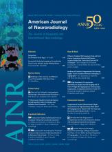
- Fig 1.
Examples of extrinsic compression by the styloid process and the posterior belly of the digastric muscle. A, In this axial CT image, there is bilateral compression of the internal jugular veins (dashed arrows) by the styloid processes (solid arrows). The vein takes a pancake-like configuration at this level. B, Similar extrinsic compression, caused by the posterior belly of the digastric muscle (solid arrows) of both internal jugular veins (dashed arrows), though to a lesser degree. In both cases, the vein is compressed anterolaterally by the described structure (styloid process or digastric muscle). Along the medial and posteromedial aspect of the vein, the adjacent vertebra is the most rigid structure and will provide the other side of the extrinsic compressive “pincer.”
- Fig 2.
Example of bilateral collateral vein filling. Four axial images from a CTA of the neck demonstrate bilateral posterior condylar and upper deep cervical vein filling (arrows). Note how contrast in these veins is isoattenuated with the jugular bulb. The left (+) and right (*) internal jugular veins are marked.
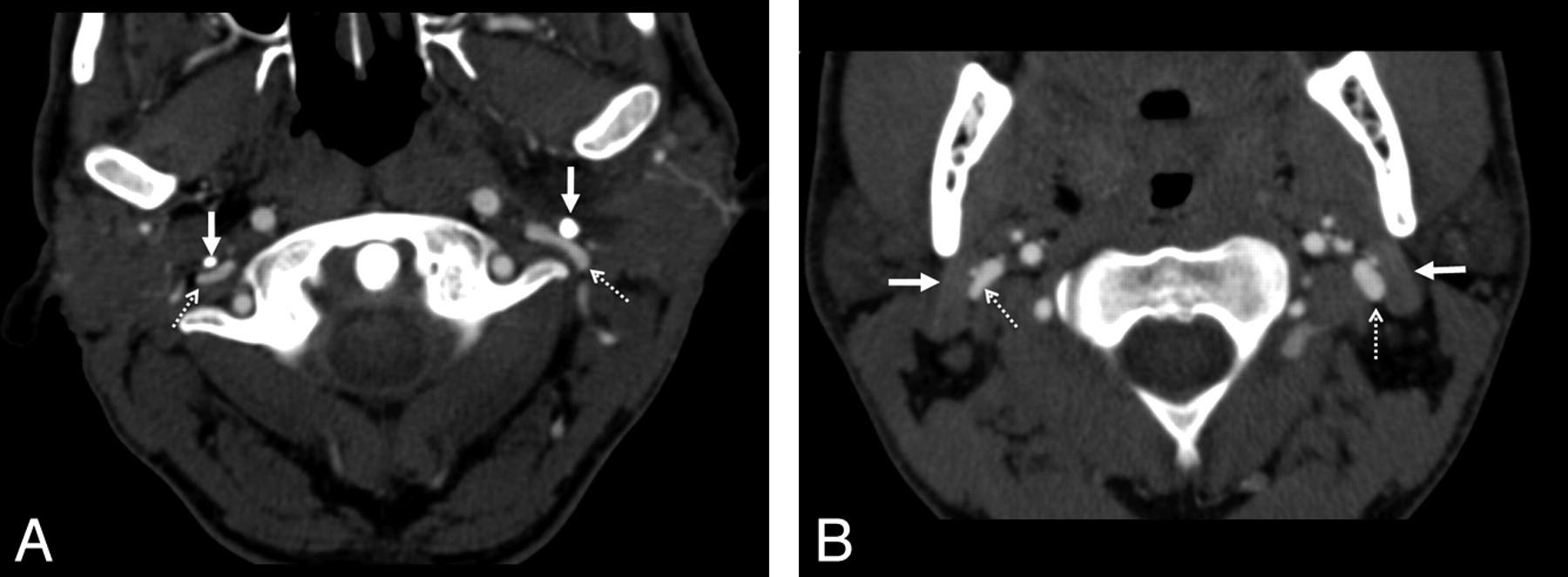
- Fig 3.
Examples of condylar veins with and without collateral flow. Two axial images from a CTA of the neck demonstrate differential contrast attenuation in the condylar veins. The left condylar veins (solid arrows) are isoattenuated with the right internal jugular vein (arrowhead). The left internal jugular vein is markedly attenuated. Note that the right-sided condylar veins (dashed arrows) have similar caliber to the left but are not filled with contrast of the same attenuation. This would suggest that the venous drainage of the brain in this patient does use the left condylar veins but not the right.
- Fig 4.
Example of severe bilateral jugular venous stenosis without collateral formation. Two axial images from a CTA of the neck demonstrate a dominant left internal jugular vein (arrow, left image). The more caudal image (right) shows severe pancake-like flattening of the internal jugular veins bilaterally (arrows) but with no condylar collateral filling seen.


Tables
- Table 1:
Relationship between the presence or absence of collaterals based on the degree of cross-sectional narrowing of the 108 jugular veins analyzed on each side
Collaterals Present No Collaterals Present Right internal jugular vein, degree of stenosis None (n = 46; 42.6%) 9/46 (19.6%) 37/46 (80.4%) Moderate (n = 36; 33.3%) 14/36 (38.9%) 22/36 (61.1%) Severe (n = 26; 24.1%) 14/26 (53.8%) 12/26 (46.2%) Left internal jugular vein, degree of stenosis None (n = 60; 55.6%) 13/60 (21.7%) 47/60 (78.3%) Moderate (n = 28; 25.9%) 10/28 (35.7%) 18/28 (64.3%) Severe (n = 20; 18.5%) 11/20 (55%) 9/20 (45%) - Table 2:
Per patient analysis of flow pattern, and the presence or absence of collaterals on the side of the dominant jugular vein (or bilateral, if codominant)
Overall (n = 108) Collaterals Present No Collaterals Present No stenosis bilaterally 35 (32.4%) Mild or moderate stenosis of nondominant jugular vein 24 (22.2%) Moderate stenosis of dominant jugular vein 31 (28.7%) 11/31 (35.5%) 20/31 (64.5%) Severe stenosis of dominant jugular vein 18 (16.7%) 10/18 (55.6%) 8/18 (44.4%) Cause Right Side (n = 108) Left Side (n = 108) Styloid process 26/108 (24.1%) 33/108 (30.6%) Digastric muscle 26/108 (24.1%) 12/108 (11.1%) Artery (ECA, ICA, or branches thereof) 3/108 (2.8%) 2/108 (1.8%) Styloid process and digastric muscle 5/108 (4.6%) 0 Other 2/108 (1.9%) 1/108 (0.9%) None 46/108 (42.6%) 60/108 (55.6%) Note:—See Figure 1 for examples of narrowing by the styloid process and posterior belly of the digastric muscle. ECA indicates external carotid artery; ICA, internal carotid artery.
In this issue


Jump to section
Related Articles
Cited By...
- Dynamic internal jugular vein venography: a descriptive study in 89 patients with suspected cerebral venous outflow disorders
- Mechanical disorders of the cervicocerebral circulation in children and young adults
- Interventional and surgical management of internal jugular venous stenosis: a narrative review
- Republished: Moving target: transient rotational stenosis precipitating jugular bow hunter's syndrome
- Moving target: transient rotational stenosis precipitating jugular bow hunter's syndrome