
Article Figures & Data
Figures
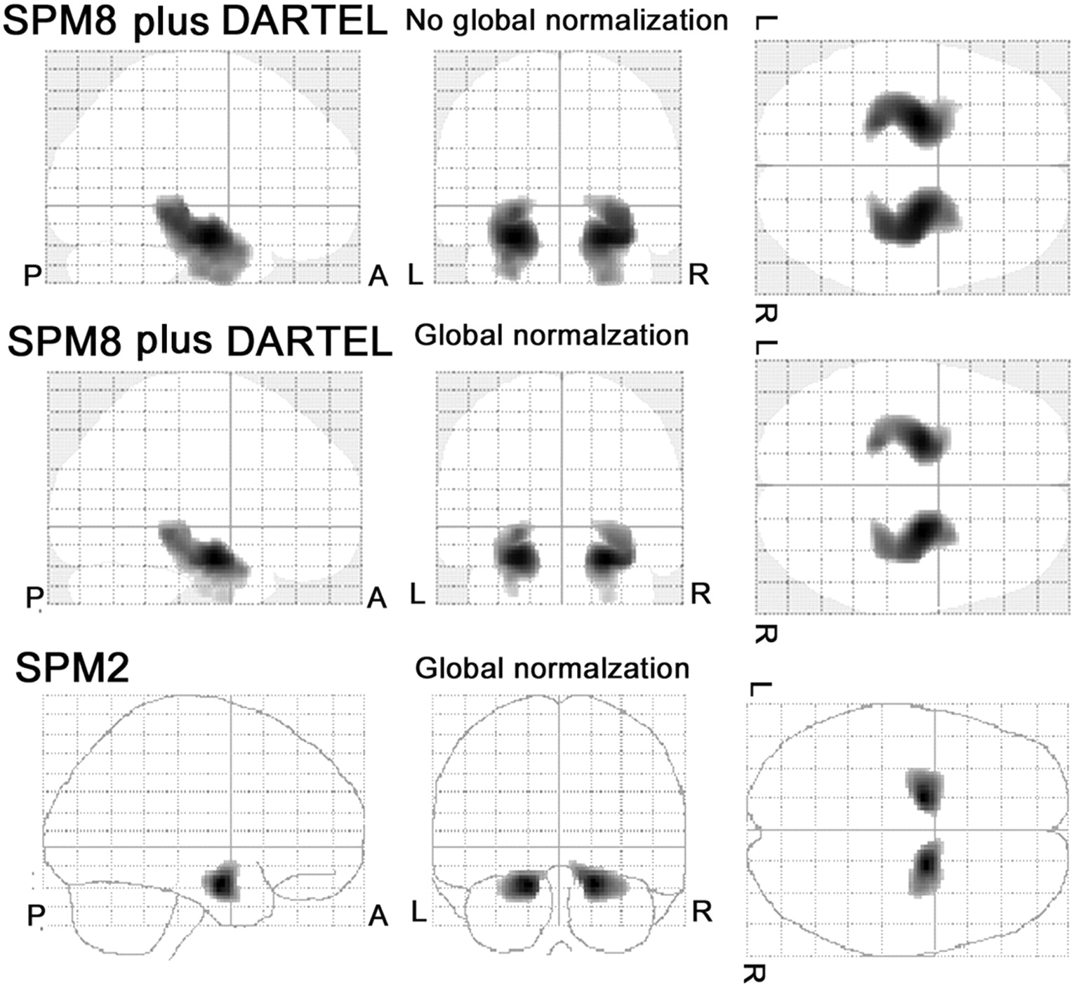
- Fig 1.
Group comparison of gray matter volume by SPM8 plus DARTEL and SPM2 between 30 patients with very mild AD and 40 healthy age-matched volunteers. The SPM8 plus DARTEL analysis demonstrates significant decline of gray matter volume in the bilateral medial temporal structures both with and without global normalization in patients with very mild AD. The cluster shape is very close to the anatomic configuration of the medial temporal structures involving the entorhinal cortex, amygdala, and hippocampal formation from head to tail. Although the SPM 2 analysis demonstrates a significant decline of gray matter volume in the bilateral medial temporal structures, the cluster is confined to the anterior parts of the medial temporal structures.
- Fig 2.
Cross-sectional VBM study by using SPM8 plus DARTEL. A, MR image of a 52-year-old woman with an MMSE score of 27. One year later the MMSE score declined to 19. B, SPM8 plus DARTEL analysis with global normalization reveals a significant decrease of gray matter volume in the right entorhinal area. Colored areas with z scores of >2 are overlaid as significantly atrophied regions on tomographic sections and cortical surface of the standardized MRI template. A target VOI in the medial temporal structures is demarcated with purple lines. The right temporoparietal cortex also shows extensive significant atrophy.
- Fig 3.
Longitudinal VBM studies by using SPM8 plus DARTEL. A 63-year-old woman with an MMSE score of 27 at the first visit was followed up for 6 years. One year later, the MMSE score decreased to 20 and gradually decreased thereafter. VBM analysis with global normalization reveals significant atrophy in the bilateral medial temporal areas even at the time of the initial study. Then the z score in a target VOI increased step by step with time. In contrast, analysis without global normalization does not demonstrate significant atrophy in the medial temporal areas for the first 3 years. Severity scores as an indicator for characterizing atrophy in the medial temporal structures are shown.


Tables
Group Global Normalization SPM8 plus DARTEL SPM2 Target VOI Whole-Brain Extent (%) Target VOI Whole-Brain Extent (%) Severity Extent (%) Ratio Severity Extent (%) Ratio Healthy controls − 0.7 ± 0.5 4.4 ± 9.8 0.8 ± 1.5 2.5 ± 4.7 NA NA NA NA + 0.7 ± 0.3 2.0 ± 4.9 1.3 ± 2.8 1.4 ± 0.9 0.5 ± 0.3 1.8 ± 7.3 0.5 ± 1.8 2.6± 3.1 Very mild AD − 1.8 ± 0.9b 39.0 ± 35.5b 9.9 ± 8.9b 5.4 ± 7.6b NA NA NA NA + 2.2 ± 0.9b 49.2 ± 30.2b 12.9 ± 7.8b 4.1 ± 2.5b 1.6 ± 1.0b 30.8 ± 32.1b 6.7 ± 7.8b 5.4 ± 3.7b Mild AD − 2.2 ± 0.7b 53.7 ± 29.8b 12.8 ± 8.8b 5.5 ± 5.1b NA NA NA NA + 2.7 ± 0.8b 63.7 ± 25.8b 15.4 ± 7.8b 4.3 ± 1.9b 2.1 ± 1.1b 42.0 ± 32.3b 9.6 ± 9.2b 5.4 ± 3.0b Moderate-to-advanced AD − 2.8 ± 1.0b,c,d 72.2 ± 26.5b,c,d 8.4 ± 7.2b 15.1 ± 14.0b,c,d NA NA NA NA + 3.0 ± 1.0b,c 68.7 ± 24.1b,c 11.7 ± 6.7b,d 7.1 ± 3.7b,c,d 2.6 ± 1.4b,c 56.3 ± 33.2b,c 7.6 ± 6.2b 9.0 ± 5.0b,c,d Global Normalization Onset SPM8 plus DARTEL SPM2 Target VOI Whole-Brain Extent (%) Target VOI Whole-Brain Extent (%) Severity Extent (%) Ratio Severity Extent (%) Ratio − Early 1.5 ± 0.7 25.3 ± 31.1 3.8 ± 3.8 5.1 ± 4.9 NA NA NA NA − Late 1.9 ± 0.9 43.8 ± 35.6 5.9 ± 8.4 11.6 ± 9.5 NA NA NA NA + Early 1.9 ± 0.7 37.4 ± 26.2 9.5 ± 6.0 4.1 ± 1.7 1.4 ± 1.0 24.4 ± 34.6 4.7 ± 5.5 3.8 ± 2.4 + Late 2.3 ± 0.9 53.6 ± 30.7 14.1 ± 8.1 4.1 ± 2.7 1.7 ± 1.0 33.2 ± 31.2 7.5 ± 8.5 6.0 ± 3.9 Note:—NA indicates not applicable; +, presence; −, absence.
In this issue


Jump to section
Related Articles
Cited By...
- Neurophysiological trajectories in Alzheimers disease progression
- Neurophysiological trajectories in Alzheimers disease progression
- A systematic comparison of VBM pipelines and their application to age prediction
- Emotional responsiveness task in emotional distress: correlated of functional neuroimaging in anorexia and bulimia
- Optimization of DARTEL Settings for the Detection of Alzheimer Disease
- Association between naturally occurring anti-amyloid {beta} autoantibodies and medial temporal lobe atrophy in Alzheimer's disease
- Chronic Kidney Disease in Patients With Lacunar Stroke: Association With Enlarged Perivascular Spaces and Total Magnetic Resonance Imaging Burden of Cerebral Small Vessel Disease