Advertisement

Main menu

  • Home
  • Content
    • Current Issue
    • Accepted Manuscripts
    • Article Preview
    • Past Issue Archive
    • Video Articles
    • AJNR Case Collection
    • Case of the Week Archive
    • Case of the Month Archive
    • Classic Case Archive
  • Special Collections
    • AJNR Awards
    • Low-Field MRI
    • Alzheimer Disease
    • ASNR Foundation Special Collection
    • Photon-Counting CT
    • View All
  • Multimedia
    • AJNR Podcasts
    • AJNR SCANtastic
    • Trainee Corner
    • MRI Safety Corner
    • Imaging Protocols
  • For Authors
    • Submit a Manuscript
    • Submit a Video Article
    • Submit an eLetter to the Editor/Response
    • Manuscript Submission Guidelines
    • Statistical Tips
    • Fast Publishing of Accepted Manuscripts
    • Graphical Abstract Preparation
    • Imaging Protocol Submission
    • Author Policies
  • About Us
    • About AJNR
    • Editorial Board
    • Editorial Board Alumni
  • More
    • Become a Reviewer/Academy of Reviewers
    • Subscribers
    • Permissions
    • Alerts
    • Feedback
    • Advertisers
    • ASNR Home

User menu

  • Alerts
  • Log in

Search

  • Advanced search
American Journal of Neuroradiology

American Journal of Neuroradiology

ASHNR American Society of Functional Neuroradiology ASHNR American Society of Pediatric Neuroradiology ASSR
  • Alerts
  • Log in

Advanced Search

  • Home
  • Content
    • Current Issue
    • Accepted Manuscripts
    • Article Preview
    • Past Issue Archive
    • Video Articles
    • AJNR Case Collection
    • Case of the Week Archive
    • Case of the Month Archive
    • Classic Case Archive
  • Special Collections
    • AJNR Awards
    • Low-Field MRI
    • Alzheimer Disease
    • ASNR Foundation Special Collection
    • Photon-Counting CT
    • View All
  • Multimedia
    • AJNR Podcasts
    • AJNR SCANtastic
    • Trainee Corner
    • MRI Safety Corner
    • Imaging Protocols
  • For Authors
    • Submit a Manuscript
    • Submit a Video Article
    • Submit an eLetter to the Editor/Response
    • Manuscript Submission Guidelines
    • Statistical Tips
    • Fast Publishing of Accepted Manuscripts
    • Graphical Abstract Preparation
    • Imaging Protocol Submission
    • Author Policies
  • About Us
    • About AJNR
    • Editorial Board
    • Editorial Board Alumni
  • More
    • Become a Reviewer/Academy of Reviewers
    • Subscribers
    • Permissions
    • Alerts
    • Feedback
    • Advertisers
    • ASNR Home
  • Follow AJNR on Twitter
  • Visit AJNR on Facebook
  • Follow AJNR on Instagram
  • Join AJNR on LinkedIn
  • RSS Feeds

AJNR Awards, New Junior Editors, and more. Read the latest AJNR updates

Research ArticleBrain
Open Access

The Role of Preload and Leakage Correction in Gadolinium-Based Cerebral Blood Volume Estimation Determined by Comparison with MION as a Criterion Standard

J. L. Boxerman, D.E. Prah, E.S. Paulson, J.T. Machan, D. Bedekar and K.M. Schmainda
American Journal of Neuroradiology June 2012, 33 (6) 1081-1087; DOI: https://doi.org/10.3174/ajnr.A2934
J. L. Boxerman
  • Find this author on Google Scholar
  • Find this author on PubMed
  • Search for this author on this site
D.E. Prah
  • Find this author on Google Scholar
  • Find this author on PubMed
  • Search for this author on this site
E.S. Paulson
  • Find this author on Google Scholar
  • Find this author on PubMed
  • Search for this author on this site
J.T. Machan
  • Find this author on Google Scholar
  • Find this author on PubMed
  • Search for this author on this site
D. Bedekar
  • Find this author on Google Scholar
  • Find this author on PubMed
  • Search for this author on this site
K.M. Schmainda
  • Find this author on Google Scholar
  • Find this author on PubMed
  • Search for this author on this site
  • Article
  • Figures & Data
  • Info & Metrics
  • Responses
  • References
  • PDF
Loading

Article Figures & Data

Figures

  • Tables
  • Fig 1.
    • Download figure
    • Open in new tab
    • Download powerpoint
    Fig 1.

    Summary of Gd-based DSC methodology: staged injection technique. Gradient-echo DSC-MRI (0.1-mmol/kg gadodiamide bolus) provides no preload (P−) rCBV data and serves as a preload (P+) for subsequent DSC-MRI (0.2 mmol/kg bolus). Gd-rCBV without (C−) and with (C+) application of postprocessing leakage correction are computed for both P− and P+ data, thereby testing P−C−, P−C+, P+C−, and P+C+ permutations of the 2 leakage-correction techniques.

  • Fig 2.
    • Download figure
    • Open in new tab
    • Download powerpoint
    Fig 2.

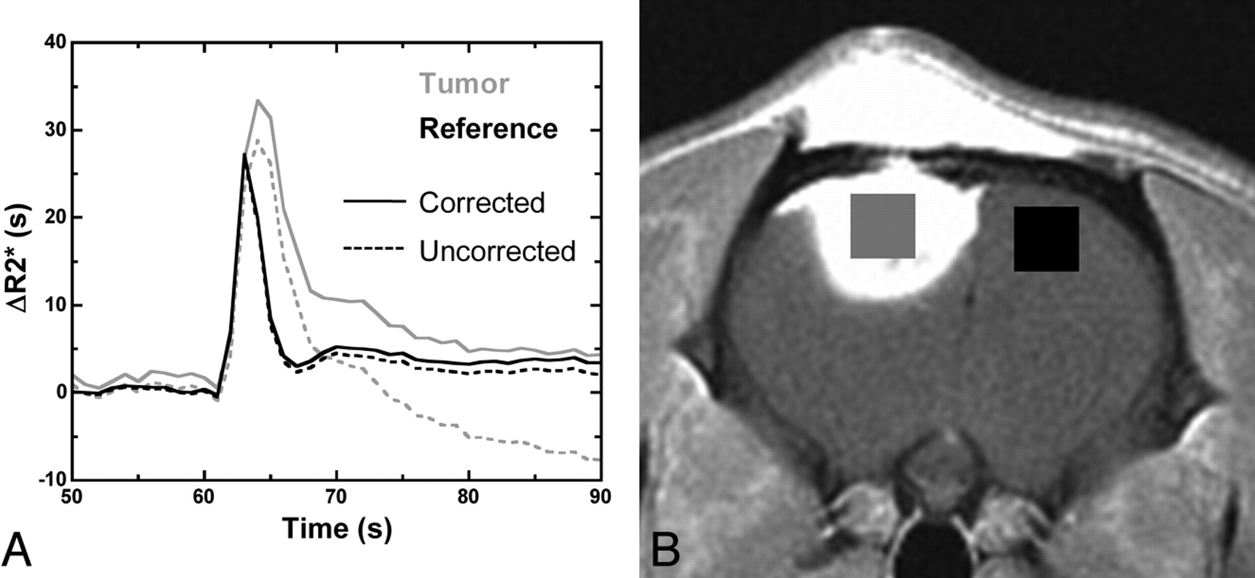
    Gd-rCBV for initial (P−) and second (P+) injection data was computed by using voxelwise trapezoidal integration of ΔR2* (t) without (C−) and with (C+) postprocessing leakage correction. A, Examples of P+C− and P+C+ curves in tumor and reference brain. B, The ratio of mean rCBV from tumor (gray) and contralateral brain (black) ROIs was computed for all injections, providing normalized rCBV values.

  • Fig 3.
    • Download figure
    • Open in new tab
    • Download powerpoint
    Fig 3.

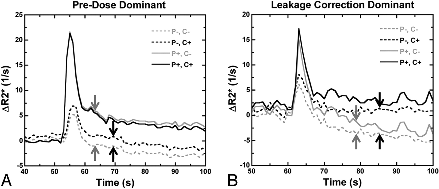
    Comparison of preload-dominant and postprocessing-dominant corrective effects on relaxivity time curves for 2 different tumors. A, Preload alone (solid gray line) eliminates most of the T1 leakage contamination (dashed gray line with blunted peak relaxivity and negative ΔR2* values; shift between gray arrows), whereas postprocessing correction does not (dashed black line; shift between black arrows). B, Postprocessing algorithm has more substantial corrective effect than preload in the tail portion of the curves (dashed black versus dashed gray lines; shift between black versus gray arrows), but the converse is true during the first pass (solid versus dashed lines), demonstrating synergy between the 2 correction schemes. Combining preload and postprocessing (solid black lines) yields the greatest peak ΔR2* without negative relaxivity values.

  • Fig 4.
    • Download figure
    • Open in new tab
    • Download powerpoint
    Fig 4.

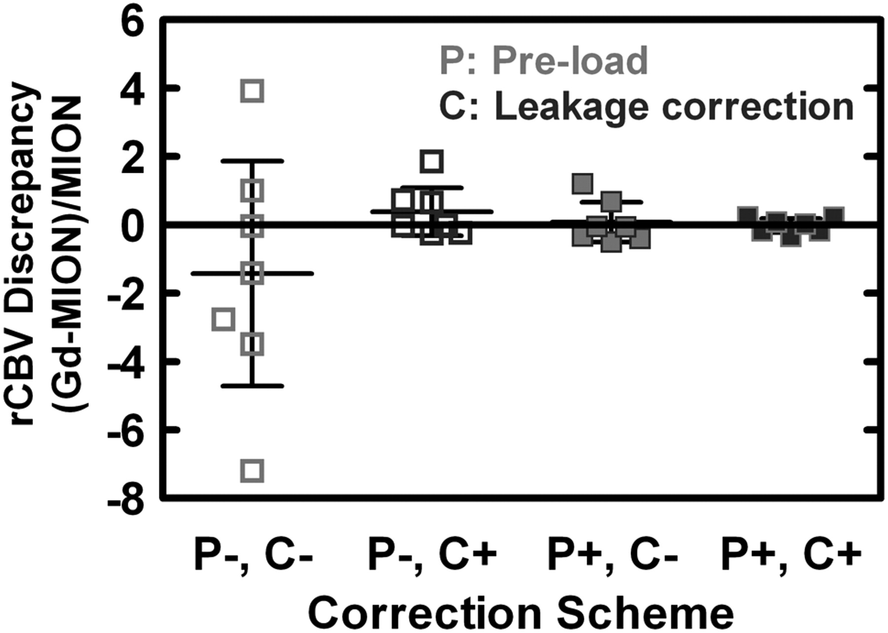
    Comparison of rCBV discrepancies ([Gd − MION] / MION) for each correction scheme permutation (mean and 95% CI). Although there is no statistically significant intrascheme or interscheme bias, mean discrepancy is closest to zero for P+C+ (−1.8%), followed by P+C− (+7.6%), P−C+ (+38.3%), and P−C− (−142.8%). The variance of rCBV discrepancies differed substantially between correction schemes, with P+C− (22-fold), P−C+ (32-fold), and P+C+ (267-fold) all statistically significantly lower compared with P−C−. The use of both correction techniques (P+C+) further significantly reduced the variance compared with that for each individually (12-fold versus P+C−, 8-fold versus P−C+).

  • Fig 5.
    • Download figure
    • Open in new tab
    • Download powerpoint
    Fig 5.

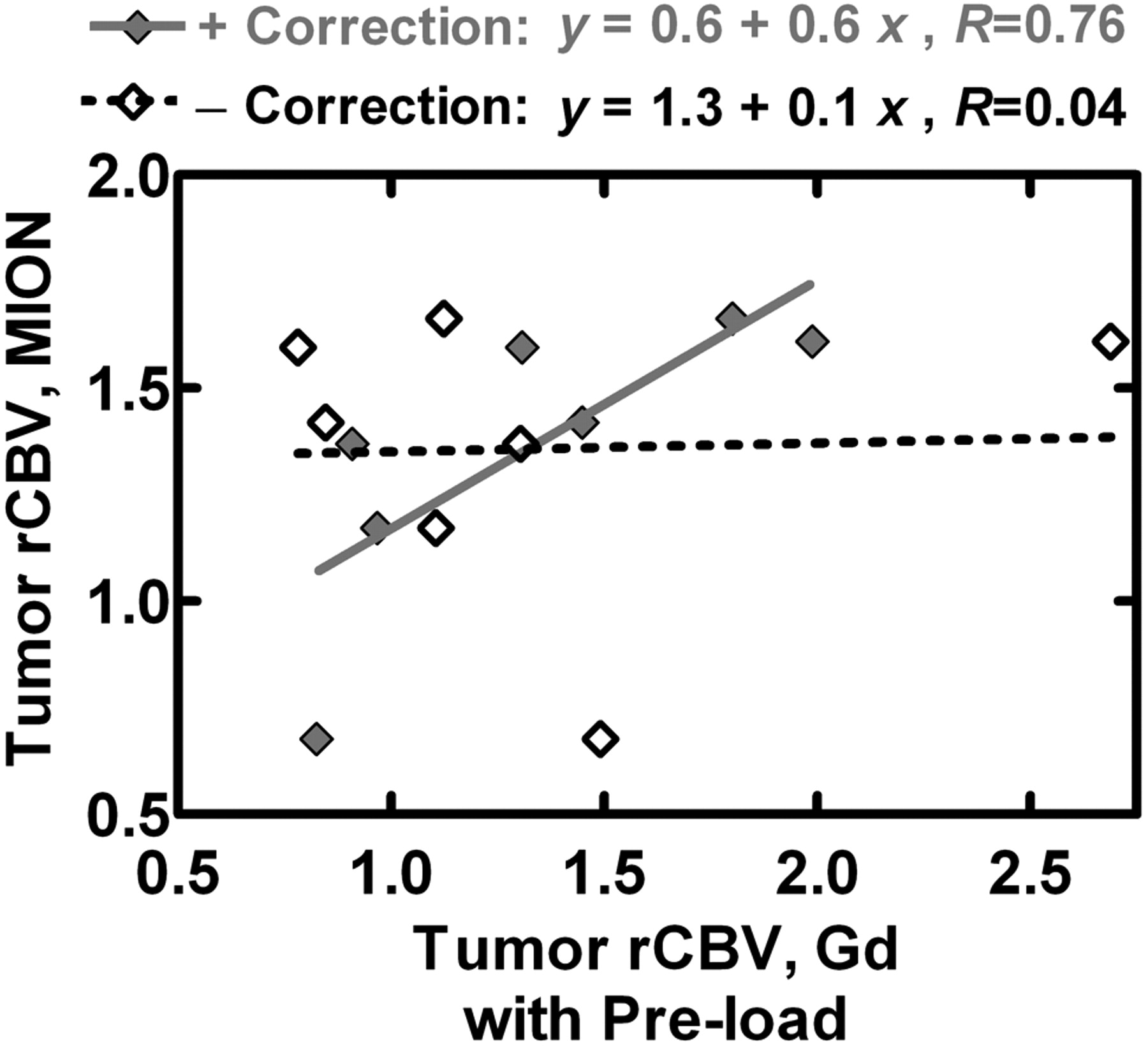
    Gd-rCBV with preload and with (P+C+) and without (P+C−) leakage correction is plotted against MION-rCBV. The linear fit for P+C+ data is much closer to the ideal line of identity than the linear fit for P+C−, arguing that the use of both correction schemes outperforms the use of only preload.

Tables

  • Figures
    • View popup
    Table 1:

    Techniques for reducing T1 leakage effects

    CategoryTechniqueExamples
    Image acquisition Low flip angle, long TE, double-echo T2*-weightedKnopp et al,11 Cha et al38 Vonken et al,27 Uematsu et al28
    Contrast agentUse of loading dosesaDonahue et al,3 Schmainda et al,21 Simonsen et al35
    Intravascular agents (eg, ferumoxytol)Gahramanov et al20
    PostprocessingLinear fit + leakage modelaWeisskoff et al,31 Schmainda et al21 Boxerman et al22
    γ-Variate fitLaw et al4
    Limited integrationWong et al30
    Baseline subtractionWetzel et al29
    • ↵a Designates the 2 techniques investigated in this article.

    • View popup
    Table 2:

    Summary of discrepancy between Gd-rCBV and MION-rCBV for each correction scheme permutation

    SchemerCBV DiscrepancyInterceptSlope
    Mean (%)95% CI (%)Mean95% CIMean95% CI
    P−C−−142.8−471.5, +186.0+1.4+1.1, +1.6−0.01−0.04, +0.02
    P−C++38.3−32.0, +108.6+1.3+0.8, +1.8+0.16+0.02, +0.31
    P+C−+7.6−50.1, +65.9+1.0+0.5, +1.5+0.02−0.29, +0.33
    P+C+−1.8−22.0, +18.3+0.6−0.2, +1.4+0.58+0.17, +1.00
    • Note:—rCBV discrepancy indicates (Gd-rCBV − MION-rCBV) / MION-rCBV (ideal equals zero); intercept and slope, the linear fit of MION-rCBV versus Gd-rCBV (ideal equals zero intercept with unity slope).

PreviousNext
Back to top

In this issue

American Journal of Neuroradiology: 33 (6)
American Journal of Neuroradiology
Vol. 33, Issue 6
1 Jun 2012
  • Table of Contents
  • Index by author
Advertisement
Print
Download PDF
Email Article

Thank you for your interest in spreading the word on American Journal of Neuroradiology.

NOTE: We only request your email address so that the person you are recommending the page to knows that you wanted them to see it, and that it is not junk mail. We do not capture any email address.

Enter multiple addresses on separate lines or separate them with commas.
The Role of Preload and Leakage Correction in Gadolinium-Based Cerebral Blood Volume Estimation Determined by Comparison with MION as a Criterion Standard
(Your Name) has sent you a message from American Journal of Neuroradiology
(Your Name) thought you would like to see the American Journal of Neuroradiology web site.
CAPTCHA
This question is for testing whether or not you are a human visitor and to prevent automated spam submissions.
Cite this article
J. L. Boxerman, D.E. Prah, E.S. Paulson, J.T. Machan, D. Bedekar, K.M. Schmainda
The Role of Preload and Leakage Correction in Gadolinium-Based Cerebral Blood Volume Estimation Determined by Comparison with MION as a Criterion Standard
American Journal of Neuroradiology Jun 2012, 33 (6) 1081-1087; DOI: 10.3174/ajnr.A2934

Citation Manager Formats

  • BibTeX
  • Bookends
  • EasyBib
  • EndNote (tagged)
  • EndNote 8 (xml)
  • Medlars
  • Mendeley
  • Papers
  • RefWorks Tagged
  • Ref Manager
  • RIS
  • Zotero
0 Responses
Respond to this article
Share
Bookmark this article
The Role of Preload and Leakage Correction in Gadolinium-Based Cerebral Blood Volume Estimation Determined by Comparison with MION as a Criterion Standard
J. L. Boxerman, D.E. Prah, E.S. Paulson, J.T. Machan, D. Bedekar, K.M. Schmainda
American Journal of Neuroradiology Jun 2012, 33 (6) 1081-1087; DOI: 10.3174/ajnr.A2934
del.icio.us logo Twitter logo Facebook logo Mendeley logo
  • Tweet Widget
  • Facebook Like
  • Google Plus One
Purchase

Jump to section

  • Article
    • Abstract
    • ABBREVIATIONS:
    • Materials and Methods
    • Results
    • Discussion
    • Conclusions
    • Acknowledgments
    • Footnotes
    • References
  • Figures & Data
  • Info & Metrics
  • Responses
  • References
  • PDF

Related Articles

  • No related articles found.
  • PubMed
  • Google Scholar

Cited By...

  • Differentiating Low-Grade from High-Grade Intracranial Ependymomas: Comparison of Dynamic Contrast-Enhanced MRI and Diffusion-Weighted Imaging
  • Discrimination between Glioblastoma and Solitary Brain Metastasis: Comparison of Inflow-Based Vascular-Space-Occupancy and Dynamic Susceptibility Contrast MR Imaging
  • Utility of Percentage Signal Recovery and Baseline Signal in DSC-MRI Optimized for Relative CBV Measurement for Differentiating Glioblastoma, Lymphoma, Metastasis, and Meningioma
  • Moving Toward a Consensus DSC-MRI Protocol: Validation of a Low-Flip Angle Single-Dose Option as a Reference Standard for Brain Tumors
  • Effects of MRI Protocol Parameters, Preload Injection Dose, Fractionation Strategies, and Leakage Correction Algorithms on the Fidelity of Dynamic-Susceptibility Contrast MRI Estimates of Relative Cerebral Blood Volume in Gliomas
  • MRI Evaluation of Non-Necrotic T2-Hyperintense Foci in Pediatric Diffuse Intrinsic Pontine Glioma
  • Improved Leakage Correction for Single-Echo Dynamic Susceptibility Contrast Perfusion MRI Estimates of Relative Cerebral Blood Volume in High-Grade Gliomas by Accounting for Bidirectional Contrast Agent Exchange
  • Differentiating Tumor Progression from Pseudoprogression in Patients with Glioblastomas Using Diffusion Tensor Imaging and Dynamic Susceptibility Contrast MRI
  • The Added Prognostic Value of Preoperative Dynamic Contrast-Enhanced MRI Histogram Analysis in Patients with Glioblastoma: Analysis of Overall and Progression-Free Survival
  • Comparison of the Diagnostic Accuracy of DSC- and Dynamic Contrast-Enhanced MRI in the Preoperative Grading of Astrocytomas
  • Repeatability of Standardized and Normalized Relative CBV in Patients with Newly Diagnosed Glioblastoma
  • ASFNR Recommendations for Clinical Performance of MR Dynamic Susceptibility Contrast Perfusion Imaging of the Brain
  • Preoperative Prognostic Value of Dynamic Contrast-Enhanced MRI-Derived Contrast Transfer Coefficient and Plasma Volume in Patients with Cerebral Gliomas
  • Evaluation of Microvascular Permeability with Dynamic Contrast-Enhanced MRI for the Differentiation of Primary CNS Lymphoma and Glioblastoma: Radiologic-Pathologic Correlation
  • Differentiation of Tumor Progression from Pseudoprogression in Patients with Posttreatment Glioblastoma Using Multiparametric Histogram Analysis
  • A Prognostic Model Based on Preoperative MRI Predicts Overall Survival in Patients with Diffuse Gliomas
  • The Effect of Pulse Sequence Parameters and Contrast Agent Dose on Percentage Signal Recovery in DSC-MRI: Implications for Clinical Applications
  • Differentiation of Primary Central Nervous System Lymphomas and Glioblastomas: Comparisons of Diagnostic Performance of Dynamic Susceptibility Contrast-Enhanced Perfusion MR Imaging without and with Contrast-Leakage Correction
  • Crossref
  • Google Scholar

This article has not yet been cited by articles in journals that are participating in Crossref Cited-by Linking.

More in this TOC Section

  • Evaluating the Effects of White Matter Multiple Sclerosis Lesions on the Volume Estimation of 6 Brain Tissue Segmentation Methods
  • Quiet PROPELLER MRI Techniques Match the Quality of Conventional PROPELLER Brain Imaging Techniques
  • Predictors of Reperfusion in Patients with Acute Ischemic Stroke
Show more Brain

Similar Articles

Advertisement

Indexed Content

  • Current Issue
  • Accepted Manuscripts
  • Article Preview
  • Past Issues
  • Editorials
  • Editor's Choice
  • Fellows' Journal Club
  • Letters to the Editor
  • Video Articles

Cases

  • Case Collection
  • Archive - Case of the Week
  • Archive - Case of the Month
  • Archive - Classic Case

More from AJNR

  • Trainee Corner
  • Imaging Protocols
  • MRI Safety Corner
  • Book Reviews

Multimedia

  • AJNR Podcasts
  • AJNR Scantastics

Resources

  • Turnaround Time
  • Submit a Manuscript
  • Submit a Video Article
  • Submit an eLetter to the Editor/Response
  • Manuscript Submission Guidelines
  • Statistical Tips
  • Fast Publishing of Accepted Manuscripts
  • Graphical Abstract Preparation
  • Imaging Protocol Submission
  • Evidence-Based Medicine Level Guide
  • Publishing Checklists
  • Author Policies
  • Become a Reviewer/Academy of Reviewers
  • News and Updates

About Us

  • About AJNR
  • Editorial Board
  • Editorial Board Alumni
  • Alerts
  • Permissions
  • Not an AJNR Subscriber? Join Now
  • Advertise with Us
  • Librarian Resources
  • Feedback
  • Terms and Conditions
  • AJNR Editorial Board Alumni

American Society of Neuroradiology

  • Not an ASNR Member? Join Now

© 2025 by the American Society of Neuroradiology All rights, including for text and data mining, AI training, and similar technologies, are reserved.
Print ISSN: 0195-6108 Online ISSN: 1936-959X

Powered by HighWire