
Article Figures & Data
Figures
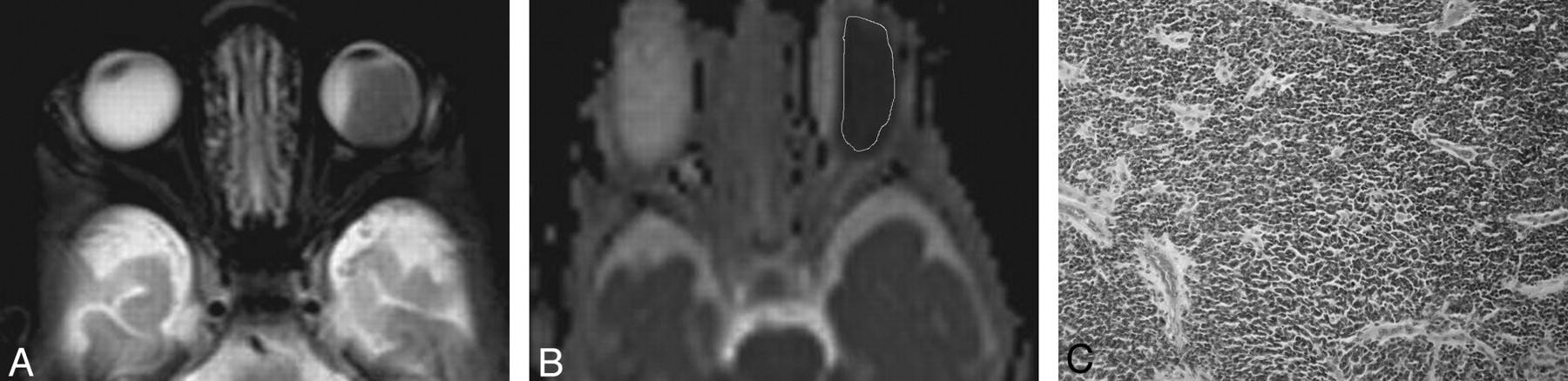
- Fig 1.
Unilateral large poorly differentiated retinoblastoma. A, Axial contrast T2-weighted image shows large tumor in the left globe. B, ADC map shows ROI localization of the tumor. There is restricted diffusion with low ADC value (0.44 × 10⁻³ mm2/s) of the tumor. C, Pathologic specimen shows poorly differentiated retinoblastoma with proliferating small round to polygonal cells that show hyperchromatic nuclei and minimal cytoplasm ×100 H&E.
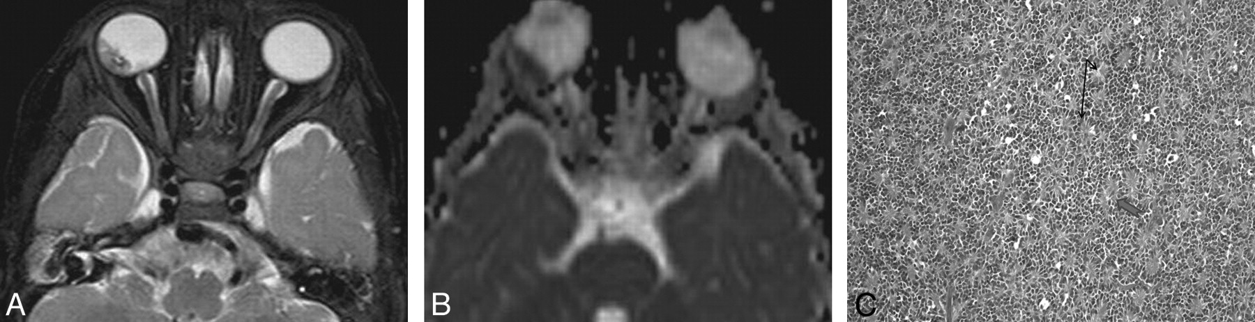
- Fig 2.
Unilateral medium-sized well-differentiated retinoblastoma. A, Axial T2-weighted image shows hypointense mass in the right globe, measuring 10 mm. B, ADC map shows restricted diffusion of the tumor with low ADC value (0.47 × 10⁻³ mm2/s). C, Pathologic specimen shows Flexner-Wintersteiner rosettes (black arrows) and fleurettes (large blue arrow), which indicate differentiation in retinoblastoma ×400 PAS.
- Fig 3.
Bilateral retinoblastoma. A, Axial T2-weighted image shows bilateral retinoblastomas with large tumor on left side and small tumor on the right side. B, ADC map shows restricted diffusion of both lesions. The ADC value of the left-side large tumor is 0.36 × 10⁻³ mm2/s and of right-side small tumor is 0.41 × 10⁻³ mm2/s. C, Pathologic specimen of large left-sided tumor shows poorly differentiated retinoblastoma with proliferating small, round to polygonal cells, which show hyperchromatic nuclei and minimal cytoplasm ×400 H&E.
- Fig 4.
Retinoblastoma with optic nerve invasion. A, Sagittal oblique contrast T1-weighted image shows retinoblastoma with extension into the optic nerve. B, ADC map shows restricted diffusion with low ADC value of retinoblastoma from the solid part of the tumor (0.33 × 10⁻³ mm2/s). Note extension of the tumor into the left optic nerve with restricted diffusion. C, Pathologic specimen shows optic nerve resection margin with invasion of retinoblastoma (red arrow) into the substance of the optic nerve (black arrow) ×25 PAS.


Tables
The minimum, maximum, mean, and standard deviation of ADC value of retinoblastoma in relation to prognostic parameters
Prognostic Parameter ADC Value P Value Pathologic grade Well differentiated (n = 16) 0.54 ± 0.20 (0.41–0.86) 0.007* Moderately differentiated (n = 20) 0.51 ± 0.07 (0.33–0.70) Poorly differentiated (n = 7) 0.44 ± 0.07 (0.23–0.57) Undifferentiated (n = 29) 0.41 ± 0.01 (0.20–0.59) Size Small (less than 10 mm; n = 11) 0.55 ± 0.09 (0.36–0.72) 0.015* Medium (10–15 mm; n = 36) 0.48 ± 0.08 (0.34–0.57) Large (more than 15 mm; n = 25) 0.38 ± 0.11 (0.20–0.86) Side Unilateral (n = 49) 0.53 ± 0.11 (0.34–0.86) 0.001* Bilateral (n = 23) 0.35 ± 0.11 (0.20–0.51) Growth pattern Endophytic (n = 18) 0.49 ± 0.16 (0.23–0.72) 0.640 Exophytic (n = 19) 0.49 ± 0.11 (0.25–0.70) Combined (n = 35) 0.46 ± 0.15 (0.20–0.86) Choroidal invasion Choroidal invasion (n = 34) 0.48 ± 0.13 (0.20–0.86) 0.661 No invasion (n = 38) 0.47 ± 0.15 (0.23–0.84) Optic nerve invasion Postlaminar (n = 8) 0.38 ± 0.11 (0.20–0.55) 0.003* Prelaminar (n = 15) 0.39 ± 0.23 (0.25–0.60) No invasion (n = 49) 0.52 ± 0.10 (0.40–0.86) -
↵* indicates significant P values.
-


