Advertisement

Main menu

  • Home
  • Content
    • Current Issue
    • Accepted Manuscripts
    • Article Preview
    • Past Issue Archive
    • Video Articles
    • AJNR Case Collection
    • Case of the Week Archive
    • Case of the Month Archive
    • Classic Case Archive
  • Special Collections
    • AJNR Awards
    • Low-Field MRI
    • Alzheimer Disease
    • ASNR Foundation Special Collection
    • Photon-Counting CT
    • View All
  • Multimedia
    • AJNR Podcasts
    • AJNR SCANtastic
    • Trainee Corner
    • MRI Safety Corner
    • Imaging Protocols
  • For Authors
    • Submit a Manuscript
    • Submit a Video Article
    • Submit an eLetter to the Editor/Response
    • Manuscript Submission Guidelines
    • Statistical Tips
    • Fast Publishing of Accepted Manuscripts
    • Graphical Abstract Preparation
    • Imaging Protocol Submission
    • Author Policies
  • About Us
    • About AJNR
    • Editorial Board
    • Editorial Board Alumni
  • More
    • Become a Reviewer/Academy of Reviewers
    • Subscribers
    • Permissions
    • Alerts
    • Feedback
    • Advertisers
    • ASNR Home

User menu

  • Alerts
  • Log in

Search

  • Advanced search
American Journal of Neuroradiology

American Journal of Neuroradiology

ASHNR American Society of Functional Neuroradiology ASHNR American Society of Pediatric Neuroradiology ASSR
  • Alerts
  • Log in

Advanced Search

  • Home
  • Content
    • Current Issue
    • Accepted Manuscripts
    • Article Preview
    • Past Issue Archive
    • Video Articles
    • AJNR Case Collection
    • Case of the Week Archive
    • Case of the Month Archive
    • Classic Case Archive
  • Special Collections
    • AJNR Awards
    • Low-Field MRI
    • Alzheimer Disease
    • ASNR Foundation Special Collection
    • Photon-Counting CT
    • View All
  • Multimedia
    • AJNR Podcasts
    • AJNR SCANtastic
    • Trainee Corner
    • MRI Safety Corner
    • Imaging Protocols
  • For Authors
    • Submit a Manuscript
    • Submit a Video Article
    • Submit an eLetter to the Editor/Response
    • Manuscript Submission Guidelines
    • Statistical Tips
    • Fast Publishing of Accepted Manuscripts
    • Graphical Abstract Preparation
    • Imaging Protocol Submission
    • Author Policies
  • About Us
    • About AJNR
    • Editorial Board
    • Editorial Board Alumni
  • More
    • Become a Reviewer/Academy of Reviewers
    • Subscribers
    • Permissions
    • Alerts
    • Feedback
    • Advertisers
    • ASNR Home
  • Follow AJNR on Twitter
  • Visit AJNR on Facebook
  • Follow AJNR on Instagram
  • Join AJNR on LinkedIn
  • RSS Feeds

AJNR Awards, New Junior Editors, and more. Read the latest AJNR updates

Review ArticleReview Articles
Open Access

The New World Health Organization Classification of Central Nervous System Tumors: What Can the Neuroradiologist Really Say?

A.G. Osborn, K.L. Salzman, M.M. Thurnher, J.H. Rees and M. Castillo
American Journal of Neuroradiology May 2012, 33 (5) 795-802; DOI: https://doi.org/10.3174/ajnr.A2583
A.G. Osborn
  • Find this author on Google Scholar
  • Find this author on PubMed
  • Search for this author on this site
K.L. Salzman
  • Find this author on Google Scholar
  • Find this author on PubMed
  • Search for this author on this site
M.M. Thurnher
  • Find this author on Google Scholar
  • Find this author on PubMed
  • Search for this author on this site
J.H. Rees
  • Find this author on Google Scholar
  • Find this author on PubMed
  • Search for this author on this site
M. Castillo
  • Find this author on Google Scholar
  • Find this author on PubMed
  • Search for this author on this site
  • Article
  • Figures & Data
  • Info & Metrics
  • Responses
  • References
  • PDF
Loading

Article Figures & Data

Figures

  • Fig 1.
    • Download figure
    • Open in new tab
    • Download powerpoint
    Fig 1.

    A young adult with intractable seizures. A, Axial fluid-attenuated inversion recovery image shows a hyperintense cortically based mass with subcortical stalk-like extension toward the ventricle (arrow). B, Axial T1C+ image shows an expanded gyrus with a hyperintense rim and hypointense center (arrow). Note subtle erosion of the inner calvaria. Angiocentric glioma was found at histologic examination.

  • Fig 2.
    • Download figure
    • Open in new tab
    • Download powerpoint
    Fig 2.

    A 20-month-old child with failure to thrive. A, Axial T2WI shows a massive inhomogeneously hyperintense suprasellar mass with extension into the left temporal lobe. B, Axial T1C+ image shows strong but heterogeneous enhancement. Pilomyxoid variant of pilocytic astrocytoma was documented at surgery. Case courtesy of R. Hewlett, MD.

  • Fig 3.
    • Download figure
    • Open in new tab
    • Download powerpoint
    Fig 3.

    Sagittal T1C+ image in an infant with hydrocephalus shows an intensely enhancing lobulated mass in the third and lateral ventricles. Histologic diagnosis was aCPP (WHO grade II).

  • Fig 4.
    • Download figure
    • Open in new tab
    • Download powerpoint
    Fig 4.

    A child with long-standing seizures. A, Sagittal T1WI shows a mixed cystic-solid mass. B, Axial T2WI shows that the nodule is inhomogeneously hyperintense. No enhancement was seen on contrast-enhanced images (not shown). Histologic diagnosis was PGNT.

  • Fig 5.
    • Download figure
    • Open in new tab
    • Download powerpoint
    Fig 5.

    A 21-year-old man with headaches. A, Axial T2* image shows a mixed cystic-solid fourth ventricular mass with fluid-fluid levels and hemorrhage. B, Postcontrast axial T1WI shows inhomogeneous enhancement. Histology proved RGNT of the fourth ventricle.

  • Fig 6.
    • Download figure
    • Open in new tab
    • Download powerpoint
    Fig 6.

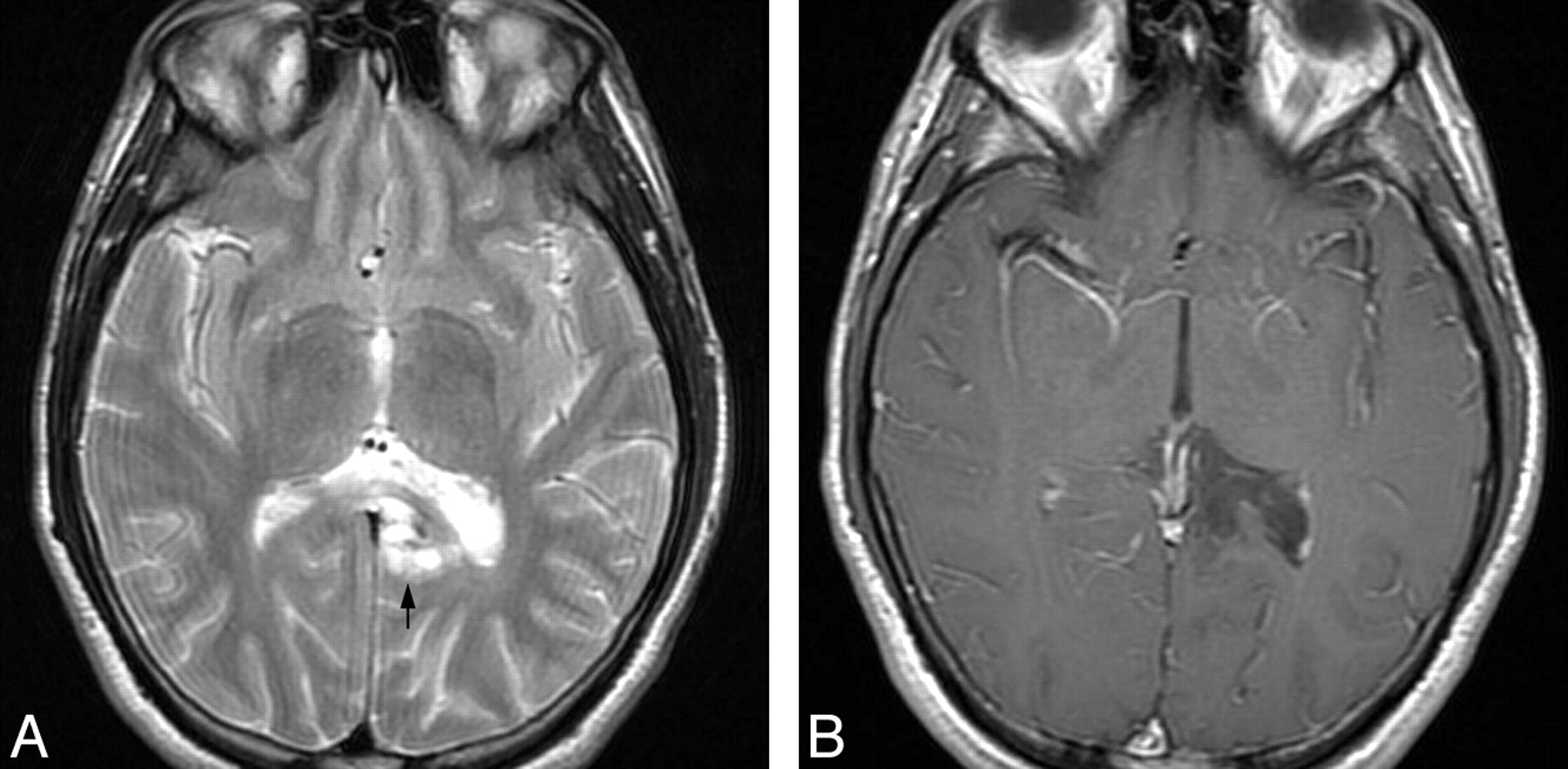
    A 24-year-old man presented with seizures. A, Axial T2WI shows a bubbly-appearing mass in the left parahippocampal gyrus (arrow). B, T1C+ image shows no enhancement. Histologic examination disclosed extraventricular neurocytoma. Case courtesy of C. Glastonbury, MBBS.

  • Fig 7.
    • Download figure
    • Open in new tab
    • Download powerpoint
    Fig 7.

    A 66-year-old patient who presented with headache and neck stiffness. A, Axial T2WI shows a well-delineated mixed iso-/hyperintense lobulated mass in the pineal region (arrow). B, Sagittal postcontrast T1WI shows that the mass enhances intensely. Papillary tumor of the pineal region was documented at pathologic examination.

  • Fig 8.
    • Download figure
    • Open in new tab
    • Download powerpoint
    Fig 8.

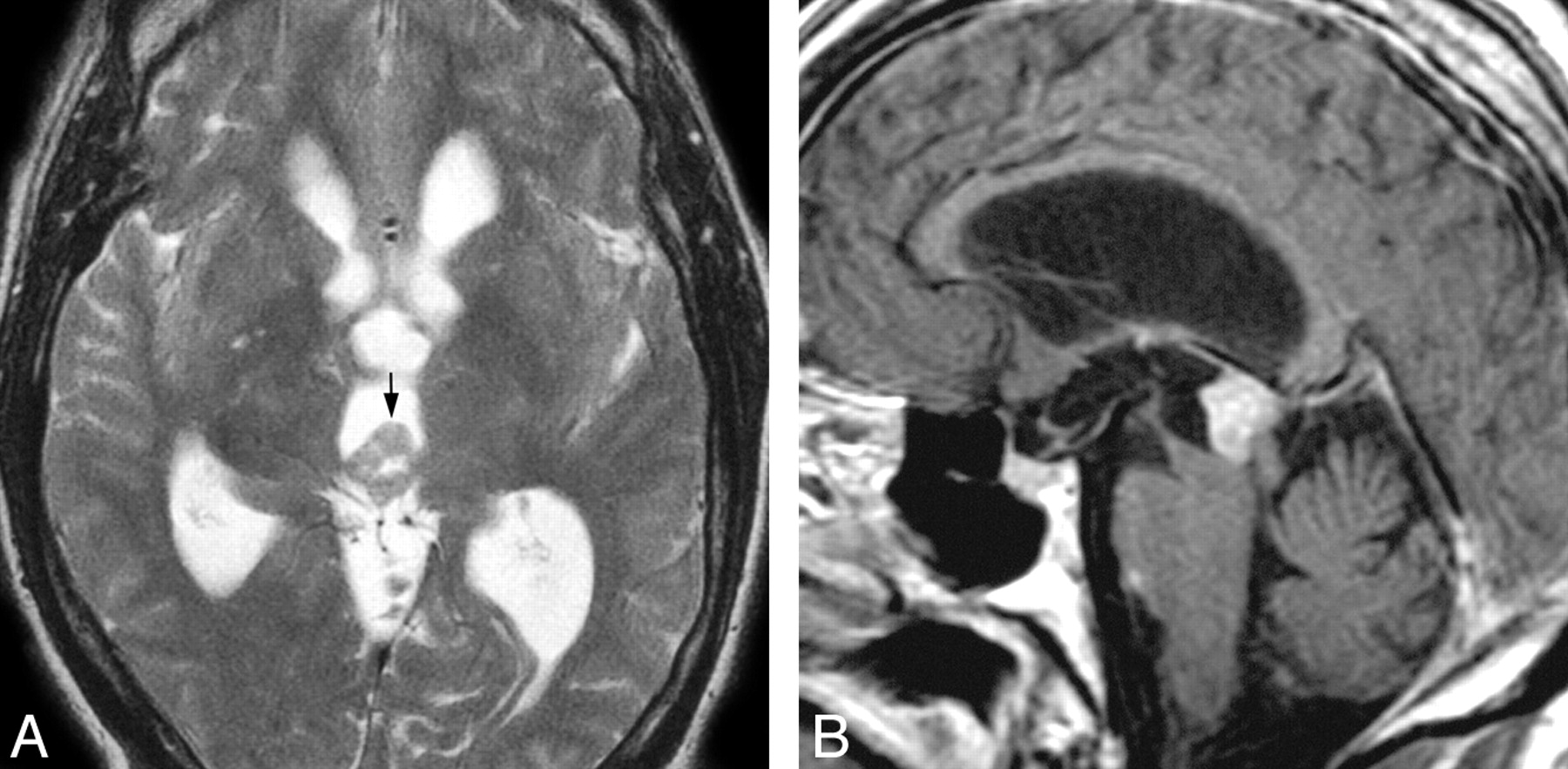
    A 57-year-old woman who presented with Parinaud syndrome. A, Axial T2WI shows a large mixed hyperintense mass with focal invasion of the right thalamus (arrow). B, Axial T1WI after contrast administration shows that the mass enhances strongly but inhomogeneously. Preoperative diagnosis of PPTID was confirmed at histologic examination.

  • Fig 9.
    • Download figure
    • Open in new tab
    • Download powerpoint
    Fig 9.

    Two different cases of medulloblastoma with extensive nodularity are illustrated. A, Coronal T1C+ image in a 10-month-old infant shows strong enhancement of grapelike nodules in a huge posterior fossa mass. MBEN was diagnosed at pathology. Case courtesy of B. Jones, MD. B, A child with headaches and papilledema has a grossly nodular enhancing mass in the right cerebellum. Histologic diagnosis was MBEN.

  • Fig 10.
    • Download figure
    • Open in new tab
    • Download powerpoint
    Fig 10.

    A 5-year-old child with surgically proved anaplastic MB. A, Axial T2WI shows a large mostly isointense mass in the left cerebellar hemisphere. B, T1C+ image the mass enhances intensely but somewhat heterogeneously. Case courtesy of S. Blaser, MD.

  • Fig 11.
    • Download figure
    • Open in new tab
    • Download powerpoint
    Fig 11.

    Newly recognized sellar region tumors are illustrated. A, Sagittal T1C+ image in a 22-year-old woman with hypopituitarism shows a well-delineated strongly enhancing infundibular mass that is clearly separate from the pituitary gland below. Pituicytoma. B, Sagittal T1C+ scan in a 69-year-old patient with headaches and bitemporal hemianopsia has an enhancing sellar/suprasellar mass that has enlarged the sella. Preoperative diagnosis was macroadenoma. Histologic diagnosis was SCO. Case courtesy of P. Hildenbrand, MD.

PreviousNext
Back to top

In this issue

American Journal of Neuroradiology: 33 (5)
American Journal of Neuroradiology
Vol. 33, Issue 5
1 May 2012
  • Table of Contents
  • Index by author
Advertisement
Print
Download PDF
Email Article

Thank you for your interest in spreading the word on American Journal of Neuroradiology.

NOTE: We only request your email address so that the person you are recommending the page to knows that you wanted them to see it, and that it is not junk mail. We do not capture any email address.

Enter multiple addresses on separate lines or separate them with commas.
The New World Health Organization Classification of Central Nervous System Tumors: What Can the Neuroradiologist Really Say?
(Your Name) has sent you a message from American Journal of Neuroradiology
(Your Name) thought you would like to see the American Journal of Neuroradiology web site.
CAPTCHA
This question is for testing whether or not you are a human visitor and to prevent automated spam submissions.
Cite this article
A.G. Osborn, K.L. Salzman, M.M. Thurnher, J.H. Rees, M. Castillo
The New World Health Organization Classification of Central Nervous System Tumors: What Can the Neuroradiologist Really Say?
American Journal of Neuroradiology May 2012, 33 (5) 795-802; DOI: 10.3174/ajnr.A2583

Citation Manager Formats

  • BibTeX
  • Bookends
  • EasyBib
  • EndNote (tagged)
  • EndNote 8 (xml)
  • Medlars
  • Mendeley
  • Papers
  • RefWorks Tagged
  • Ref Manager
  • RIS
  • Zotero
0 Responses
Respond to this article
Share
Bookmark this article
The New World Health Organization Classification of Central Nervous System Tumors: What Can the Neuroradiologist Really Say?
A.G. Osborn, K.L. Salzman, M.M. Thurnher, J.H. Rees, M. Castillo
American Journal of Neuroradiology May 2012, 33 (5) 795-802; DOI: 10.3174/ajnr.A2583
del.icio.us logo Twitter logo Facebook logo Mendeley logo
  • Tweet Widget
  • Facebook Like
  • Google Plus One
Purchase

Jump to section

  • Article
    • Abstract
    • ABBREVIATIONS:
    • Conclusions
    • Footnotes
    • References
  • Figures & Data
  • Info & Metrics
  • Responses
  • References
  • PDF

Related Articles

  • No related articles found.
  • PubMed
  • Google Scholar

Cited By...

  • No citing articles found.
  • Crossref
  • Google Scholar

This article has not yet been cited by articles in journals that are participating in Crossref Cited-by Linking.

More in this TOC Section

  • An Atlas of Neonatal Neurovascular Imaging Anatomy as Depicted with Microvascular Imaging: The Intracranial Arteries
  • An Atlas of Neonatal Neurovascular Imaging Anatomy as Depicted with Microvascular Imaging: The Intracranial Veins
  • Clinical Translation of Hyperpolarized 13C Metabolic Probes for Glioma Imaging
Show more Review Articles

Similar Articles

Advertisement

Indexed Content

  • Current Issue
  • Accepted Manuscripts
  • Article Preview
  • Past Issues
  • Editorials
  • Editor's Choice
  • Fellows' Journal Club
  • Letters to the Editor
  • Video Articles

Cases

  • Case Collection
  • Archive - Case of the Week
  • Archive - Case of the Month
  • Archive - Classic Case

More from AJNR

  • Trainee Corner
  • Imaging Protocols
  • MRI Safety Corner
  • Book Reviews

Multimedia

  • AJNR Podcasts
  • AJNR Scantastics

Resources

  • Turnaround Time
  • Submit a Manuscript
  • Submit a Video Article
  • Submit an eLetter to the Editor/Response
  • Manuscript Submission Guidelines
  • Statistical Tips
  • Fast Publishing of Accepted Manuscripts
  • Graphical Abstract Preparation
  • Imaging Protocol Submission
  • Evidence-Based Medicine Level Guide
  • Publishing Checklists
  • Author Policies
  • Become a Reviewer/Academy of Reviewers
  • News and Updates

About Us

  • About AJNR
  • Editorial Board
  • Editorial Board Alumni
  • Alerts
  • Permissions
  • Not an AJNR Subscriber? Join Now
  • Advertise with Us
  • Librarian Resources
  • Feedback
  • Terms and Conditions
  • AJNR Editorial Board Alumni

American Society of Neuroradiology

  • Not an ASNR Member? Join Now

© 2025 by the American Society of Neuroradiology All rights, including for text and data mining, AI training, and similar technologies, are reserved.
Print ISSN: 0195-6108 Online ISSN: 1936-959X

Powered by HighWire