
Article Figures & Data
Figures
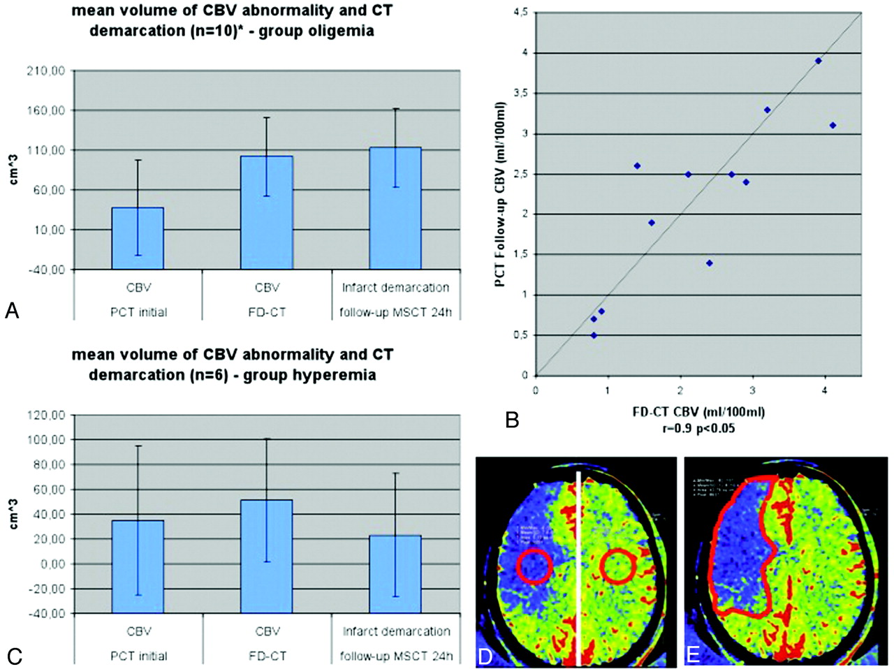
- Fig 1.
In the oligemia group (A), CBV abnormality volume increased from the initial measurement to the FPCT CBV measurement performed immediately at the end of treatment. The FPCT CBV abnormality volume was nearly identical to the infarct volume, as determined on the 24-hour follow-up CT. Comparison of CBV values (B) of the oligemia group revealed a high correlation on FPCT and follow-up PCT. In the hyperemia group (C), the abnormality volume increased only slightly, but not significantly, from initial to posttreatment FPCT CBV measurement. Infarct volume was lower than FPCT CBV abnormality volume. Only 2 patients in this group of 6 patients presented with stroke demarcation. (D) The region of interest for measurement of the absolute CBV values is displayed. (E) The region of interest for measurement of the volume is displayed.
- Fig 2.
Patient 9. The initial CT (A) and PCT CBV map (D) showed no abnormality. After successful revascularization, there was a small lesion (oligemia) identified on the FPCT CBV map (E, black arrows). This corresponded well with the lesion identified on the follow-up PCT CBV map (F, black arrows). The CBV abnormality observed on the FPCT CBV map (E) matches the sizes of the infarct observed on the follow-up MSCT (C, black arrows). Brain parenchyma reconstruction of the FPCT (B) was without findings.
- Fig 3.
Patient 2. A CBV lesion (oligemia) is observed on the initial PCT CBV map (D, red arrows); the CT scan (A) was without findings. Revascularization was not successful, and the abnormality had increased in size on the FPCT CBV map perform at the end of treatment (E). No evidence of hemorrhage or contrast extravasation was seen on the corresponding FPCT (B). On the 24-hour follow-up, the infarct observed on the MSCT (C, red arrows) corresponds to the CBV abnormality identified on both the FPCT CBV map (E) and the follow-up PCT CBV map (F).
- Fig 4.
Patient 11. The initial PCT CBV map (D) shows a large area of CBV abnormality (oligemia). No clearly defined infarct was identified on the initial CT (A). Revascularization was not successful. On the FPCT CBV map generated immediately after treatment, the CBV abnormality (E) was unchanged from that observed on the inital PCT study. A small area of hyperattenuation was seen on the FPCT performed immediately after treatment (B, black arrow). This was felt to be due to contrast medium extravasation. The follow-up PCT CBV map showed an abnormality corresponding to those observed the initial study and in the study performed immediately after treatment (F). The follow-up CT showed no evidence of the previously observed area of hyperattenuation. The area of infarction seen on the follow-up MSCT (C) matches that seen on the 3 CBV studies.
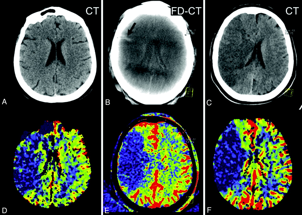
- Fig 5.
Patient 10. On the initial PCT CBV map, a small area of decreased CBV (oligemia) is present, adjacent to the lateral aspect of the right frontal horn (D, black arrows). After successful treatment, a hyperattenuated lesion (B, red arrow) within the basal ganglia, without space occupying effect, is obvious on the FPCT. The FPCT CBV map generated immediately after treatment shows an area of increased CBV (hyperemia) in this same area (E, black arrows). The follow-up PCT CBV map (F, black arrows) shows persistence of the area of elevated CBV. No clear area of infarct is seen on the corresponding noncontrast CT (C). In addition, the hyperattenuated abnormality seen on the FPCT (B, red arrow) is no longer visible. This represented contrast extravasation.


In this issue


Jump to section
Related Articles
Cited By...
- Clinical Applications of Conebeam CTP Imaging in Cerebral Disease: A Systematic Review
- Evaluation of time-resolved whole brain flat panel detector perfusion imaging using RAPID ANGIO in patients with acute stroke: comparison with CT perfusion imaging
- Correlation of Collateral Scores Derived from Whole-Brain Time-Resolved Flat Panel Detector Imaging in Acute Ischemic Stroke
- Multisociety Consensus Quality Improvement Revised Consensus Statement for Endovascular Therapy of Acute Ischemic Stroke
- Feasibility of Flat Panel Detector CT in Perfusion Assessment of Brain Arteriovenous Malformations: Initial Clinical Experience
- A novel reconstruction tool (syngo DynaCT Head Clear) in the post-processing of DynaCT images to reduce artefacts and improve image quality
- Time-Resolved C-Arm Computed Tomographic Angiography Derived From Computed Tomographic Perfusion Acquisition: New Capability for One-Stop-Shop Acute Ischemic Stroke Treatment in the Angiosuite
- Dynamic Angiography and Perfusion Imaging Using Flat Detector CT in the Angiography Suite: A Pilot Study in Patients with Acute Middle Cerebral Artery Occlusions
- C-Arm Flat Detector CT Parenchymal Blood Volume Thresholds for Identification of Infarcted Parenchyma in the Neurointerventional Suite
- Exploring the Value of Using Color-Coded Quantitative DSA Evaluation on Bilateral Common Carotid Arteries in Predicting the Reliability of Intra-Ascending Aorta Flat Detector CT-CBV Maps
- A Novel Technique for the Measurement of CBF and CBV with Robot-Arm-Mounted Flat Panel CT in a Large-Animal Model
- A prospective, multicenter pilot study investigating the utility of flat detector derived parenchymal blood volume maps to estimate cerebral blood volume in stroke patients
- Radiation Doses of Cerebral Blood Volume Measurements Using C-Arm CT: A Phantom Study
- Advances in Stroke: Advances in Interventional Radiology 2013