Advertisement

Main menu

  • Home
  • Content
    • Current Issue
    • Accepted Manuscripts
    • Article Preview
    • Past Issue Archive
    • Video Articles
    • AJNR Case Collection
    • Case of the Week Archive
    • Case of the Month Archive
    • Classic Case Archive
  • Special Collections
    • AJNR Awards
    • Low-Field MRI
    • Alzheimer Disease
    • ASNR Foundation Special Collection
    • Photon-Counting CT
    • View All
  • Multimedia
    • AJNR Podcasts
    • AJNR SCANtastic
    • Trainee Corner
    • MRI Safety Corner
    • Imaging Protocols
  • For Authors
    • Submit a Manuscript
    • Submit a Video Article
    • Submit an eLetter to the Editor/Response
    • Manuscript Submission Guidelines
    • Statistical Tips
    • Fast Publishing of Accepted Manuscripts
    • Graphical Abstract Preparation
    • Imaging Protocol Submission
    • Author Policies
  • About Us
    • About AJNR
    • Editorial Board
    • Editorial Board Alumni
  • More
    • Become a Reviewer/Academy of Reviewers
    • Subscribers
    • Permissions
    • Alerts
    • Feedback
    • Advertisers
    • ASNR Home

User menu

  • Alerts
  • Log in

Search

  • Advanced search
American Journal of Neuroradiology

American Journal of Neuroradiology

ASHNR American Society of Functional Neuroradiology ASHNR American Society of Pediatric Neuroradiology ASSR
  • Alerts
  • Log in

Advanced Search

  • Home
  • Content
    • Current Issue
    • Accepted Manuscripts
    • Article Preview
    • Past Issue Archive
    • Video Articles
    • AJNR Case Collection
    • Case of the Week Archive
    • Case of the Month Archive
    • Classic Case Archive
  • Special Collections
    • AJNR Awards
    • Low-Field MRI
    • Alzheimer Disease
    • ASNR Foundation Special Collection
    • Photon-Counting CT
    • View All
  • Multimedia
    • AJNR Podcasts
    • AJNR SCANtastic
    • Trainee Corner
    • MRI Safety Corner
    • Imaging Protocols
  • For Authors
    • Submit a Manuscript
    • Submit a Video Article
    • Submit an eLetter to the Editor/Response
    • Manuscript Submission Guidelines
    • Statistical Tips
    • Fast Publishing of Accepted Manuscripts
    • Graphical Abstract Preparation
    • Imaging Protocol Submission
    • Author Policies
  • About Us
    • About AJNR
    • Editorial Board
    • Editorial Board Alumni
  • More
    • Become a Reviewer/Academy of Reviewers
    • Subscribers
    • Permissions
    • Alerts
    • Feedback
    • Advertisers
    • ASNR Home
  • Follow AJNR on Twitter
  • Visit AJNR on Facebook
  • Follow AJNR on Instagram
  • Join AJNR on LinkedIn
  • RSS Feeds

AJNR Awards, New Junior Editors, and more. Read the latest AJNR updates

Research ArticleBrain
Open Access

CT Perfusion Mean Transit Time Maps Optimally Distinguish Benign Oligemia from True “At-Risk” Ischemic Penumbra, but Thresholds Vary by Postprocessing Technique

Shervin Kamalian, Shahmir Kamalian, A.A. Konstas, M.B. Maas, S. Payabvash, S.R. Pomerantz, P.W. Schaefer, K.L. Furie, R.G. González and M.H. Lev
American Journal of Neuroradiology March 2012, 33 (3) 545-549; DOI: https://doi.org/10.3174/ajnr.A2809
Shervin Kamalian
  • Find this author on Google Scholar
  • Find this author on PubMed
  • Search for this author on this site
Shahmir Kamalian
  • Find this author on Google Scholar
  • Find this author on PubMed
  • Search for this author on this site
A.A. Konstas
  • Find this author on Google Scholar
  • Find this author on PubMed
  • Search for this author on this site
M.B. Maas
  • Find this author on Google Scholar
  • Find this author on PubMed
  • Search for this author on this site
S. Payabvash
  • Find this author on Google Scholar
  • Find this author on PubMed
  • Search for this author on this site
S.R. Pomerantz
  • Find this author on Google Scholar
  • Find this author on PubMed
  • Search for this author on this site
P.W. Schaefer
  • Find this author on Google Scholar
  • Find this author on PubMed
  • Search for this author on this site
K.L. Furie
  • Find this author on Google Scholar
  • Find this author on PubMed
  • Search for this author on this site
R.G. González
  • Find this author on Google Scholar
  • Find this author on PubMed
  • Search for this author on this site
M.H. Lev
  • Find this author on Google Scholar
  • Find this author on PubMed
  • Search for this author on this site
  • Article
  • Figures & Data
  • Info & Metrics
  • Responses
  • References
  • PDF
Loading

Article Figures & Data

Figures

  • Tables
  • Fig 1.
    • Download figure
    • Open in new tab
    • Download powerpoint
    Fig 1.

    Expansion of core infarct size (area of selected axial section) between the admission DWI scan and the coregistered follow-up CT or MR imaging was an inclusion criterion, and was present in all patients (left). Sample ROC curves (right) showing the sensitivity/specificity of different CTP parameter thresholds used to define “at-risk” penumbra destined to infarct, comparing maps processed by using standard software. Green curves represent rMTT; blue curves, rCBF; orange, rCBV; and purple, the rCBF*rCBV voxel product value maps.
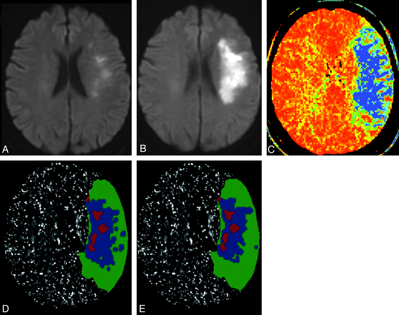
  • Fig 2.
    • Download figure
    • Open in new tab
    • Download powerpoint
    Fig 2.

    Example of thresholded MTT map prediction of penumbra destined to infarct in a 70-year-old woman presenting with left hemispheric stroke symptoms. Ictus-to-CTP imaging time was 5 hours 33 minutes, admission NIHSS score was 6, and follow-up MR imaging was performed 44 hours after admission CTP scanning; NIHSS score was 12. Infarct core is segmented on the admission DWI scan (A, and red overlays on D, E), and final infarct volume is segmented on follow-up DWI scan (B). CT-MTT map shows blue/green regions with increased mean transit time (C). D and E, respectively, show the optimally thresholded absolute-MTT (12 seconds threshold) and relative-MTT (249% threshold) maps, both postprocessed by using standard algorithm, which distinguish benign oligemia (green overlays) from true “at-risk” ischemic penumbra (blue overlays).

Tables

  • Figures
  • Optimal absolute and rMTT thresholds for identification of penumbra destined to infarct

    aMTTAUCaT (s)SensSpecrMTTAUCrT (%)SensSpec
    Std softwareAll0.7612.00.6500.761All0.782490.6460.796
    GM0.7612.00.6620.750GM0.782500.6570.785
    WM0.7713.10.6480.778WM0.782500.6760.778
    BG0.6113.30.2890.938BG0.631980.3260.922
    DC softwareAll0.7213.50.6390.704All0.711500.6490.692
    GM0.7312.00.7320.636GM0.731420.7130.663
    WM0.7114.40.6150.718WM0.701670.5990.725
    BG0.6313.80.3770.796BG0.631140.5420.641
    • Note:—All whole-brain optimal thresholds. GM indicates gray matter; BG, basal ganglia region-specific thresholds; aT, absolute thresholds (sec); rT, relative thresholds; sens, sensitivity; spec, specificity; std, standard algorithm; DC, delay-corrected algorithm; aMTT, absolute MTT.

PreviousNext
Back to top

In this issue

American Journal of Neuroradiology: 33 (3)
American Journal of Neuroradiology
Vol. 33, Issue 3
1 Mar 2012
  • Table of Contents
  • Index by author
Advertisement
Print
Download PDF
Email Article

Thank you for your interest in spreading the word on American Journal of Neuroradiology.

NOTE: We only request your email address so that the person you are recommending the page to knows that you wanted them to see it, and that it is not junk mail. We do not capture any email address.

Enter multiple addresses on separate lines or separate them with commas.
CT Perfusion Mean Transit Time Maps Optimally Distinguish Benign Oligemia from True “At-Risk” Ischemic Penumbra, but Thresholds Vary by Postprocessing Technique
(Your Name) has sent you a message from American Journal of Neuroradiology
(Your Name) thought you would like to see the American Journal of Neuroradiology web site.
CAPTCHA
This question is for testing whether or not you are a human visitor and to prevent automated spam submissions.
Cite this article
Shervin Kamalian, Shahmir Kamalian, A.A. Konstas, M.B. Maas, S. Payabvash, S.R. Pomerantz, P.W. Schaefer, K.L. Furie, R.G. González, M.H. Lev
CT Perfusion Mean Transit Time Maps Optimally Distinguish Benign Oligemia from True “At-Risk” Ischemic Penumbra, but Thresholds Vary by Postprocessing Technique
American Journal of Neuroradiology Mar 2012, 33 (3) 545-549; DOI: 10.3174/ajnr.A2809

Citation Manager Formats

  • BibTeX
  • Bookends
  • EasyBib
  • EndNote (tagged)
  • EndNote 8 (xml)
  • Medlars
  • Mendeley
  • Papers
  • RefWorks Tagged
  • Ref Manager
  • RIS
  • Zotero
0 Responses
Respond to this article
Share
Bookmark this article
CT Perfusion Mean Transit Time Maps Optimally Distinguish Benign Oligemia from True “At-Risk” Ischemic Penumbra, but Thresholds Vary by Postprocessing Technique
Shervin Kamalian, Shahmir Kamalian, A.A. Konstas, M.B. Maas, S. Payabvash, S.R. Pomerantz, P.W. Schaefer, K.L. Furie, R.G. González, M.H. Lev
American Journal of Neuroradiology Mar 2012, 33 (3) 545-549; DOI: 10.3174/ajnr.A2809
del.icio.us logo Twitter logo Facebook logo Mendeley logo
  • Tweet Widget
  • Facebook Like
  • Google Plus One
Purchase

Jump to section

  • Article
    • Abstract
    • ABBREVIATIONS:
    • Materials and Methods
    • Results
    • Discussion
    • Conclusions
    • Footnotes
    • References
  • Figures & Data
  • Info & Metrics
  • Responses
  • References
  • PDF

Related Articles

  • No related articles found.
  • PubMed
  • Google Scholar

Cited By...

  • Excellence is a habit: Enhancing predictions of language impairment by identifying stable features in clinical perfusion scans
  • Detecting CTP Truncation Artifacts in Acute Stroke Imaging from the Arterial Input and the Vascular Output Functions
  • Endovascular thrombectomy beyond 12 hours of stroke onset: a stroke networks experience of late intervention
  • Perfusion Computed Tomography for the Evaluation of Acute Ischemic Stroke: Strengths and Pitfalls
  • C-Arm Flat Detector CT Parenchymal Blood Volume Thresholds for Identification of Infarcted Parenchyma in the Neurointerventional Suite
  • Limited Reliability of Computed Tomographic Perfusion Acute Infarct Volume Measurements Compared With Diffusion-Weighted Imaging in Anterior Circulation Stroke
  • Optimal Perfusion Computed Tomographic Thresholds for Ischemic Core and Penumbra Are Not Time Dependent in the Clinically Relevant Time Window
  • Pretreatment Advanced Imaging in Patients with Stroke Treated with IV Thrombolysis: Evaluation of a Multihospital Data Base
  • Toward Patient-Tailored Perfusion Thresholds for Prediction of Stroke Outcome
  • CT Brain Perfusion Protocol to Eliminate the Need for Selecting a Venous Output Function
  • Imaging-based selection for intra-arterial stroke therapies
  • Location of the Clot and Outcome of Perfusion Defects in Acute Anterior Circulation Stroke Treated with Intravenous Thrombolysis
  • Reply:
  • Comments on an Article by Kamalian et al
  • Acute Stroke Imaging: CT with CT Angiography and CT Perfusion before Management Decisions
  • Crossref
  • Google Scholar

This article has not yet been cited by articles in journals that are participating in Crossref Cited-by Linking.

More in this TOC Section

  • Multimodal CT Provides Improved Performance for Lacunar Infarct Detection
  • Optimal MRI Sequence for Identifying Occlusion Location in Acute Stroke: Which Value of Time-Resolved Contrast-Enhanced MRA?
  • Evaluating the Effects of White Matter Multiple Sclerosis Lesions on the Volume Estimation of 6 Brain Tissue Segmentation Methods
Show more Brain

Similar Articles

Advertisement

Indexed Content

  • Current Issue
  • Accepted Manuscripts
  • Article Preview
  • Past Issues
  • Editorials
  • Editor's Choice
  • Fellows' Journal Club
  • Letters to the Editor
  • Video Articles

Cases

  • Case Collection
  • Archive - Case of the Week
  • Archive - Case of the Month
  • Archive - Classic Case

More from AJNR

  • Trainee Corner
  • Imaging Protocols
  • MRI Safety Corner
  • Book Reviews

Multimedia

  • AJNR Podcasts
  • AJNR Scantastics

Resources

  • Turnaround Time
  • Submit a Manuscript
  • Submit a Video Article
  • Submit an eLetter to the Editor/Response
  • Manuscript Submission Guidelines
  • Statistical Tips
  • Fast Publishing of Accepted Manuscripts
  • Graphical Abstract Preparation
  • Imaging Protocol Submission
  • Evidence-Based Medicine Level Guide
  • Publishing Checklists
  • Author Policies
  • Become a Reviewer/Academy of Reviewers
  • News and Updates

About Us

  • About AJNR
  • Editorial Board
  • Editorial Board Alumni
  • Alerts
  • Permissions
  • Not an AJNR Subscriber? Join Now
  • Advertise with Us
  • Librarian Resources
  • Feedback
  • Terms and Conditions
  • AJNR Editorial Board Alumni

American Society of Neuroradiology

  • Not an ASNR Member? Join Now

© 2025 by the American Society of Neuroradiology All rights, including for text and data mining, AI training, and similar technologies, are reserved.
Print ISSN: 0195-6108 Online ISSN: 1936-959X

Powered by HighWire