
Article Figures & Data
Figures
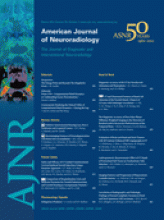
- Fig 1.
T2-weighted MR imaging appearance of a healthy 60-year-old woman (A), a 66-year-old woman with idiopathic Parkinson disease (B), and a 16-year-old female patient with idiopathic NBIA (C) obtained on a 1.5T scanner by using standard clinical TEs and TRs.
- Fig 2.
A clinical- and neuroimaging-based algorithm for evaluating patients with suspected NBIA. BG indicates basal ganglia; WM, white matter; FFF, facial-faucial-finger.
- Fig 3.
PKAN. A and B, The eye-of-the-tiger sign begins with T2 hyperintensity within the globus pallidus. C and D, Iron subsequently accumulates with time. Cerebral and/or cerebellar atrophy and white matter hyperintensity are not typical features.
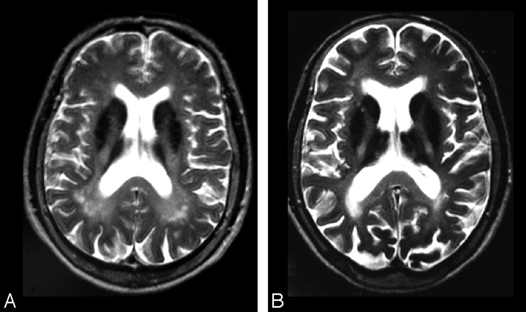
- Fig 4.
NFT. A, Patchy hypointensity is typically seen within multiple deep gray nuclei, including the caudate, putamen, globus pallidus, and thalamus in symptomatic cases. B, Concurrent T2 hyperintensities (cavitation) may be seen within regions of hypointensity. Images courtesy of P.F. Chinnery.
- Fig 5.
NAD. Iron deposition may be seen in the globus pallidus (A) and the substantia nigra (B) on T2* and T2 images. C, Confluent white matter hyperintensities may be seen on fluid-attenuated inversion recovery sequences as well. D, Global cerebellar atrophy is a frequent feature.
- Fig 6.
ACP. A and B, More homogeneous iron deposition is seen within the basal ganglia, with juxtaposed confluent white matter hyperintensities on T2-weighted sequences. Images courtesy of H. Miyajima.
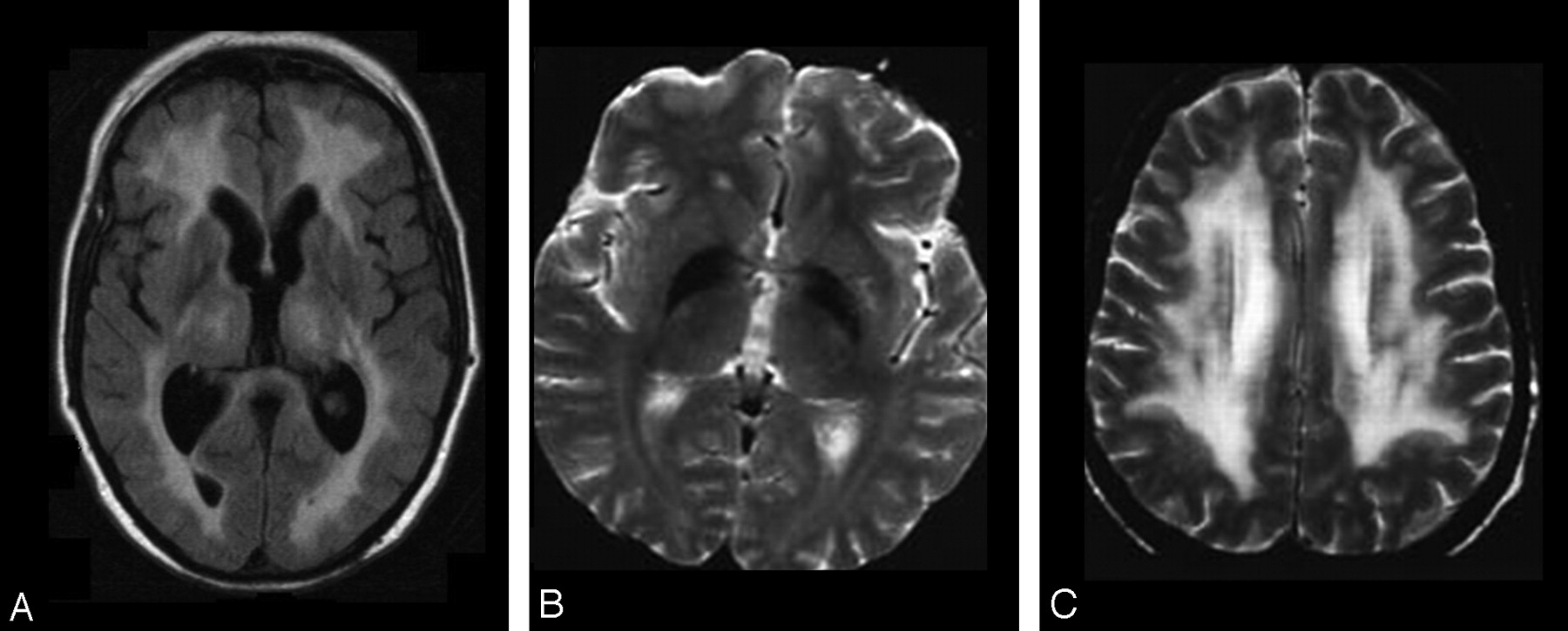
- Fig 7.
FAHN. Evidence of iron deposition in the globus pallidus (A) and, to a lesser extent, the substantia nigra (B) may be seen on T2-weighted images. C, Confluent white matter abnormalities may be apparent on T2/fluid-attenuated inversion recovery sequences. D, Mild cerebral atrophy may occur, along with significant pontocerebellar atrophy and thinning of the corpus callosum (A).
- Fig 8.
KRS. Globus pallidus, caudate, and putamen hypointensity may be seen on T2-weighted images (A and B), in addition to generalized cerebral and cerebellar atrophy (A and C).
- Fig 9.
WSS. Extensive confluent white matter T2 hyperintensity is typical of the disorder (A and C), while hypointensity of the globus pallidus on T2 sequences is an inconsistent feature (B). Images courtesy of S. Bohlega.
- Fig 10.
SENDA. Hypointensity of the globus pallidus (A) is overshadowed by that of the substantia nigra and cerebral peduncles (B) on T2-weighted imaging. C, T1 sequences demonstrate hyperintensity of the substantia nigra and cerebral peduncles with central linear hypointensity. D, Global cerebral atrophy is also a feature.


Tables
Metal T1 Appearance T2 Appearance Other Features Ca2+ Hypo-/hyperintense Hypointense Hyperdense on CT Fe3+ Isointense Hypointense Isodense on CT Mn2+ Hyperintense Isointense Cu2+ Iso-/hyperintense Hypo-/hyperintense -
Note:—Mn2+ indicates manganese ions; Cu2+, copper ions.
-
Disorder Iron Deposition White Matter Involvement Other Findings PKAN Globus pallidus, substantia nigra (mild) No Eye-of-the-tiger sign PLAN Globus pallidus,a substantia nigraa Mild Moderate cerebellar atrophy NFT “Patchy” globus pallidus, putamen, caudate, dentate, thalamus Mild, moderate Cystic cavitation, mild cerebral, cerebellar atrophy ACP Globus pallidus, putamen, caudate, thalamus, red nucleus, dentate Moderate, severe Mild cerebellar atrophy FAHN Globus pallidus, substantia nigrab Moderate Pontocerebellar atrophy KRS Globus pallidus, putamen, caudateb Severe cerebral, cerebellar, brain stem atrophy WSS Globus pallidusb Severe, confluent SENDA Substantia nigra, globus pallidus Occasional Midbrain T1 hyperintensity
In this issue


Jump to section
Related Articles
Cited By...
- Early Neuroimaging Markers in {beta} Propeller Protein-Associated Neurodegeneration
- Cross-sectional Observations on the Natural History of Mucolipidosis Type IV
- Genetic Risk for Hemochromatosis is Associated with Movement Disorders
- Imaging Patterns Characterizing Mitochondrial Leukodystrophies
- Quantification of different iron forms in the aceruloplasminemia brain to explore iron-related neurodegeneration
- Autophagy in the mammalian nervous system: a primer for neuroscientists
- 13-year diagnostic delay as cerebral palsy of an Iraqi patient with NBIA type 4
- Brain MR Imaging Findings in Woodhouse-Sakati Syndrome
- Transient swelling in the globus pallidus and substantia nigra in childhood suggests SENDA/BPAN
- Cortical pencil lining in neuroferritinopathy: A diagnostic clue
- Pediatric movement disorders: Five new things