Advertisement

Main menu

  • Home
  • Content
    • Current Issue
    • Accepted Manuscripts
    • Article Preview
    • Past Issue Archive
    • Video Articles
    • AJNR Case Collection
    • Case of the Week Archive
    • Case of the Month Archive
    • Classic Case Archive
  • Special Collections
    • AJNR Awards
    • Low-Field MRI
    • Alzheimer Disease
    • ASNR Foundation Special Collection
    • Photon-Counting CT
    • View All
  • Multimedia
    • AJNR Podcasts
    • AJNR SCANtastic
    • Trainee Corner
    • MRI Safety Corner
    • Imaging Protocols
  • For Authors
    • Submit a Manuscript
    • Submit a Video Article
    • Submit an eLetter to the Editor/Response
    • Manuscript Submission Guidelines
    • Statistical Tips
    • Fast Publishing of Accepted Manuscripts
    • Graphical Abstract Preparation
    • Imaging Protocol Submission
    • Author Policies
  • About Us
    • About AJNR
    • Editorial Board
    • Editorial Board Alumni
  • More
    • Become a Reviewer/Academy of Reviewers
    • Subscribers
    • Permissions
    • Alerts
    • Feedback
    • Advertisers
    • ASNR Home

User menu

  • Alerts
  • Log in

Search

  • Advanced search
American Journal of Neuroradiology

American Journal of Neuroradiology

ASHNR American Society of Functional Neuroradiology ASHNR American Society of Pediatric Neuroradiology ASSR
  • Alerts
  • Log in

Advanced Search

  • Home
  • Content
    • Current Issue
    • Accepted Manuscripts
    • Article Preview
    • Past Issue Archive
    • Video Articles
    • AJNR Case Collection
    • Case of the Week Archive
    • Case of the Month Archive
    • Classic Case Archive
  • Special Collections
    • AJNR Awards
    • Low-Field MRI
    • Alzheimer Disease
    • ASNR Foundation Special Collection
    • Photon-Counting CT
    • View All
  • Multimedia
    • AJNR Podcasts
    • AJNR SCANtastic
    • Trainee Corner
    • MRI Safety Corner
    • Imaging Protocols
  • For Authors
    • Submit a Manuscript
    • Submit a Video Article
    • Submit an eLetter to the Editor/Response
    • Manuscript Submission Guidelines
    • Statistical Tips
    • Fast Publishing of Accepted Manuscripts
    • Graphical Abstract Preparation
    • Imaging Protocol Submission
    • Author Policies
  • About Us
    • About AJNR
    • Editorial Board
    • Editorial Board Alumni
  • More
    • Become a Reviewer/Academy of Reviewers
    • Subscribers
    • Permissions
    • Alerts
    • Feedback
    • Advertisers
    • ASNR Home
  • Follow AJNR on Twitter
  • Visit AJNR on Facebook
  • Follow AJNR on Instagram
  • Join AJNR on LinkedIn
  • RSS Feeds

AJNR Awards, New Junior Editors, and more. Read the latest AJNR updates

Research ArticleFunctional

Clinical Standardized fMRI Reveals Altered Language Lateralization in Patients with Brain Tumor

S. Partovi, B. Jacobi, N. Rapps, L. Zipp, S. Karimi, F. Rengier, J.K. Lyo and C. Stippich
American Journal of Neuroradiology December 2012, 33 (11) 2151-2157; DOI: https://doi.org/10.3174/ajnr.A3137
S. Partovi
aFrom the Department of Neuroradiology (S.P., L.Z., F.R., C.S.), University Hospital of Basel, Basel, Switzerland
bDepartment of Radiology, Section of Neuroradiology (S.P., S.K., J.K.L.), Memorial Sloan-Kettering Cancer Center, New York, New York
  • Find this author on Google Scholar
  • Find this author on PubMed
  • Search for this author on this site
B. Jacobi
cClinical Cooperation Unit Molecular Hematology/Oncology (B.J.), German Cancer Research Center, Heidelberg, Germany
eDepartments of Internal Medicine V (B.J.), Neuroradiology (N.R., C.S.), and Diagnostic and Interventional Radiology (F.R.), University Hospital of Heidelberg, Heidelberg, Germany.
  • Find this author on Google Scholar
  • Find this author on PubMed
  • Search for this author on this site
N. Rapps
dDepartment of Psychosomatic Medicine and Psychotherapy (N.R.), University Hospital of Tuebingen, Tuebingen, Germany
eDepartments of Internal Medicine V (B.J.), Neuroradiology (N.R., C.S.), and Diagnostic and Interventional Radiology (F.R.), University Hospital of Heidelberg, Heidelberg, Germany.
  • Find this author on Google Scholar
  • Find this author on PubMed
  • Search for this author on this site
L. Zipp
aFrom the Department of Neuroradiology (S.P., L.Z., F.R., C.S.), University Hospital of Basel, Basel, Switzerland
  • Find this author on Google Scholar
  • Find this author on PubMed
  • Search for this author on this site
S. Karimi
bDepartment of Radiology, Section of Neuroradiology (S.P., S.K., J.K.L.), Memorial Sloan-Kettering Cancer Center, New York, New York
  • Find this author on Google Scholar
  • Find this author on PubMed
  • Search for this author on this site
F. Rengier
aFrom the Department of Neuroradiology (S.P., L.Z., F.R., C.S.), University Hospital of Basel, Basel, Switzerland
eDepartments of Internal Medicine V (B.J.), Neuroradiology (N.R., C.S.), and Diagnostic and Interventional Radiology (F.R.), University Hospital of Heidelberg, Heidelberg, Germany.
  • Find this author on Google Scholar
  • Find this author on PubMed
  • Search for this author on this site
J.K. Lyo
bDepartment of Radiology, Section of Neuroradiology (S.P., S.K., J.K.L.), Memorial Sloan-Kettering Cancer Center, New York, New York
  • Find this author on Google Scholar
  • Find this author on PubMed
  • Search for this author on this site
C. Stippich
aFrom the Department of Neuroradiology (S.P., L.Z., F.R., C.S.), University Hospital of Basel, Basel, Switzerland
eDepartments of Internal Medicine V (B.J.), Neuroradiology (N.R., C.S.), and Diagnostic and Interventional Radiology (F.R.), University Hospital of Heidelberg, Heidelberg, Germany.
  • Find this author on Google Scholar
  • Find this author on PubMed
  • Search for this author on this site
  • Article
  • Figures & Data
  • Info & Metrics
  • Responses
  • References
  • PDF
Loading

Article Figures & Data

Figures

  • Tables
  • Fig 1.
    • Download figure
    • Open in new tab
    • Download powerpoint
    Fig 1.

    Applied visually triggered paradigms in clinical language fMRI. A, Two examples of the sentence generation paradigm are shown. Comic-like pictures such as the lion or the elephant are shown and the subject has to verbalize a standard simple sentence, such as “The lion is dangerous” or “The elephant is heavy.” Altogether, this paradigm consists of a set of 12 visual comic-like stimuli, with 12 corresponding standard simple sentences. B, Two examples of the word-generation paradigm are shown. Generic terms like “cars” or “countries” are shown and the subject is asked to generate as many words as possible, such as, “Ford, Porsche, Mercedes,” or “United States, Switzerland, Canada.” Altogether, this paradigm consists of a set of 12 generic terms.
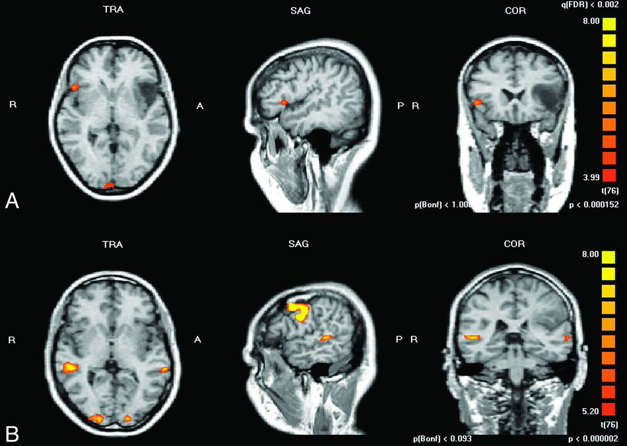
  • Fig 2.
    • Download figure
    • Open in new tab
    • Download powerpoint
    Fig 2.

    Representative instances of the patient group are demonstrated. A, A right-handed 62-year-old patient with left hemispheric frontal astrocytoma (WHO II) is depicted. The word-generation paradigm was applied and a lateralization index of Broca area versus its right hemisphere homolog of −0.85 was calculated (lateralization toward right hemisphere). B, A right-handed 49-year-old patient with left hemispheric parietal astrocytoma (WHO II) is depicted. A sentence-generation paradigm was applied and a lateralization index of Wernicke area versus its right hemisphere homolog of −0.84 was calculated (lateralization toward right hemisphere).

  • Fig 3.
    • Download figure
    • Open in new tab
    • Download powerpoint
    Fig 3.

    Local lateralization indexes in the patient group compared with healthy volunteers are depicted as box-and-whisker plots. A, A significant decrease of the lateralization index in Broca area versus its right hemisphere homolog, when performing the word generation paradigm for brain tumors affecting Broca area, is shown (P = .017). B, A significant reduction of lateralization index in Wernicke area versus its right hemisphere homolog, when performing sentence generation paradigm for brain tumors affecting Wernicke area, is shown (P = .007).

Tables

  • Figures
  • Baseline characteristics of the control (healthy volunteers) and patient groups

    Healthy volunteers (n = 14)
        Age, years (± SD)26 ± 5
        Male sex (n [%])7 (50%)
    Patient population (n = 57)
        Age, years (± SD)44 ± 16
        Male sex (n [%])32 (56%)
    Tumor localization (n [%])
        Affecting Broca area19 (33%)
        Affecting Wernicke area38 (67%)
    WHO tumor grade (n [%])
        I9 (16%)
        II12 (21%)
        III4 (4%)
        IV12 (21%)
        No WHO grade16 (28%)
        No histology available4 (7%)
    • Note:—For tumor data, n = 57.

PreviousNext
Back to top

In this issue

American Journal of Neuroradiology: 33 (11)
American Journal of Neuroradiology
Vol. 33, Issue 11
1 Dec 2012
  • Table of Contents
  • Index by author
Advertisement
Print
Download PDF
Email Article

Thank you for your interest in spreading the word on American Journal of Neuroradiology.

NOTE: We only request your email address so that the person you are recommending the page to knows that you wanted them to see it, and that it is not junk mail. We do not capture any email address.

Enter multiple addresses on separate lines or separate them with commas.
Clinical Standardized fMRI Reveals Altered Language Lateralization in Patients with Brain Tumor
(Your Name) has sent you a message from American Journal of Neuroradiology
(Your Name) thought you would like to see the American Journal of Neuroradiology web site.
CAPTCHA
This question is for testing whether or not you are a human visitor and to prevent automated spam submissions.
Cite this article
S. Partovi, B. Jacobi, N. Rapps, L. Zipp, S. Karimi, F. Rengier, J.K. Lyo, C. Stippich
Clinical Standardized fMRI Reveals Altered Language Lateralization in Patients with Brain Tumor
American Journal of Neuroradiology Dec 2012, 33 (11) 2151-2157; DOI: 10.3174/ajnr.A3137

Citation Manager Formats

  • BibTeX
  • Bookends
  • EasyBib
  • EndNote (tagged)
  • EndNote 8 (xml)
  • Medlars
  • Mendeley
  • Papers
  • RefWorks Tagged
  • Ref Manager
  • RIS
  • Zotero
0 Responses
Respond to this article
Share
Bookmark this article
Clinical Standardized fMRI Reveals Altered Language Lateralization in Patients with Brain Tumor
S. Partovi, B. Jacobi, N. Rapps, L. Zipp, S. Karimi, F. Rengier, J.K. Lyo, C. Stippich
American Journal of Neuroradiology Dec 2012, 33 (11) 2151-2157; DOI: 10.3174/ajnr.A3137
del.icio.us logo Twitter logo Facebook logo Mendeley logo
  • Tweet Widget
  • Facebook Like
  • Google Plus One
Purchase

Jump to section

  • Article
    • Abstract
    • ABBREVIATIONS:
    • Materials and Methods
    • Results
    • Discussion
    • Conclusions
    • Footnotes
    • References
  • Figures & Data
  • Info & Metrics
  • Responses
  • References
  • PDF

Related Articles

  • No related articles found.
  • PubMed
  • Google Scholar

Cited By...

  • Grey matter reshaping of language-related regions depends on tumor lateralization
  • A new fMRI localizer for preoperative language mapping using a sentence completion task: Validity, choice of baseline condition, and test-retest reliability
  • Resting-State Functional Connectivity of the Middle Frontal Gyrus Can Predict Language Lateralization in Patients with Brain Tumors
  • Crossref
  • Google Scholar

This article has not yet been cited by articles in journals that are participating in Crossref Cited-by Linking.

More in this TOC Section

  • Kurtosis and Epileptogenic Tubers: A Pilot Study
  • Glutaric Aciduria Type 1: DK vs. Conventional MRI
  • Multiparametric MRI in PEDS Pontine Glioma
Show more Functional

Similar Articles

Advertisement

Indexed Content

  • Current Issue
  • Accepted Manuscripts
  • Article Preview
  • Past Issues
  • Editorials
  • Editor's Choice
  • Fellows' Journal Club
  • Letters to the Editor
  • Video Articles

Cases

  • Case Collection
  • Archive - Case of the Week
  • Archive - Case of the Month
  • Archive - Classic Case

More from AJNR

  • Trainee Corner
  • Imaging Protocols
  • MRI Safety Corner
  • Book Reviews

Multimedia

  • AJNR Podcasts
  • AJNR Scantastics

Resources

  • Turnaround Time
  • Submit a Manuscript
  • Submit a Video Article
  • Submit an eLetter to the Editor/Response
  • Manuscript Submission Guidelines
  • Statistical Tips
  • Fast Publishing of Accepted Manuscripts
  • Graphical Abstract Preparation
  • Imaging Protocol Submission
  • Evidence-Based Medicine Level Guide
  • Publishing Checklists
  • Author Policies
  • Become a Reviewer/Academy of Reviewers
  • News and Updates

About Us

  • About AJNR
  • Editorial Board
  • Editorial Board Alumni
  • Alerts
  • Permissions
  • Not an AJNR Subscriber? Join Now
  • Advertise with Us
  • Librarian Resources
  • Feedback
  • Terms and Conditions
  • AJNR Editorial Board Alumni

American Society of Neuroradiology

  • Not an ASNR Member? Join Now

© 2025 by the American Society of Neuroradiology All rights, including for text and data mining, AI training, and similar technologies, are reserved.
Print ISSN: 0195-6108 Online ISSN: 1936-959X

Powered by HighWire