Advertisement

Main menu

  • Home
  • Content
    • Current Issue
    • Accepted Manuscripts
    • Article Preview
    • Past Issue Archive
    • Video Articles
    • AJNR Case Collection
    • Case of the Week Archive
    • Case of the Month Archive
    • Classic Case Archive
  • Special Collections
    • AJNR Awards
    • Low-Field MRI
    • Alzheimer Disease
    • ASNR Foundation Special Collection
    • Photon-Counting CT
    • View All
  • Multimedia
    • AJNR Podcasts
    • AJNR SCANtastic
    • Trainee Corner
    • MRI Safety Corner
    • Imaging Protocols
  • For Authors
    • Submit a Manuscript
    • Submit a Video Article
    • Submit an eLetter to the Editor/Response
    • Manuscript Submission Guidelines
    • Statistical Tips
    • Fast Publishing of Accepted Manuscripts
    • Graphical Abstract Preparation
    • Imaging Protocol Submission
    • Author Policies
  • About Us
    • About AJNR
    • Editorial Board
    • Editorial Board Alumni
  • More
    • Become a Reviewer/Academy of Reviewers
    • Subscribers
    • Permissions
    • Alerts
    • Feedback
    • Advertisers
    • ASNR Home

User menu

  • Alerts
  • Log in

Search

  • Advanced search
American Journal of Neuroradiology

American Journal of Neuroradiology

ASHNR American Society of Functional Neuroradiology ASHNR American Society of Pediatric Neuroradiology ASSR
  • Alerts
  • Log in

Advanced Search

  • Home
  • Content
    • Current Issue
    • Accepted Manuscripts
    • Article Preview
    • Past Issue Archive
    • Video Articles
    • AJNR Case Collection
    • Case of the Week Archive
    • Case of the Month Archive
    • Classic Case Archive
  • Special Collections
    • AJNR Awards
    • Low-Field MRI
    • Alzheimer Disease
    • ASNR Foundation Special Collection
    • Photon-Counting CT
    • View All
  • Multimedia
    • AJNR Podcasts
    • AJNR SCANtastic
    • Trainee Corner
    • MRI Safety Corner
    • Imaging Protocols
  • For Authors
    • Submit a Manuscript
    • Submit a Video Article
    • Submit an eLetter to the Editor/Response
    • Manuscript Submission Guidelines
    • Statistical Tips
    • Fast Publishing of Accepted Manuscripts
    • Graphical Abstract Preparation
    • Imaging Protocol Submission
    • Author Policies
  • About Us
    • About AJNR
    • Editorial Board
    • Editorial Board Alumni
  • More
    • Become a Reviewer/Academy of Reviewers
    • Subscribers
    • Permissions
    • Alerts
    • Feedback
    • Advertisers
    • ASNR Home
  • Follow AJNR on Twitter
  • Visit AJNR on Facebook
  • Follow AJNR on Instagram
  • Join AJNR on LinkedIn
  • RSS Feeds

AJNR Awards, New Junior Editors, and more. Read the latest AJNR updates

Getting new auth cookie, if you see this message a lot, tell someone!
Research ArticleBrain

Comparison of Conventional CTA and Volume Perfusion CTA in Evaluation of Cerebral Arterial Vasculature in Acute Stroke

M. Saake, P. Goelitz, T. Struffert, L. Breuer, B. Volbers, A. Doerfler and S. Kloska
American Journal of Neuroradiology December 2012, 33 (11) 2068-2073; DOI: https://doi.org/10.3174/ajnr.A3155
M. Saake
aFrom the Departments of Neuroradiology (M.S., P.G., T.S., A.D., S.K.)
  • Find this author on Google Scholar
  • Find this author on PubMed
  • Search for this author on this site
P. Goelitz
aFrom the Departments of Neuroradiology (M.S., P.G., T.S., A.D., S.K.)
  • Find this author on Google Scholar
  • Find this author on PubMed
  • Search for this author on this site
T. Struffert
aFrom the Departments of Neuroradiology (M.S., P.G., T.S., A.D., S.K.)
  • Find this author on Google Scholar
  • Find this author on PubMed
  • Search for this author on this site
L. Breuer
aFrom the Departments of Neuroradiology (M.S., P.G., T.S., A.D., S.K.)
  • Find this author on Google Scholar
  • Find this author on PubMed
  • Search for this author on this site
B. Volbers
bNeurology (L.B., B.V.), University of Erlangen-Nuremberg, Erlangen, Germany.
  • Find this author on Google Scholar
  • Find this author on PubMed
  • Search for this author on this site
A. Doerfler
bNeurology (L.B., B.V.), University of Erlangen-Nuremberg, Erlangen, Germany.
  • Find this author on Google Scholar
  • Find this author on PubMed
  • Search for this author on this site
S. Kloska
aFrom the Departments of Neuroradiology (M.S., P.G., T.S., A.D., S.K.)
  • Find this author on Google Scholar
  • Find this author on PubMed
  • Search for this author on this site
  • Article
  • Figures & Data
  • Info & Metrics
  • Responses
  • References
  • PDF
Loading

Article Figures & Data

Figures

  • Tables
  • Fig 1.
    • Download figure
    • Open in new tab
    • Download powerpoint
    Fig 1.

    A and B, CTA, transversal and coronal MIP reconstructions; image thickness 10 mm. C and D, VPCTA, transversal and coronal MIP reconstructions; image thickness 10 mm. The same window settings were used for all reconstructions. Occlusion of the distal main branch of the right middle cerebral artery is demonstrated in CTA and VPCTA (arrows). In VPCTA, veins are less contrasted.

  • Fig 2.
    • Download figure
    • Open in new tab
    • Download powerpoint
    Fig 2.

    A and B, CTA, coronal and sagittal MIP reconstructions; image thickness 10 mm. C and D, VPCTA, coronal and sagittal MIP reconstructions; image thickness 10 mm. The same window settings were used for all reconstructions. Occlusion of the A2 segment of the left anterior cerebral artery is clearly visible in CTA and VPCTA (arrows).
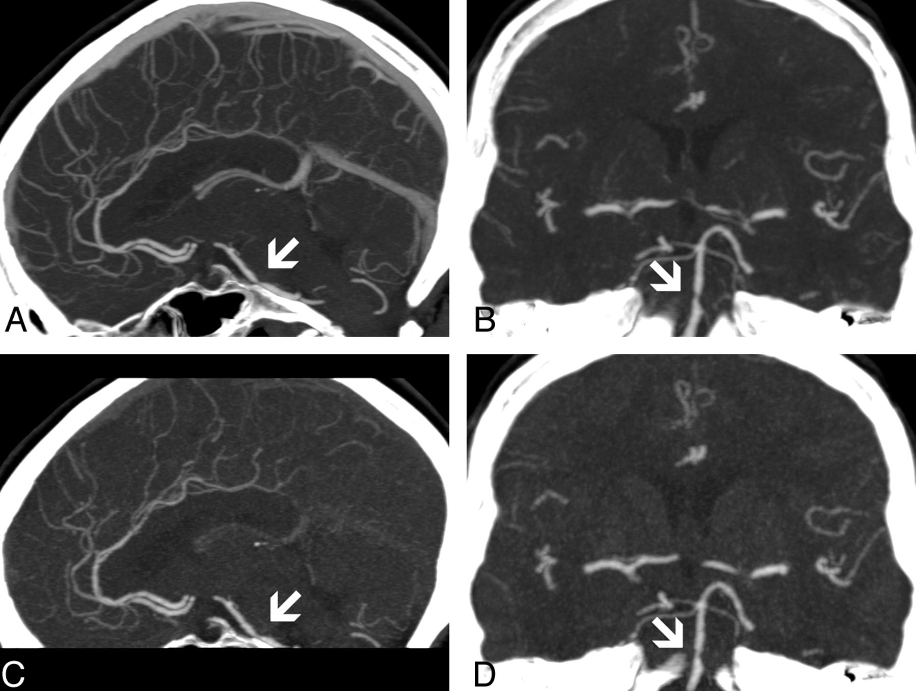
  • Fig 3.
    • Download figure
    • Open in new tab
    • Download powerpoint
    Fig 3.

    A and B, CTA, sagittal and coronal MIP reconstructions; image thickness 10 mm. C and D, VPCTA, sagittal and coronal MIP reconstructions; image thickness 10 mm. The same window settings were used for all reconstructions. Stenosis >50% of the basilar artery is pictured in CTA and VPCTA (arrows). Generally, the most caudal part of the basilar artery is not covered in VPCTA, as direct irradiation of the eye lenses has to be avoided.

  • Fig 4.
    • Download figure
    • Open in new tab
    • Download powerpoint
    Fig 4.

    A, CTA, transversal MIP reconstruction; image thickness 10 mm. B, VPCTA, transversal MIP reconstruction; image thickness 10 mm. The same window settings were used for both reconstructions. While CTA was scored normal, in VPCTA, stenosis >50% of a proximal right M2 segment was rated (arrows).

Tables

  • Figures
    • View popup
    Table 1:

    Image quality scorings

    ModalityImage Quality
    1234
        CTA (n [%])0 (0.0)3 (1.8)90 (55.2)70 (43.0)
        VPCTA (n [%])0 (0.0)1 (0.6)135 (82.8)27 (16.6)
    • Note:—P < .01.

    • View popup
    Table 2:

    Dichotomized image quality scorings

    CTATotal
    DiagnosticNondiagnostic
    VPCTA
        Diagnostic1593162
        Nondiagnostic101
    Total1603163
    • Note:—P = 1.00.

    • View popup
    Table 3:

    Vascular pathology scorings

    Vascular Segment(1) Not Pictured in VPCTACTAVPCTANumber of DiscrepanciesP
    (2) Normal or Stenosis <50 %(3) Stenosis >50 %(4) Occlusion(2) Normal or Stenosis <50 %(3) Stenosis >50 %(4) Occlusion
    Right ICA3152041520401
    Left ICA5148061480601
    Right proximal M11152061520601
    Right distal M11150081500801
    Left proximal M10154051540501
    Left distal M10150181501801
    Right M2–301480111471111.32
    Left M2–3014701214701221
    Right ACA0159001590001
    Left ACA0155041550401
    BA1151341513401
    Right PCA0155221552201
    Left PCA0153151531501
    • Note:—In CTA, all vessel segments were pictured. ACA indicates anterior cerebral artery; PCA, posterior cerebral artery; BA, basilar artery.

PreviousNext
Back to top

In this issue

American Journal of Neuroradiology: 33 (11)
American Journal of Neuroradiology
Vol. 33, Issue 11
1 Dec 2012
  • Table of Contents
  • Index by author
Advertisement
Print
Download PDF
Email Article

Thank you for your interest in spreading the word on American Journal of Neuroradiology.

NOTE: We only request your email address so that the person you are recommending the page to knows that you wanted them to see it, and that it is not junk mail. We do not capture any email address.

Enter multiple addresses on separate lines or separate them with commas.
Comparison of Conventional CTA and Volume Perfusion CTA in Evaluation of Cerebral Arterial Vasculature in Acute Stroke
(Your Name) has sent you a message from American Journal of Neuroradiology
(Your Name) thought you would like to see the American Journal of Neuroradiology web site.
CAPTCHA
This question is for testing whether or not you are a human visitor and to prevent automated spam submissions.
Cite this article
M. Saake, P. Goelitz, T. Struffert, L. Breuer, B. Volbers, A. Doerfler, S. Kloska
Comparison of Conventional CTA and Volume Perfusion CTA in Evaluation of Cerebral Arterial Vasculature in Acute Stroke
American Journal of Neuroradiology Dec 2012, 33 (11) 2068-2073; DOI: 10.3174/ajnr.A3155

Citation Manager Formats

  • BibTeX
  • Bookends
  • EasyBib
  • EndNote (tagged)
  • EndNote 8 (xml)
  • Medlars
  • Mendeley
  • Papers
  • RefWorks Tagged
  • Ref Manager
  • RIS
  • Zotero
0 Responses
Respond to this article
Share
Bookmark this article
Comparison of Conventional CTA and Volume Perfusion CTA in Evaluation of Cerebral Arterial Vasculature in Acute Stroke
M. Saake, P. Goelitz, T. Struffert, L. Breuer, B. Volbers, A. Doerfler, S. Kloska
American Journal of Neuroradiology Dec 2012, 33 (11) 2068-2073; DOI: 10.3174/ajnr.A3155
del.icio.us logo Twitter logo Facebook logo Mendeley logo
  • Tweet Widget
  • Facebook Like
  • Google Plus One
Purchase

Jump to section

  • Article
    • Abstract
    • ABBREVIATIONS:
    • Materials and Methods
    • Results
    • Discussion
    • Conclusions
    • References
  • Figures & Data
  • Info & Metrics
  • Responses
  • References
  • PDF

Related Articles

  • No related articles found.
  • PubMed
  • Google Scholar

Cited By...

  • Visualization of Lenticulostriate Arteries on CT Angiography Using Ultra-High-Resolution CT Compared with Conventional-Detector CT
  • Dynamic Angiography and Perfusion Imaging Using Flat Detector CT in the Angiography Suite: A Pilot Study in Patients with Acute Middle Cerebral Artery Occlusions
  • 4D-CTA in Neurovascular Disease: A Review
  • Evaluation of a Metal Artifacts Reduction Algorithm Applied to Postinterventional Flat Panel Detector CT Imaging
  • 4D CT Angiography More Closely Defines Intracranial Thrombus Burden Than Single-Phase CT Angiography
  • Computed Tomography Angiography in Hyperacute Ischemic Stroke: Prognostic Implications and Role in Decision-Making
  • Crossref
  • Google Scholar

This article has not yet been cited by articles in journals that are participating in Crossref Cited-by Linking.

More in this TOC Section

  • White Matter Alterations in the Brains of Patients with Active, Remitted, and Cured Cushing Syndrome: A DTI Study
  • Qualitative and Quantitative Analysis of MR Imaging Findings in Patients with Middle Cerebral Artery Stroke Implanted with Mesenchymal Stem Cells
  • Fast Contrast-Enhanced 4D MRA and 4D Flow MRI Using Constrained Reconstruction (HYPRFlow): Potential Applications for Brain Arteriovenous Malformations
Show more Brain

Similar Articles

Advertisement

Indexed Content

  • Current Issue
  • Accepted Manuscripts
  • Article Preview
  • Past Issues
  • Editorials
  • Editor's Choice
  • Fellows' Journal Club
  • Letters to the Editor
  • Video Articles

Cases

  • Case Collection
  • Archive - Case of the Week
  • Archive - Case of the Month
  • Archive - Classic Case

More from AJNR

  • Trainee Corner
  • Imaging Protocols
  • MRI Safety Corner
  • Book Reviews

Multimedia

  • AJNR Podcasts
  • AJNR Scantastics

Resources

  • Turnaround Time
  • Submit a Manuscript
  • Submit a Video Article
  • Submit an eLetter to the Editor/Response
  • Manuscript Submission Guidelines
  • Statistical Tips
  • Fast Publishing of Accepted Manuscripts
  • Graphical Abstract Preparation
  • Imaging Protocol Submission
  • Evidence-Based Medicine Level Guide
  • Publishing Checklists
  • Author Policies
  • Become a Reviewer/Academy of Reviewers
  • News and Updates

About Us

  • About AJNR
  • Editorial Board
  • Editorial Board Alumni
  • Alerts
  • Permissions
  • Not an AJNR Subscriber? Join Now
  • Advertise with Us
  • Librarian Resources
  • Feedback
  • Terms and Conditions
  • AJNR Editorial Board Alumni

American Society of Neuroradiology

  • Not an ASNR Member? Join Now

© 2025 by the American Society of Neuroradiology All rights, including for text and data mining, AI training, and similar technologies, are reserved.
Print ISSN: 0195-6108 Online ISSN: 1936-959X

Powered by HighWire