Advertisement

Main menu

  • Home
  • Content
    • Current Issue
    • Accepted Manuscripts
    • Article Preview
    • Past Issue Archive
    • Video Articles
    • AJNR Case Collection
    • Case of the Week Archive
    • Case of the Month Archive
    • Classic Case Archive
  • Special Collections
    • AJNR Awards
    • Low-Field MRI
    • Alzheimer Disease
    • ASNR Foundation Special Collection
    • Photon-Counting CT
    • View All
  • Multimedia
    • AJNR Podcasts
    • AJNR SCANtastic
    • Trainee Corner
    • MRI Safety Corner
    • Imaging Protocols
  • For Authors
    • Submit a Manuscript
    • Submit a Video Article
    • Submit an eLetter to the Editor/Response
    • Manuscript Submission Guidelines
    • Statistical Tips
    • Fast Publishing of Accepted Manuscripts
    • Graphical Abstract Preparation
    • Imaging Protocol Submission
    • Author Policies
  • About Us
    • About AJNR
    • Editorial Board
    • Editorial Board Alumni
  • More
    • Become a Reviewer/Academy of Reviewers
    • Subscribers
    • Permissions
    • Alerts
    • Feedback
    • Advertisers
    • ASNR Home

User menu

  • Alerts
  • Log in

Search

  • Advanced search
American Journal of Neuroradiology

American Journal of Neuroradiology

ASHNR American Society of Functional Neuroradiology ASHNR American Society of Pediatric Neuroradiology ASSR
  • Alerts
  • Log in

Advanced Search

  • Home
  • Content
    • Current Issue
    • Accepted Manuscripts
    • Article Preview
    • Past Issue Archive
    • Video Articles
    • AJNR Case Collection
    • Case of the Week Archive
    • Case of the Month Archive
    • Classic Case Archive
  • Special Collections
    • AJNR Awards
    • Low-Field MRI
    • Alzheimer Disease
    • ASNR Foundation Special Collection
    • Photon-Counting CT
    • View All
  • Multimedia
    • AJNR Podcasts
    • AJNR SCANtastic
    • Trainee Corner
    • MRI Safety Corner
    • Imaging Protocols
  • For Authors
    • Submit a Manuscript
    • Submit a Video Article
    • Submit an eLetter to the Editor/Response
    • Manuscript Submission Guidelines
    • Statistical Tips
    • Fast Publishing of Accepted Manuscripts
    • Graphical Abstract Preparation
    • Imaging Protocol Submission
    • Author Policies
  • About Us
    • About AJNR
    • Editorial Board
    • Editorial Board Alumni
  • More
    • Become a Reviewer/Academy of Reviewers
    • Subscribers
    • Permissions
    • Alerts
    • Feedback
    • Advertisers
    • ASNR Home
  • Follow AJNR on Twitter
  • Visit AJNR on Facebook
  • Follow AJNR on Instagram
  • Join AJNR on LinkedIn
  • RSS Feeds

AJNR Awards, New Junior Editors, and more. Read the latest AJNR updates

Getting new auth cookie, if you see this message a lot, tell someone!
Research ArticleTechnical Note
Open Access

Volume-of-Interest Imaging of the Inner Ear in a Human Temporal Bone Specimen Using a Robot- Driven C-Arm Flat Panel Detector CT System

D. Kolditz, T. Struffert, Y. Kyriakou, A. Bozzato, A. Dörfler and W.A. Kalender
American Journal of Neuroradiology November 2012, 33 (10) E124-E128; DOI: https://doi.org/10.3174/ajnr.A2577
D. Kolditz
aFrom the Institute of Medical Physics (D.K., Y.K., W.A.K.)
  • Find this author on Google Scholar
  • Find this author on PubMed
  • Search for this author on this site
T. Struffert
bDepartments of Neuroradiology (T.S., A.D.)
  • Find this author on Google Scholar
  • Find this author on PubMed
  • Search for this author on this site
Y. Kyriakou
aFrom the Institute of Medical Physics (D.K., Y.K., W.A.K.)
  • Find this author on Google Scholar
  • Find this author on PubMed
  • Search for this author on this site
A. Bozzato
cOtorhinolaryngology, Head and Neck Surgery (A.B.), University of Erlangen-Nürnberg, Erlangen, Germany.
  • Find this author on Google Scholar
  • Find this author on PubMed
  • Search for this author on this site
A. Dörfler
bDepartments of Neuroradiology (T.S., A.D.)
  • Find this author on Google Scholar
  • Find this author on PubMed
  • Search for this author on this site
W.A. Kalender
aFrom the Institute of Medical Physics (D.K., Y.K., W.A.K.)
  • Find this author on Google Scholar
  • Find this author on PubMed
  • Search for this author on this site
  • Article
  • Figures & Data
  • Info & Metrics
  • Responses
  • References
  • PDF
Loading

Article Figures & Data

Figures

  • Tables
  • Fig 1.
    • Download figure
    • Open in new tab
    • Download powerpoint
    Fig 1.

    Definition of the C-arm FPCT geometry. A, OV scan with a wide open collimation and a large SFOV covering the complete patient cross-section. B, VOI scan with a narrowed collimation and a small SFOV only covering the region of interest.
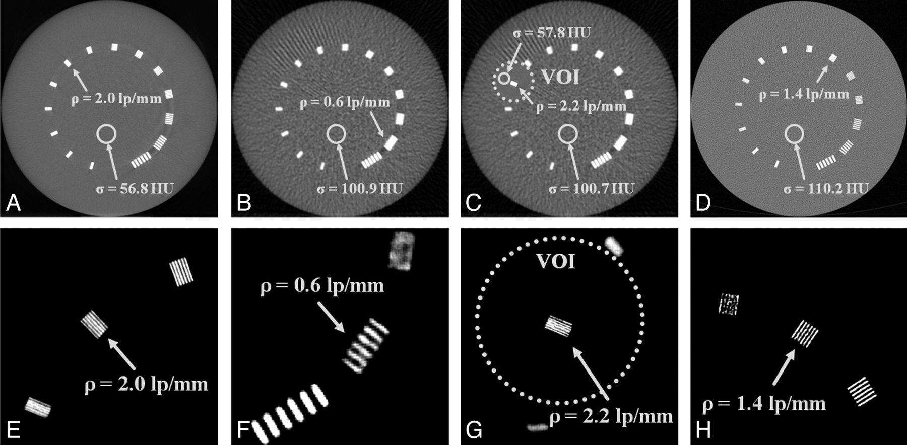
  • Fig 2.
    • Download figure
    • Open in new tab
    • Download powerpoint
    Fig 2.

    Measurements of a bar pattern phantom to determine spatial resolution (ρ) and image noise (σ). A−D, Complete images of the phantom (center = 0 HU, width = 1000 HU). E−H, Detailed images of the respective resolution bar pattern (center = 1000 HU, width = 500 HU) for the HD scan (A and E), the OV scan (B and F), the combination of OV and VOI scans (C and G), and the MSCT scan (D and H).

  • Fig 3.
    • Download figure
    • Open in new tab
    • Download powerpoint
    Fig 3.

    Measurements of an inner ear specimen. Complete transversal views (A−D), complete coronal views (E−H), detailed transversal views (I−L), and detailed coronal views (M−P). The first column shows the HD scan; the second column, the OV scan; the third column, the combination of the OV and VOI scans; and the last column, the MSCT scan (center = 0 HU, width = 2000 HU).

  • Fig 4.
    • Download figure
    • Open in new tab
    • Download powerpoint
    Fig 4.

    Monte Carlo dose simulations for the different scanning approaches in transversal (A−D) and coronal views (E−H) for the HD scan (A and E), the OV scan (B and F), the combination of OV and VOI scans (C and G), and the MSCT scan (D and H). All distributions are scaled according to the color scale shown in I.

Tables

  • Figures
  • Summary of spatial resolution, image noise, and dose values

    ScanSpatial Resolution (lp/mm)Image Noise (HU)Dose (mGy)
    OV0.61010.2
    VOI2.2581.4
    OV + VOI2.2581.6
    HD2.05714.7
    MSCT1.41108.2
PreviousNext
Back to top

In this issue

American Journal of Neuroradiology: 33 (10)
American Journal of Neuroradiology
Vol. 33, Issue 10
1 Nov 2012
  • Table of Contents
  • Index by author
Advertisement
Print
Download PDF
Email Article

Thank you for your interest in spreading the word on American Journal of Neuroradiology.

NOTE: We only request your email address so that the person you are recommending the page to knows that you wanted them to see it, and that it is not junk mail. We do not capture any email address.

Enter multiple addresses on separate lines or separate them with commas.
Volume-of-Interest Imaging of the Inner Ear in a Human Temporal Bone Specimen Using a Robot- Driven C-Arm Flat Panel Detector CT System
(Your Name) has sent you a message from American Journal of Neuroradiology
(Your Name) thought you would like to see the American Journal of Neuroradiology web site.
CAPTCHA
This question is for testing whether or not you are a human visitor and to prevent automated spam submissions.
Cite this article
D. Kolditz, T. Struffert, Y. Kyriakou, A. Bozzato, A. Dörfler, W.A. Kalender
Volume-of-Interest Imaging of the Inner Ear in a Human Temporal Bone Specimen Using a Robot- Driven C-Arm Flat Panel Detector CT System
American Journal of Neuroradiology Nov 2012, 33 (10) E124-E128; DOI: 10.3174/ajnr.A2577

Citation Manager Formats

  • BibTeX
  • Bookends
  • EasyBib
  • EndNote (tagged)
  • EndNote 8 (xml)
  • Medlars
  • Mendeley
  • Papers
  • RefWorks Tagged
  • Ref Manager
  • RIS
  • Zotero
0 Responses
Respond to this article
Share
Bookmark this article
Volume-of-Interest Imaging of the Inner Ear in a Human Temporal Bone Specimen Using a Robot- Driven C-Arm Flat Panel Detector CT System
D. Kolditz, T. Struffert, Y. Kyriakou, A. Bozzato, A. Dörfler, W.A. Kalender
American Journal of Neuroradiology Nov 2012, 33 (10) E124-E128; DOI: 10.3174/ajnr.A2577
del.icio.us logo Twitter logo Facebook logo Mendeley logo
  • Tweet Widget
  • Facebook Like
  • Google Plus One
Purchase

Jump to section

  • Article
    • Abstract
    • ABBREVIATIONS:
    • Materials and Methods
    • Results
    • Discussion
    • Conclusions
    • Footnotes
    • References
  • Figures & Data
  • Info & Metrics
  • Responses
  • References
  • PDF

Related Articles

  • No related articles found.
  • PubMed
  • Google Scholar

Cited By...

  • The Added Value of Volume-of-Interest C-Arm CT Imaging during Endovascular Treatment of Intracranial Aneurysms
  • Low-Dose Volume-of-Interest C-Arm CT Imaging of Intracranial Stents and Flow Diverters
  • Crossref
  • Google Scholar

This article has not yet been cited by articles in journals that are participating in Crossref Cited-by Linking.

More in this TOC Section

  • Decubitus Myelography for Spinal Extradural Arachnoid Cyst—Better Classified as a Lateral Dural CSF Leak?
  • Mitigating brain MR imaging degradation due to ferumoxytol therapy in patients with iron deficiency anemia
  • Advanced Imaging of Type 2 Spinal CSF Leaks with Ultrahigh-Resolution Cone-Beam CT Myelography
Show more TECHNICAL NOTE

Similar Articles

Advertisement

Indexed Content

  • Current Issue
  • Accepted Manuscripts
  • Article Preview
  • Past Issues
  • Editorials
  • Editor's Choice
  • Fellows' Journal Club
  • Letters to the Editor
  • Video Articles

Cases

  • Case Collection
  • Archive - Case of the Week
  • Archive - Case of the Month
  • Archive - Classic Case

More from AJNR

  • Trainee Corner
  • Imaging Protocols
  • MRI Safety Corner
  • Book Reviews

Multimedia

  • AJNR Podcasts
  • AJNR Scantastics

Resources

  • Turnaround Time
  • Submit a Manuscript
  • Submit a Video Article
  • Submit an eLetter to the Editor/Response
  • Manuscript Submission Guidelines
  • Statistical Tips
  • Fast Publishing of Accepted Manuscripts
  • Graphical Abstract Preparation
  • Imaging Protocol Submission
  • Evidence-Based Medicine Level Guide
  • Publishing Checklists
  • Author Policies
  • Become a Reviewer/Academy of Reviewers
  • News and Updates

About Us

  • About AJNR
  • Editorial Board
  • Editorial Board Alumni
  • Alerts
  • Permissions
  • Not an AJNR Subscriber? Join Now
  • Advertise with Us
  • Librarian Resources
  • Feedback
  • Terms and Conditions
  • AJNR Editorial Board Alumni

American Society of Neuroradiology

  • Not an ASNR Member? Join Now

© 2025 by the American Society of Neuroradiology All rights, including for text and data mining, AI training, and similar technologies, are reserved.
Print ISSN: 0195-6108 Online ISSN: 1936-959X

Powered by HighWire