Advertisement

Main menu

  • Home
  • Content
    • Current Issue
    • Accepted Manuscripts
    • Article Preview
    • Past Issue Archive
    • Video Articles
    • AJNR Case Collection
    • Case of the Week Archive
    • Case of the Month Archive
    • Classic Case Archive
  • Special Collections
    • AJNR Awards
    • Low-Field MRI
    • Alzheimer Disease
    • ASNR Foundation Special Collection
    • Photon-Counting CT
    • View All
  • Multimedia
    • AJNR Podcasts
    • AJNR SCANtastic
    • Trainee Corner
    • MRI Safety Corner
    • Imaging Protocols
  • For Authors
    • Submit a Manuscript
    • Submit a Video Article
    • Submit an eLetter to the Editor/Response
    • Manuscript Submission Guidelines
    • Statistical Tips
    • Fast Publishing of Accepted Manuscripts
    • Graphical Abstract Preparation
    • Imaging Protocol Submission
    • Author Policies
  • About Us
    • About AJNR
    • Editorial Board
    • Editorial Board Alumni
  • More
    • Become a Reviewer/Academy of Reviewers
    • Subscribers
    • Permissions
    • Alerts
    • Feedback
    • Advertisers
    • ASNR Home

User menu

  • Alerts
  • Log in

Search

  • Advanced search
American Journal of Neuroradiology

American Journal of Neuroradiology

ASHNR American Society of Functional Neuroradiology ASHNR American Society of Pediatric Neuroradiology ASSR
  • Alerts
  • Log in

Advanced Search

  • Home
  • Content
    • Current Issue
    • Accepted Manuscripts
    • Article Preview
    • Past Issue Archive
    • Video Articles
    • AJNR Case Collection
    • Case of the Week Archive
    • Case of the Month Archive
    • Classic Case Archive
  • Special Collections
    • AJNR Awards
    • Low-Field MRI
    • Alzheimer Disease
    • ASNR Foundation Special Collection
    • Photon-Counting CT
    • View All
  • Multimedia
    • AJNR Podcasts
    • AJNR SCANtastic
    • Trainee Corner
    • MRI Safety Corner
    • Imaging Protocols
  • For Authors
    • Submit a Manuscript
    • Submit a Video Article
    • Submit an eLetter to the Editor/Response
    • Manuscript Submission Guidelines
    • Statistical Tips
    • Fast Publishing of Accepted Manuscripts
    • Graphical Abstract Preparation
    • Imaging Protocol Submission
    • Author Policies
  • About Us
    • About AJNR
    • Editorial Board
    • Editorial Board Alumni
  • More
    • Become a Reviewer/Academy of Reviewers
    • Subscribers
    • Permissions
    • Alerts
    • Feedback
    • Advertisers
    • ASNR Home
  • Follow AJNR on Twitter
  • Visit AJNR on Facebook
  • Follow AJNR on Instagram
  • Join AJNR on LinkedIn
  • RSS Feeds

AJNR Awards, New Junior Editors, and more. Read the latest AJNR updates

Research ArticleBrain

Temporal Resolution of Dynamic Angiography Using Flat Panel Volume CT: In Vivo Evaluation of Time-Dependent Vascular Pathologies

R. Gupta, A. Mehndiratta, A.P. Mitha, M. Grasruck, C. Leidecker, C. Ogilvy and T.J. Brady
American Journal of Neuroradiology October 2011, 32 (9) 1688-1696; DOI: https://doi.org/10.3174/ajnr.A2586
R. Gupta
  • Find this author on Google Scholar
  • Find this author on PubMed
  • Search for this author on this site
A. Mehndiratta
  • Find this author on Google Scholar
  • Find this author on PubMed
  • Search for this author on this site
A.P. Mitha
  • Find this author on Google Scholar
  • Find this author on PubMed
  • Search for this author on this site
M. Grasruck
  • Find this author on Google Scholar
  • Find this author on PubMed
  • Search for this author on this site
C. Leidecker
  • Find this author on Google Scholar
  • Find this author on PubMed
  • Search for this author on this site
C. Ogilvy
  • Find this author on Google Scholar
  • Find this author on PubMed
  • Search for this author on this site
T.J. Brady
  • Find this author on Google Scholar
  • Find this author on PubMed
  • Search for this author on this site
  • Article
  • Figures & Data
  • Info & Metrics
  • Responses
  • References
  • PDF
Loading

Article Figures & Data

Figures

  • Tables
  • Fig 1.
    • Download figure
    • Open in new tab
    • Download powerpoint
    Fig 1.

    Zoomed-in view of the ACA (of Macaca sylvanus) at the peak of arterial opacification. Nearly the entire course of the ACA, with a diameter <200 μm in the distal portions, is visualized because of the excellent resolution afforded by the digital flat panel technology.

  • Fig 2.
    • Download figure
    • Open in new tab
    • Download powerpoint
    Fig 2.

    Time-resolved MIP images through a sagittal slab of the brain parenchyma of a Macaca sylvanus showing arterial, parenchymal, and venous phases of circulation in this animal with a heart rate of approximately 150 bps.

  • Fig 3.
    • Download figure
    • Open in new tab
    • Download powerpoint
    Fig 3.

    Six sequential frames showing opacification of the arteries, parenchyma, and the veins of the brain in a Rattus norvegicus model. In the parenchymal phase, the contrast enhancement of the brain is only minimally above the baseline value because of the contrast resolution of the scanner.

  • Fig 4.
    • Download figure
    • Open in new tab
    • Download powerpoint
    Fig 4.

    Demonstration of subclavian steal phenomenon by using dynamic CTA in a NZW rabbit model. A, An early arterial phase MIP image shows opacification of the left subclavian, left vertebral, and left common carotid arteries. B, A late arterial phase image shows opacification of the right subclavian artery via retrograde flow (reverse flow) from the right vertebral artery. Note the chemical vasculitis induced in the RCCA from an intra-arterial injection of elastase, which was used to demonstrate endothelial injury in this artery before scanning. C, Contrast intensity (HU) vs time curve showing the temporal delay in peak enhancement of right subclavian artery (yellow) with respect to aorta (blue) and left subclavian artery (pink).
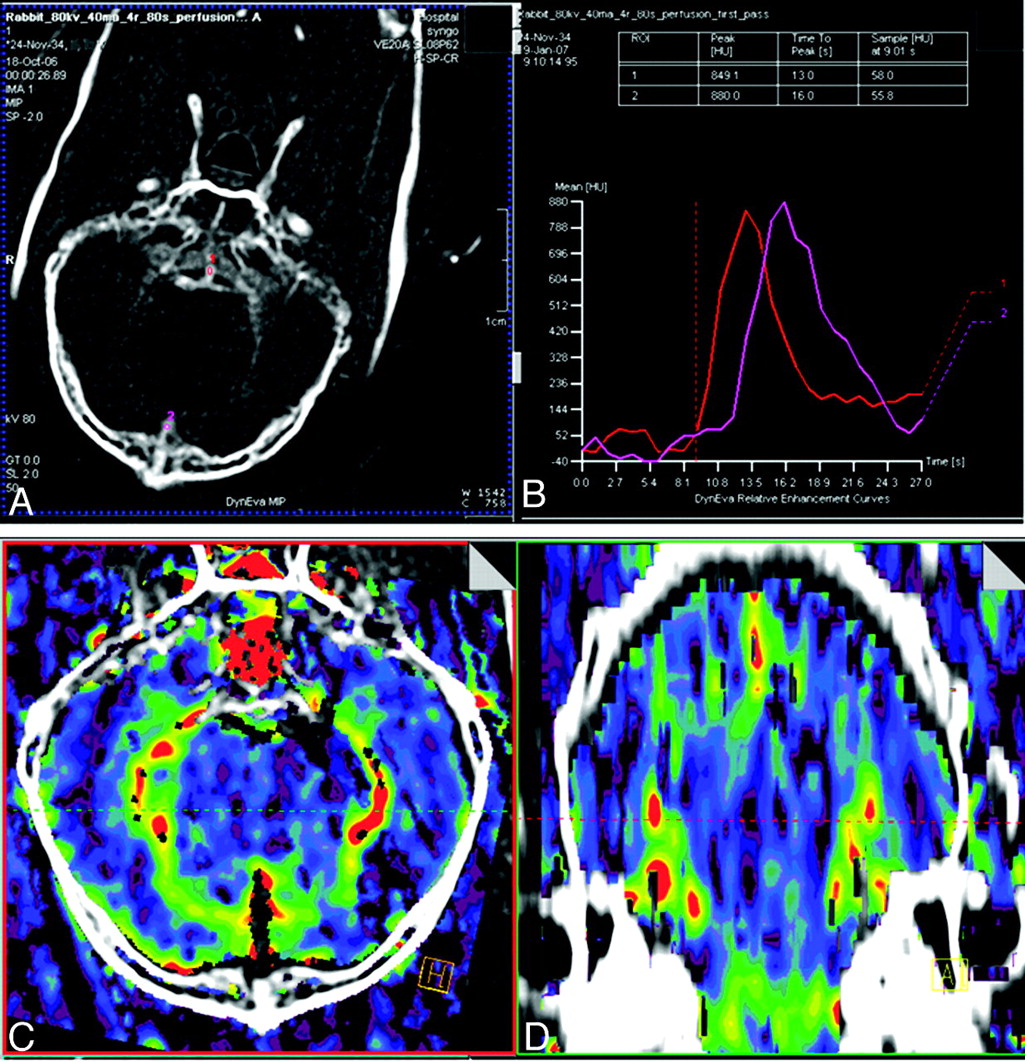
  • Fig 5.
    • Download figure
    • Open in new tab
    • Download powerpoint
    Fig 5.

    A, An axial section through a single phase of a 4D time-resolved image stack used for estimating brain perfusion. The arterial and venous inputs used for computation are marked. B, Arterial and venous time opacification curves. C and D, Axial and coronal CBV maps computed by using the Patlak model.
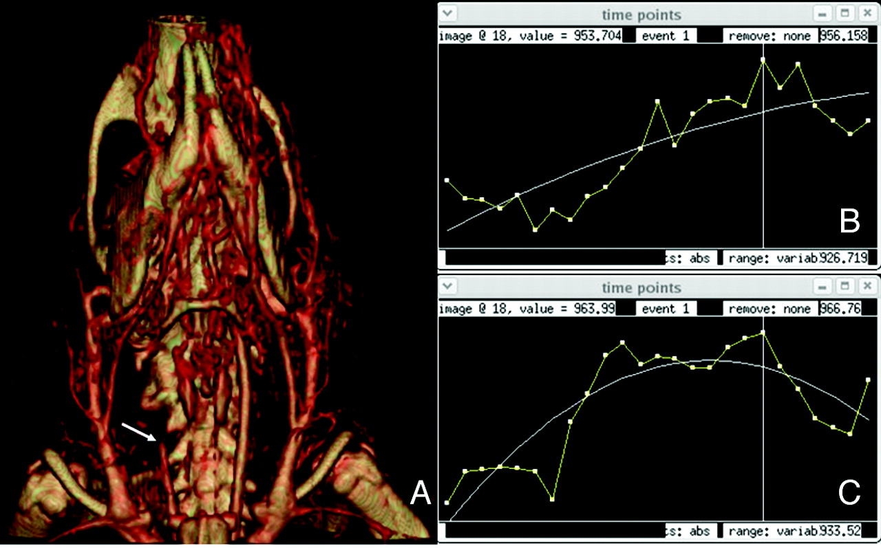
  • Fig 6.
    • Download figure
    • Open in new tab
    • Download powerpoint
    Fig 6.

    A, A frame from the arterial phase of a dynamic scan obtained on a rat with occlusion of the proximal right ICA (arrow). The right ICA was occluded via a filament inserted in the ECA and then advanced into the ICA. B and C, Time-attenuation curves of the brain parenchyma on the right (B) and left (C) side of the brain. The perfusion maps derived from this model were unable to demonstrate the transit time difference between the left and right side of the brain due to limitations of the temporal and contrast resolution of the fpVCT scanner.

  • Fig 7.
    • Download figure
    • Open in new tab
    • Download powerpoint
    Fig 7.

    Image showing aneurysm creation used in NZW rabbit models: 3D reconstructions of an aneurysm by using fpVCT (left) and MDCT (right). Despite the small size of this aneurysm, the anatomy of the aneurysm sac, including a small bleb (arrow), can be well visualized by using fpVCT. The surface anatomy is considerably smoothed, and no bleb is identified with MDCT.

Tables

  • Figures
    • View popup
    Table 1:

    Variation in heart rates of animals used for validating the temporal resolution of fpVCT in vascular pathology models

    AnimalHeart Rate (bpm)No.
    Macaca sylvanus1002
    NZW rabbit15017
    Rattus norvegicus300–4003
    Mus musculus500–60013
    • View popup
    Table 2:

    Different animals used for each dynamic study protocol and time-dependent vascular models created in each animal

    Dynamic StudyAnimalNo.Model
    Phases of blood flowMacaca sylvanus2 Healthy control
    NZW rabbit10Healthy control
    Mus musculus10Healthy control
    Aneurysm fillingNZW rabbit10Modified elastase aneurysm model
    Tumor blood flowNZW rabbit4VX2 tumor model
    Time-dependent pathologiesNZW rabbit1Subclavian steal model
    Neuroperfusion (CBV, CBF, MTT)NZW rabbit2Stroke model
    Rattus norvegicus3ECA ICA filament
    Mus musculus3ECA ICA filament
    • View popup
    Table 3:

    CT acquisition settings used for static and dynamic scanning protocol for each animal

    AnimalTube Voltage(kV[peak])Tube Current(mAs)Gantry Rotation Time (sec)Total CE Scanning Time (sec)
    Macaca sylvanus10050570–80
    NZW rabbit12050545
    Rattus norvegicus10030560
    Mus musculus10030555–60
    • View popup
    Table 4:

    Results of each dynamic process and animal model comparing the time measured using 30-fps projection acquisition and visualization in dynamic 4D datasets after image reconstruction

    Dynamic Process Being MonitoredAnatomic Location and Flow PatternActual Time from Projection Data, 30 fps (sec)Animal ModelVisualization in Dynamic 4D Datasets (gantry rotation time = 5 sec)
    Cerebral circulationCommon carotid artery to internal jugular vein15Macaca sylvanusYes
    Cerebral circulationCommon carotid artery to internal jugular vein12.3–13.3NZW rabbitYes
    SSS modelAnomalous circulation5–7.5NZW rabbitYes
    Pulmonary circulationIVC to aorta,5.3NZW rabbitYes
    right heart to left heart,3NZW rabbitYes
    tail vein to heart,2–3Mus musculusYes
    heart to abdominal aorta,2–3Mus musculusYes
    IVC to right heart2.3NZW rabbitYes
    Pulmonary circulationRight heart to left heart,<2Mus musculusNo
    left heart to common carotid artery,1.33NZW rabbitNo
    tail vein to IVC,<1Mus musculusNo
    IVC to right heart,<1Mus musculusNo
    left heart chamber to aorta<1Mus musculusNo
PreviousNext
Back to top

In this issue

American Journal of Neuroradiology: 32 (9)
American Journal of Neuroradiology
Vol. 32, Issue 9
1 Oct 2011
  • Table of Contents
  • Index by author
Advertisement
Print
Download PDF
Email Article

Thank you for your interest in spreading the word on American Journal of Neuroradiology.

NOTE: We only request your email address so that the person you are recommending the page to knows that you wanted them to see it, and that it is not junk mail. We do not capture any email address.

Enter multiple addresses on separate lines or separate them with commas.
Temporal Resolution of Dynamic Angiography Using Flat Panel Volume CT: In Vivo Evaluation of Time-Dependent Vascular Pathologies
(Your Name) has sent you a message from American Journal of Neuroradiology
(Your Name) thought you would like to see the American Journal of Neuroradiology web site.
CAPTCHA
This question is for testing whether or not you are a human visitor and to prevent automated spam submissions.
Cite this article
R. Gupta, A. Mehndiratta, A.P. Mitha, M. Grasruck, C. Leidecker, C. Ogilvy, T.J. Brady
Temporal Resolution of Dynamic Angiography Using Flat Panel Volume CT: In Vivo Evaluation of Time-Dependent Vascular Pathologies
American Journal of Neuroradiology Oct 2011, 32 (9) 1688-1696; DOI: 10.3174/ajnr.A2586

Citation Manager Formats

  • BibTeX
  • Bookends
  • EasyBib
  • EndNote (tagged)
  • EndNote 8 (xml)
  • Medlars
  • Mendeley
  • Papers
  • RefWorks Tagged
  • Ref Manager
  • RIS
  • Zotero
0 Responses
Respond to this article
Share
Bookmark this article
Temporal Resolution of Dynamic Angiography Using Flat Panel Volume CT: In Vivo Evaluation of Time-Dependent Vascular Pathologies
R. Gupta, A. Mehndiratta, A.P. Mitha, M. Grasruck, C. Leidecker, C. Ogilvy, T.J. Brady
American Journal of Neuroradiology Oct 2011, 32 (9) 1688-1696; DOI: 10.3174/ajnr.A2586
del.icio.us logo Twitter logo Facebook logo Mendeley logo
  • Tweet Widget
  • Facebook Like
  • Google Plus One
Purchase

Jump to section

  • Article
    • Abstract
    • Abbreviations
    • Materials and Methods
    • Results
    • Discussion
    • Conclusions
    • Acknowledgments
    • Footnotes
    • References
  • Figures & Data
  • Info & Metrics
  • Responses
  • References
  • PDF

Related Articles

  • No related articles found.
  • PubMed
  • Google Scholar

Cited By...

  • No citing articles found.
  • Crossref
  • Google Scholar

This article has not yet been cited by articles in journals that are participating in Crossref Cited-by Linking.

More in this TOC Section

  • Enhanced Axonal Metabolism during Early Natalizumab Treatment in Relapsing-Remitting Multiple Sclerosis
  • Progression of Microstructural Damage in Spinocerebellar Ataxia Type 2: A Longitudinal DTI Study
  • SWI or T2*: Which MRI Sequence to Use in the Detection of Cerebral Microbleeds? The Karolinska Imaging Dementia Study
Show more Brain

Similar Articles

Advertisement

Indexed Content

  • Current Issue
  • Accepted Manuscripts
  • Article Preview
  • Past Issues
  • Editorials
  • Editor's Choice
  • Fellows' Journal Club
  • Letters to the Editor
  • Video Articles

Cases

  • Case Collection
  • Archive - Case of the Week
  • Archive - Case of the Month
  • Archive - Classic Case

More from AJNR

  • Trainee Corner
  • Imaging Protocols
  • MRI Safety Corner
  • Book Reviews

Multimedia

  • AJNR Podcasts
  • AJNR Scantastics

Resources

  • Turnaround Time
  • Submit a Manuscript
  • Submit a Video Article
  • Submit an eLetter to the Editor/Response
  • Manuscript Submission Guidelines
  • Statistical Tips
  • Fast Publishing of Accepted Manuscripts
  • Graphical Abstract Preparation
  • Imaging Protocol Submission
  • Evidence-Based Medicine Level Guide
  • Publishing Checklists
  • Author Policies
  • Become a Reviewer/Academy of Reviewers
  • News and Updates

About Us

  • About AJNR
  • Editorial Board
  • Editorial Board Alumni
  • Alerts
  • Permissions
  • Not an AJNR Subscriber? Join Now
  • Advertise with Us
  • Librarian Resources
  • Feedback
  • Terms and Conditions
  • AJNR Editorial Board Alumni

American Society of Neuroradiology

  • Not an ASNR Member? Join Now

© 2025 by the American Society of Neuroradiology All rights, including for text and data mining, AI training, and similar technologies, are reserved.
Print ISSN: 0195-6108 Online ISSN: 1936-959X

Powered by HighWire