
Article Figures & Data
Figures
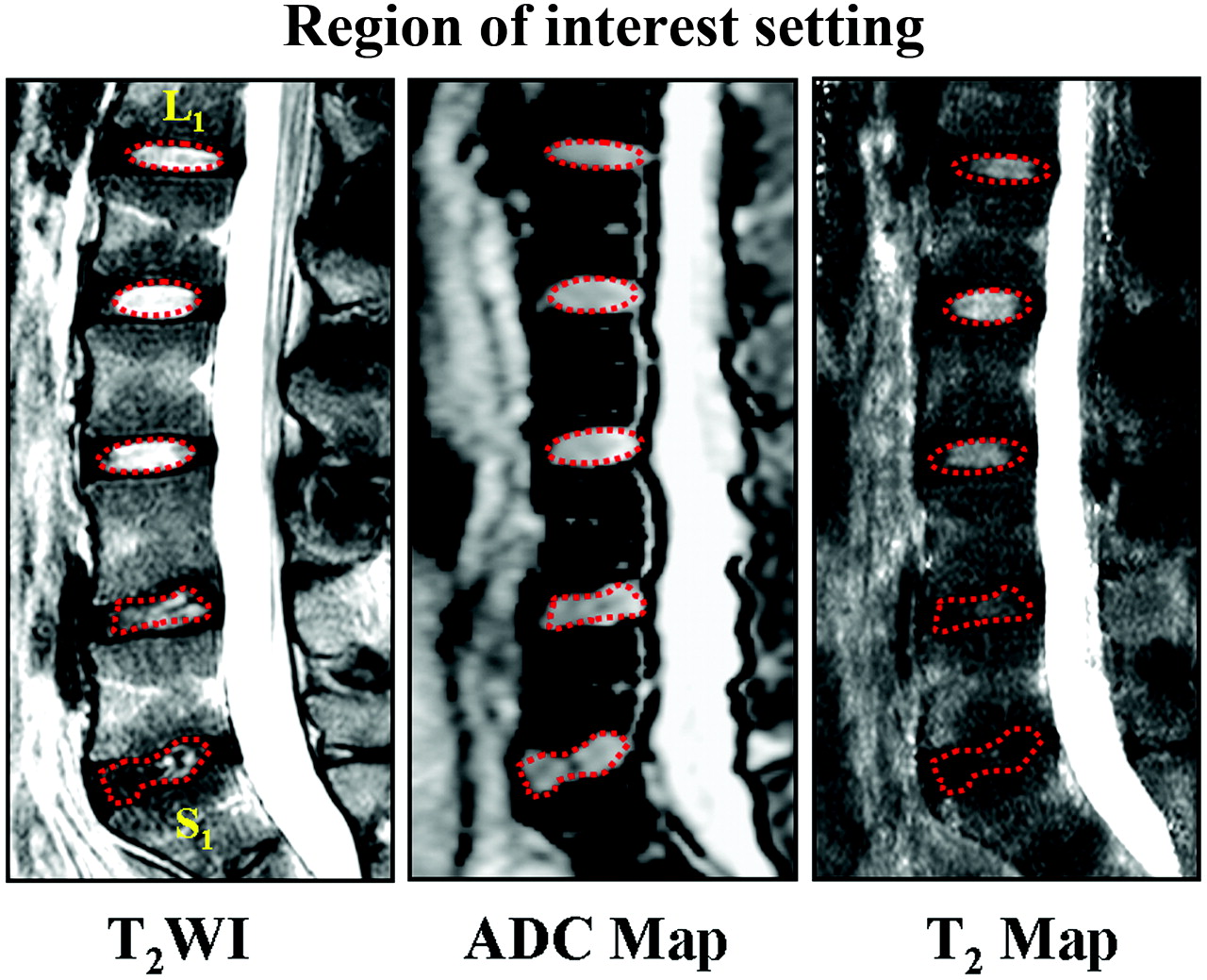
- Fig 1.
Illustration of regions of interest for T2 and ADC measurements in a 20-year-old man with low back pain. The elliptic regions of interest with red dotted lines are manually delineated in T2WI at the center of lumbar disks to cover the nucleus pulposus and inner annulus fibrosus from L1 to L4. The irregular regions of interest for L4-S1 were selected by the operator according to the expected location of inner portion of lumbar disks. Then these regions of interest in T2WI were copied to the ADC and T2 maps, and T2 and ADC values were measured.
- Fig 2.
The correlations of the T2 and ADC measurements in lumbar disks with age in asymptomatic volunteers. The solid line represents the regression line.
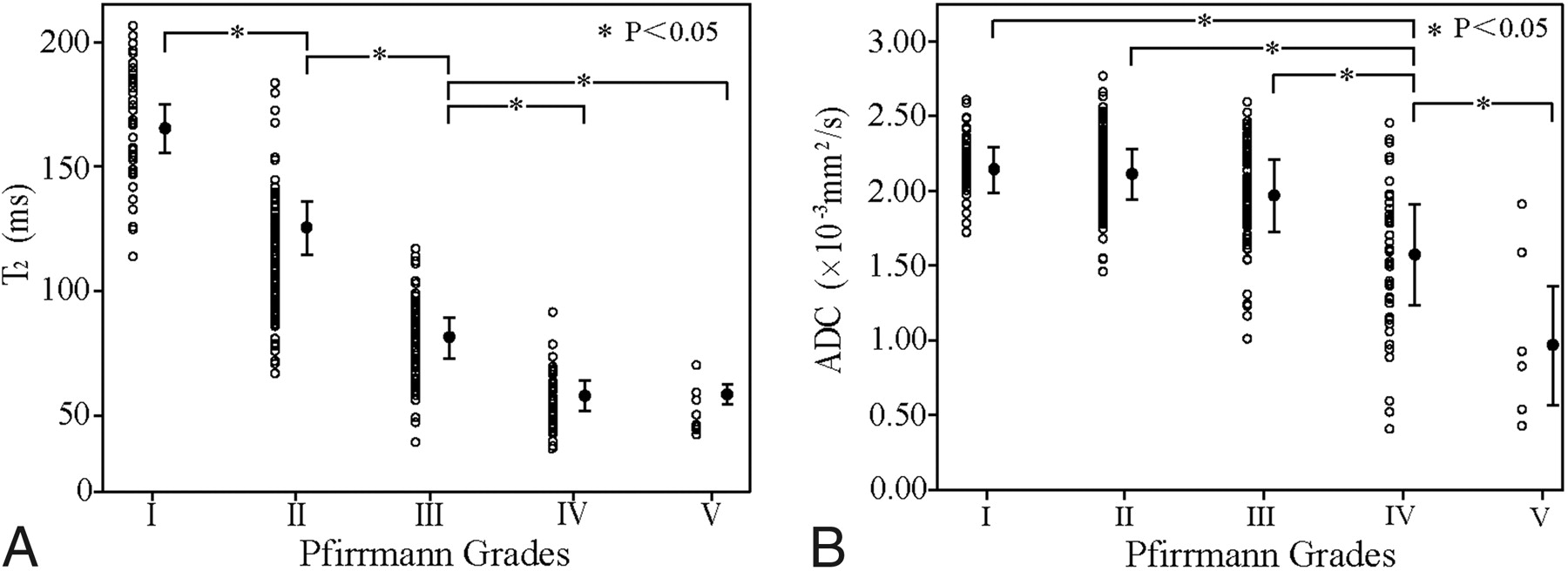
- Fig 3.
T2 (A) and ADC (B) measurements versus Pfirrmann grading.
- Fig 4.
ROC curves of T2 and ADC in assessing the Pfirrmann grading. Note that the areas under the curves are computed. Their difference is statistically significant (U-test, P < .05).


Tables
Grade Structure Distinction of Nucleus Pulposus and Annulus Fibrosus Signal Intensity Height of Intervertebral Disk I Homogeneous, bright white Clear Hyperintense Normal II Inhomogeneous with or without horizontal bands Clear Hyperintense Normal III Inhomogeneous, gray Unclear Intermediate Normal-to-slight decrease IV Inhomogeneous, gray to black Lost Intermediate to hypointense Normal-to-moderate decrease V Inhomogeneous, black Lost Hypointense Collapsed disk space - Table 2:
Intraobserver and interobserver agreement and reliability of the Pfirrmann grading
Observers Agreement(%) κ Value Grade I Grade II Grade III Grade IV Grade V Intraobserver A1-A2 76.7 89.0 83.1 71.6 87.5 0.795 B1-B2 90.7 91.2 79.8 51.0 87.5 0.822 Interobserver A1-B1 72.1 83.8 75.3 51.0 87.5 0.675 A1-B2 74.4 89.7 75.3 61.2 87.5 0.730 A2-B1 86.0 86.8 80.9 53.1 87.5 0.722 A2-B2 90.7 94.1 83.1 63.3 87.5 0.812 Age Group (yr) No. % T2 (ms) (mean) ADC (×10−3 mm2/s) (mean) 20–29 8 21.6% 164 ± 31 2.09 ± 0.25 30–39 10 27.0% 122 ± 30 2.07 ± 0.24 40–49 8 21.6% 115 ± 25 2.25 ± 0.22 50–59 5 13.5% 84 ± 26 1.96 ± 0.34 >60 6 16.2% 65 ± 16 1.64 ± 0.64 Total/average 37 100% 115 ± 42 2.03 ± 0.40 Grade No. T2 (ms) (mean) No. ADC (×10−3 mm2/s) (mean) I 43 166 ± 23 43 2.16 ± 0.19 II 135 128 ± 22 136 2.13 ± 0.23 III 89 79 ± 16 89 1.96 ± 0.32 IV 49 58 ± 12 45 1.58 ± 0.48 V 8 59 ± 10 6 0.99 ± 0.60 Total 324 107 ± 40 319 1.99 ± 0.38


