Advertisement

Main menu

  • Home
  • Content
    • Current Issue
    • Accepted Manuscripts
    • Article Preview
    • Past Issue Archive
    • Video Articles
    • AJNR Case Collection
    • Case of the Week Archive
    • Case of the Month Archive
    • Classic Case Archive
  • Special Collections
    • AJNR Awards
    • Low-Field MRI
    • Alzheimer Disease
    • ASNR Foundation Special Collection
    • Photon-Counting CT
    • View All
  • Multimedia
    • AJNR Podcasts
    • AJNR SCANtastic
    • Trainee Corner
    • MRI Safety Corner
    • Imaging Protocols
  • For Authors
    • Submit a Manuscript
    • Submit a Video Article
    • Submit an eLetter to the Editor/Response
    • Manuscript Submission Guidelines
    • Statistical Tips
    • Fast Publishing of Accepted Manuscripts
    • Graphical Abstract Preparation
    • Imaging Protocol Submission
    • Author Policies
  • About Us
    • About AJNR
    • Editorial Board
    • Editorial Board Alumni
  • More
    • Become a Reviewer/Academy of Reviewers
    • Subscribers
    • Permissions
    • Alerts
    • Feedback
    • Advertisers
    • ASNR Home

User menu

  • Alerts
  • Log in

Search

  • Advanced search
American Journal of Neuroradiology

American Journal of Neuroradiology

ASHNR American Society of Functional Neuroradiology ASHNR American Society of Pediatric Neuroradiology ASSR
  • Alerts
  • Log in

Advanced Search

  • Home
  • Content
    • Current Issue
    • Accepted Manuscripts
    • Article Preview
    • Past Issue Archive
    • Video Articles
    • AJNR Case Collection
    • Case of the Week Archive
    • Case of the Month Archive
    • Classic Case Archive
  • Special Collections
    • AJNR Awards
    • Low-Field MRI
    • Alzheimer Disease
    • ASNR Foundation Special Collection
    • Photon-Counting CT
    • View All
  • Multimedia
    • AJNR Podcasts
    • AJNR SCANtastic
    • Trainee Corner
    • MRI Safety Corner
    • Imaging Protocols
  • For Authors
    • Submit a Manuscript
    • Submit a Video Article
    • Submit an eLetter to the Editor/Response
    • Manuscript Submission Guidelines
    • Statistical Tips
    • Fast Publishing of Accepted Manuscripts
    • Graphical Abstract Preparation
    • Imaging Protocol Submission
    • Author Policies
  • About Us
    • About AJNR
    • Editorial Board
    • Editorial Board Alumni
  • More
    • Become a Reviewer/Academy of Reviewers
    • Subscribers
    • Permissions
    • Alerts
    • Feedback
    • Advertisers
    • ASNR Home
  • Follow AJNR on Twitter
  • Visit AJNR on Facebook
  • Follow AJNR on Instagram
  • Join AJNR on LinkedIn
  • RSS Feeds

AJNR Awards, New Junior Editors, and more. Read the latest AJNR updates

Research ArticleHead and Neck Imaging
Open Access

In Vivo Evaluation of Quantitative MR Angiography in a Canine Carotid Artery Stenosis Model

M. Calderon-Arnulphi, S. Amin-Hanjani, A. Alaraj, M. Zhao, X. Du, S. Ruland, X.J. Zhou, K.R. Thulborn and F.T. Charbel
American Journal of Neuroradiology September 2011, 32 (8) 1552-1559; DOI: https://doi.org/10.3174/ajnr.A2546
M. Calderon-Arnulphi
  • Find this author on Google Scholar
  • Find this author on PubMed
  • Search for this author on this site
S. Amin-Hanjani
  • Find this author on Google Scholar
  • Find this author on PubMed
  • Search for this author on this site
A. Alaraj
  • Find this author on Google Scholar
  • Find this author on PubMed
  • Search for this author on this site
M. Zhao
  • Find this author on Google Scholar
  • Find this author on PubMed
  • Search for this author on this site
X. Du
  • Find this author on Google Scholar
  • Find this author on PubMed
  • Search for this author on this site
S. Ruland
  • Find this author on Google Scholar
  • Find this author on PubMed
  • Search for this author on this site
X.J. Zhou
  • Find this author on Google Scholar
  • Find this author on PubMed
  • Search for this author on this site
K.R. Thulborn
  • Find this author on Google Scholar
  • Find this author on PubMed
  • Search for this author on this site
F.T. Charbel
  • Find this author on Google Scholar
  • Find this author on PubMed
  • Search for this author on this site
  • Article
  • Figures & Data
  • Info & Metrics
  • Responses
  • References
  • PDF
Loading

Article Figures & Data

Figures

  • Tables
  • Fig 1.
    • Download figure
    • Open in new tab
    • Download powerpoint
    Fig 1.

    Intraoperative photograph of the dissected CCA of the dog. A, Sonographic flow probe around the CCA. B, Vascular tourniquet around the CCA.
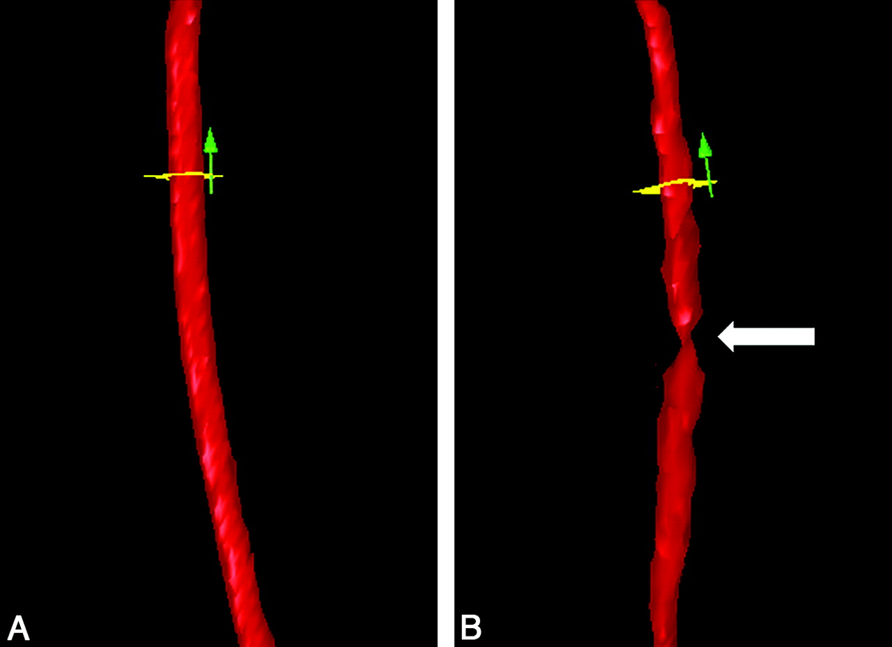
  • Fig 2.
    • Download figure
    • Open in new tab
    • Download powerpoint
    Fig 2.

    3D surface rendering of the CCA created by QMRA. A: The CCA segment before stenosis is induced. The tourniquet is in place and loose. B: The same CCA segment after stenosis is created by tightening the tourniquet. The vertical arrow indicates the direction of flow. The white arrow indicates the position of the stenosis. The flow probe is proximal to the stenosis outside the FOV.
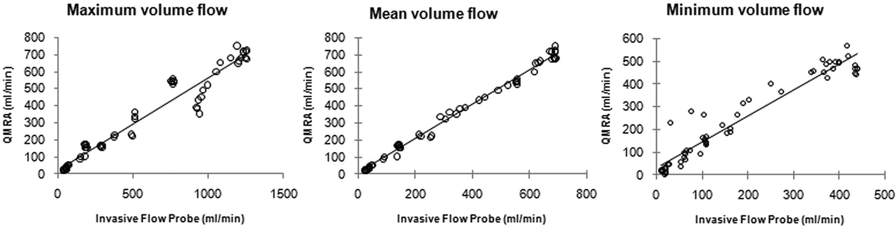
  • Fig 3.
    • Download figure
    • Open in new tab
    • Download powerpoint
    Fig 3.

    Correlations between mean, maximum, and minimum flow volume rates obtained with QMRA and those obtained with sonographic flow.

  • Fig 4.
    • Download figure
    • Open in new tab
    • Download powerpoint
    Fig 4.

    An example of simultaneous flow-volume measurements obtained with QMRA and the flow probe across time in an animal with no stenosis. The QMRA measurements closely track flow-probe measurements for maximum, mean, and minimum flow volumes. The QMRA method gives lower maxima and higher minima compared with the flow probe due to its reduced temporal resolution.

  • Fig 5.
    • Download figure
    • Open in new tab
    • Download powerpoint
    Fig 5.

    Bland-Altman plot shows the relative degree of agreement between volume flow measurements with QMRA and the invasive flow probe. Dashed lines demonstrate upper and lower limits of agreement. (±2 SDs).

  • Fig 6.
    • Download figure
    • Open in new tab
    • Download powerpoint
    Fig 6.

    Correlations of mean flow-volume rates between the flow probe and QMRA with no stenosis, proximal to the stenosis, and distal to the stenosis.

Tables

  • Figures
    • View popup
    Table 1:

    Comparison of the PD between flow measurements from QMRA and the flow probe at different flow ranges

    Flow (mL/min)No. of MeasurementsPD (± SE) (%)Mean Flow Rate (mL/min)
    20–79139.4 ± 234
    80–1791413.3 ± 2134
    180–350a89.7 ± 2274
    >350a253.2 ± 0.5584
    • a Flows obtained without induced stenosis.

    • View popup
    Table 2:

    Comparison of the PD between flow measurements from QMRA and the flow probe across the normal and stenotic CCA as a function of stenosis

    PositionPD (± SE) (%)No. of MeasurementsAverage Flow (mL/min)
    No stenosis4.8 ± 133509
    Stenosis11.4 ± 12786
        Proximal to stenosis9.6 ± 2882
        Distal to stenosis12.2 ± 21988
PreviousNext
Back to top

In this issue

American Journal of Neuroradiology: 32 (8)
American Journal of Neuroradiology
Vol. 32, Issue 8
1 Sep 2011
  • Table of Contents
  • Index by author
Advertisement
Print
Download PDF
Email Article

Thank you for your interest in spreading the word on American Journal of Neuroradiology.

NOTE: We only request your email address so that the person you are recommending the page to knows that you wanted them to see it, and that it is not junk mail. We do not capture any email address.

Enter multiple addresses on separate lines or separate them with commas.
In Vivo Evaluation of Quantitative MR Angiography in a Canine Carotid Artery Stenosis Model
(Your Name) has sent you a message from American Journal of Neuroradiology
(Your Name) thought you would like to see the American Journal of Neuroradiology web site.
CAPTCHA
This question is for testing whether or not you are a human visitor and to prevent automated spam submissions.
Cite this article
M. Calderon-Arnulphi, S. Amin-Hanjani, A. Alaraj, M. Zhao, X. Du, S. Ruland, X.J. Zhou, K.R. Thulborn, F.T. Charbel
In Vivo Evaluation of Quantitative MR Angiography in a Canine Carotid Artery Stenosis Model
American Journal of Neuroradiology Sep 2011, 32 (8) 1552-1559; DOI: 10.3174/ajnr.A2546

Citation Manager Formats

  • BibTeX
  • Bookends
  • EasyBib
  • EndNote (tagged)
  • EndNote 8 (xml)
  • Medlars
  • Mendeley
  • Papers
  • RefWorks Tagged
  • Ref Manager
  • RIS
  • Zotero
0 Responses
Respond to this article
Share
Bookmark this article
In Vivo Evaluation of Quantitative MR Angiography in a Canine Carotid Artery Stenosis Model
M. Calderon-Arnulphi, S. Amin-Hanjani, A. Alaraj, M. Zhao, X. Du, S. Ruland, X.J. Zhou, K.R. Thulborn, F.T. Charbel
American Journal of Neuroradiology Sep 2011, 32 (8) 1552-1559; DOI: 10.3174/ajnr.A2546
del.icio.us logo Twitter logo Facebook logo Mendeley logo
  • Tweet Widget
  • Facebook Like
  • Google Plus One
Purchase

Jump to section

  • Article
    • Abstract
    • Abbreviations
    • Materials and Methods
    • Results
    • Discussion
    • Conclusions
    • Appendix: QMRA Technique
    • Acknowledgments
    • Footnotes
    • References
  • Figures & Data
  • Info & Metrics
  • Responses
  • References
  • PDF

Related Articles

  • No related articles found.
  • PubMed
  • Google Scholar

Cited By...

  • Evaluation of Sex-Related Differences in Cerebrovascular Bypass Patency: An Institutional Review of 357 Direct Cerebral Bypasses
  • Validation of cerebral arteriovenous malformation hemodynamics assessed by DSA using quantitative magnetic resonance angiography: preliminary study
  • Cerebral Arteriovenous Malformation Flow Is Associated With Venous Intimal Hyperplasia
  • Hemodynamic Characteristics of Cerebral Arteriovenous Malformation Feeder Vessels With and Without Aneurysms
  • Changes in Wall Shear Stress of Cerebral Arteriovenous Malformation Feeder Arteries After Embolization and Surgery
  • Quantitative Assessment of Changes in Cerebral Arteriovenous Malformation Hemodynamics After Embolization
  • Crossref
  • Google Scholar

This article has not yet been cited by articles in journals that are participating in Crossref Cited-by Linking.

More in this TOC Section

  • MRI of the Parasellar Ligaments
  • ASL Sensitivity for Head and Neck Paraganglioma
  • Post SRS Peritumoral Hyperintense Signal of VSs
Show more Head and Neck Imaging

Similar Articles

Advertisement

Indexed Content

  • Current Issue
  • Accepted Manuscripts
  • Article Preview
  • Past Issues
  • Editorials
  • Editor's Choice
  • Fellows' Journal Club
  • Letters to the Editor
  • Video Articles

Cases

  • Case Collection
  • Archive - Case of the Week
  • Archive - Case of the Month
  • Archive - Classic Case

More from AJNR

  • Trainee Corner
  • Imaging Protocols
  • MRI Safety Corner
  • Book Reviews

Multimedia

  • AJNR Podcasts
  • AJNR Scantastics

Resources

  • Turnaround Time
  • Submit a Manuscript
  • Submit a Video Article
  • Submit an eLetter to the Editor/Response
  • Manuscript Submission Guidelines
  • Statistical Tips
  • Fast Publishing of Accepted Manuscripts
  • Graphical Abstract Preparation
  • Imaging Protocol Submission
  • Evidence-Based Medicine Level Guide
  • Publishing Checklists
  • Author Policies
  • Become a Reviewer/Academy of Reviewers
  • News and Updates

About Us

  • About AJNR
  • Editorial Board
  • Editorial Board Alumni
  • Alerts
  • Permissions
  • Not an AJNR Subscriber? Join Now
  • Advertise with Us
  • Librarian Resources
  • Feedback
  • Terms and Conditions
  • AJNR Editorial Board Alumni

American Society of Neuroradiology

  • Not an ASNR Member? Join Now

© 2025 by the American Society of Neuroradiology All rights, including for text and data mining, AI training, and similar technologies, are reserved.
Print ISSN: 0195-6108 Online ISSN: 1936-959X

Powered by HighWire