Advertisement

Main menu

  • Home
  • Content
    • Current Issue
    • Accepted Manuscripts
    • Article Preview
    • Past Issue Archive
    • Video Articles
    • AJNR Case Collection
    • Case of the Week Archive
    • Case of the Month Archive
    • Classic Case Archive
  • Special Collections
    • AJNR Awards
    • Low-Field MRI
    • Alzheimer Disease
    • ASNR Foundation Special Collection
    • Photon-Counting CT
    • View All
  • Multimedia
    • AJNR Podcasts
    • AJNR SCANtastic
    • Trainee Corner
    • MRI Safety Corner
    • Imaging Protocols
  • For Authors
    • Submit a Manuscript
    • Submit a Video Article
    • Submit an eLetter to the Editor/Response
    • Manuscript Submission Guidelines
    • Statistical Tips
    • Fast Publishing of Accepted Manuscripts
    • Graphical Abstract Preparation
    • Imaging Protocol Submission
    • Author Policies
  • About Us
    • About AJNR
    • Editorial Board
    • Editorial Board Alumni
  • More
    • Become a Reviewer/Academy of Reviewers
    • Subscribers
    • Permissions
    • Alerts
    • Feedback
    • Advertisers
    • ASNR Home

User menu

  • Alerts
  • Log in

Search

  • Advanced search
American Journal of Neuroradiology

American Journal of Neuroradiology

ASHNR American Society of Functional Neuroradiology ASHNR American Society of Pediatric Neuroradiology ASSR
  • Alerts
  • Log in

Advanced Search

  • Home
  • Content
    • Current Issue
    • Accepted Manuscripts
    • Article Preview
    • Past Issue Archive
    • Video Articles
    • AJNR Case Collection
    • Case of the Week Archive
    • Case of the Month Archive
    • Classic Case Archive
  • Special Collections
    • AJNR Awards
    • Low-Field MRI
    • Alzheimer Disease
    • ASNR Foundation Special Collection
    • Photon-Counting CT
    • View All
  • Multimedia
    • AJNR Podcasts
    • AJNR SCANtastic
    • Trainee Corner
    • MRI Safety Corner
    • Imaging Protocols
  • For Authors
    • Submit a Manuscript
    • Submit a Video Article
    • Submit an eLetter to the Editor/Response
    • Manuscript Submission Guidelines
    • Statistical Tips
    • Fast Publishing of Accepted Manuscripts
    • Graphical Abstract Preparation
    • Imaging Protocol Submission
    • Author Policies
  • About Us
    • About AJNR
    • Editorial Board
    • Editorial Board Alumni
  • More
    • Become a Reviewer/Academy of Reviewers
    • Subscribers
    • Permissions
    • Alerts
    • Feedback
    • Advertisers
    • ASNR Home
  • Follow AJNR on Twitter
  • Visit AJNR on Facebook
  • Follow AJNR on Instagram
  • Join AJNR on LinkedIn
  • RSS Feeds

AJNR Awards, New Junior Editors, and more. Read the latest AJNR updates

Research ArticleBrain
Open Access

Cerebral CT Perfusion Using an Interventional C-Arm Imaging System: Cerebral Blood Flow Measurements

A. Ganguly, A. Fieselmann, M. Marks, J. Rosenberg, J. Boese, Y. Deuerling-Zheng, M. Straka, G. Zaharchuk, R. Bammer and R. Fahrig
American Journal of Neuroradiology September 2011, 32 (8) 1525-1531; DOI: https://doi.org/10.3174/ajnr.A2518
A. Ganguly
  • Find this author on Google Scholar
  • Find this author on PubMed
  • Search for this author on this site
A. Fieselmann
  • Find this author on Google Scholar
  • Find this author on PubMed
  • Search for this author on this site
M. Marks
  • Find this author on Google Scholar
  • Find this author on PubMed
  • Search for this author on this site
J. Rosenberg
  • Find this author on Google Scholar
  • Find this author on PubMed
  • Search for this author on this site
J. Boese
  • Find this author on Google Scholar
  • Find this author on PubMed
  • Search for this author on this site
Y. Deuerling-Zheng
  • Find this author on Google Scholar
  • Find this author on PubMed
  • Search for this author on this site
M. Straka
  • Find this author on Google Scholar
  • Find this author on PubMed
  • Search for this author on this site
G. Zaharchuk
  • Find this author on Google Scholar
  • Find this author on PubMed
  • Search for this author on this site
R. Bammer
  • Find this author on Google Scholar
  • Find this author on PubMed
  • Search for this author on this site
R. Fahrig
  • Find this author on Google Scholar
  • Find this author on PubMed
  • Search for this author on this site
  • Article
  • Figures & Data
  • Info & Metrics
  • Responses
  • References
  • PDF
Loading

Article Figures & Data

Figures

  • Tables
  • Fig 1.
    • Download figure
    • Open in new tab
    • Download powerpoint
    Fig 1.

    Temporal spacing of different scan sets to allow higher temporal sampling. The function is a typical TDC extracted at 1-voxel position within a vessel, and the markers define the center time point of each rotation of the C-arm.

  • Fig 2.
    • Download figure
    • Open in new tab
    • Download powerpoint
    Fig 2.

    CBF maps for 6-scan sets (A-B-C-D-E-F) from different injection-protocol C-arm CBCT (A−C) and CTP images (D−F). The 3 injection protocols used are 3 mL/s 67% (A and D), 6 mL/s 50% (B and E), and 3 mL/s 100% (C and F). All injections had a duration of 8 seconds.

  • Fig 3.
    • Download figure
    • Open in new tab
    • Download powerpoint
    Fig 3.

    Results of CBF values for the 6-mL/s 50% contrast injection. (A), CTP, C-arm CBCT for scan sets (B), A-B-C-D-E-F (C), B-D-F (D), A-C (E), B-D and (F) B.
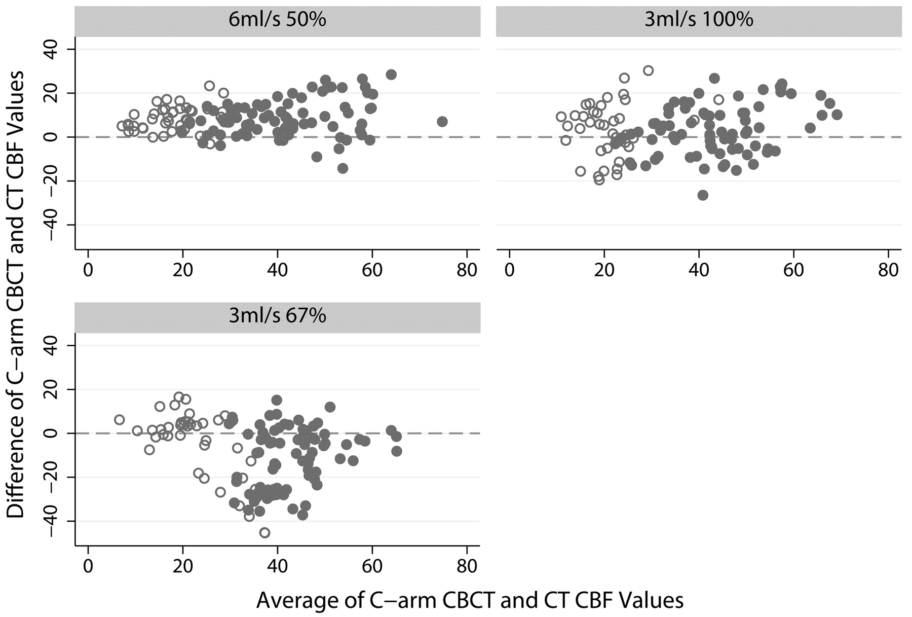
  • Fig 4.
    • Download figure
    • Open in new tab
    • Download powerpoint
    Fig 4.

    Bland-Altman plots of C-arm CBCT versus CT CBF values for the 3 different injection protocols. Units for both axes are milliliters/100 g/min of tissue. Filled gray circles represent average values in gray matter regions of interest; open circles, white matter regions of interest.

Tables

  • Figures
    • View popup
    Table 1:

    Difference in CBF between right and left hemispheres calculated from the clinical CT images only, for ROIs located in located in grey or white matter in the 2 hemispheresa

    Injection MethodDifference in CBF between Left and Right Hemispheres (mL/100 g/min)
    White MatterGrey Matter
    6 mL/s 50%
        Mean2.041.98
        SD6.036.77
        Overall ρ0.92
        Overall r0.92
    3 mL/s 100%
        Mean0.750.26
        SD7.869.49
        Overall ρ0.84
        Overall r0.84
    3 mL/s 67%
        Mean0.730.61
        SD9.246.74
        Overall ρ0.90
        Overall r0.90
    • a Mean and SD values are given in milliliters/100 g/min. The overall CCCs and the Pearson correlation coefficients between the left and right hemispheres (without grey/white matter discrimination) are also included and were found to be identical, implying no underlying bias due to position of the catheter or otherwise.

    • View popup
    Table 2:

    Statistical results comparing the CBF values from CT and CBCT from different scan sets for the 6-mL/s 50% contrast-concentration injection protocola

    Scan SetCCC (ρ)Correlation Coefficient (r)Difference (CT-CBCT) (mL/100 g/min)Bland-Altman 95% Limits
    MeanSD
    B-D-F0.7960.876.568.34−9.78–22.89
    A-B-C-D-E-F0.7720.888.607.65−6.40–23.59
    A-C0.7510.888.787.68−6.27–23.84
    • a The top 3 candidates are shown.

PreviousNext
Back to top

In this issue

American Journal of Neuroradiology: 32 (8)
American Journal of Neuroradiology
Vol. 32, Issue 8
1 Sep 2011
  • Table of Contents
  • Index by author
Advertisement
Print
Download PDF
Email Article

Thank you for your interest in spreading the word on American Journal of Neuroradiology.

NOTE: We only request your email address so that the person you are recommending the page to knows that you wanted them to see it, and that it is not junk mail. We do not capture any email address.

Enter multiple addresses on separate lines or separate them with commas.
Cerebral CT Perfusion Using an Interventional C-Arm Imaging System: Cerebral Blood Flow Measurements
(Your Name) has sent you a message from American Journal of Neuroradiology
(Your Name) thought you would like to see the American Journal of Neuroradiology web site.
CAPTCHA
This question is for testing whether or not you are a human visitor and to prevent automated spam submissions.
Cite this article
A. Ganguly, A. Fieselmann, M. Marks, J. Rosenberg, J. Boese, Y. Deuerling-Zheng, M. Straka, G. Zaharchuk, R. Bammer, R. Fahrig
Cerebral CT Perfusion Using an Interventional C-Arm Imaging System: Cerebral Blood Flow Measurements
American Journal of Neuroradiology Sep 2011, 32 (8) 1525-1531; DOI: 10.3174/ajnr.A2518

Citation Manager Formats

  • BibTeX
  • Bookends
  • EasyBib
  • EndNote (tagged)
  • EndNote 8 (xml)
  • Medlars
  • Mendeley
  • Papers
  • RefWorks Tagged
  • Ref Manager
  • RIS
  • Zotero
0 Responses
Respond to this article
Share
Bookmark this article
Cerebral CT Perfusion Using an Interventional C-Arm Imaging System: Cerebral Blood Flow Measurements
A. Ganguly, A. Fieselmann, M. Marks, J. Rosenberg, J. Boese, Y. Deuerling-Zheng, M. Straka, G. Zaharchuk, R. Bammer, R. Fahrig
American Journal of Neuroradiology Sep 2011, 32 (8) 1525-1531; DOI: 10.3174/ajnr.A2518
del.icio.us logo Twitter logo Facebook logo Mendeley logo
  • Tweet Widget
  • Facebook Like
  • Google Plus One
Purchase

Jump to section

  • Article
    • Abstract
    • Abbreviations
    • Materials and Methods
    • Results
    • Discussion
    • Conclusions
    • Acknowledgments
    • Footnotes
    • References
  • Figures & Data
  • Info & Metrics
  • Responses
  • References
  • PDF

Related Articles

  • No related articles found.
  • PubMed
  • Google Scholar

Cited By...

  • Clinical Applications of Conebeam CTP Imaging in Cerebral Disease: A Systematic Review
  • Dynamic Angiography and Perfusion Imaging Using Flat Detector CT in the Angiography Suite: A Pilot Study in Patients with Acute Middle Cerebral Artery Occlusions
  • Endovascular Recanalization in Acute Ischemic Stroke Using the Solitaire FR Revascularization Device with Adjunctive C-Arm CT Imaging
  • Exploring the Value of Using Color-Coded Quantitative DSA Evaluation on Bilateral Common Carotid Arteries in Predicting the Reliability of Intra-Ascending Aorta Flat Detector CT-CBV Maps
  • A Novel Technique for the Measurement of CBF and CBV with Robot-Arm-Mounted Flat Panel CT in a Large-Animal Model
  • C-Arm CT Measurement of Cerebral Blood Volume and Cerebral Blood Flow Using a Novel High-Speed Acquisition and a Single Intravenous Contrast Injection
  • 4D Digital Subtraction Angiography: Implementation and Demonstration of Feasibility
  • Initial experience with a combined multidetector CT and biplane digital subtraction angiography suite with a single interactive table for the diagnosis and treatment of neurovascular disease
  • Frameless multimodal image guidance of localized convection-enhanced delivery of therapeutics in the brain
  • Advances in Stroke: Advances in Interventional Neuroradiology
  • Crossref
  • Google Scholar

This article has not yet been cited by articles in journals that are participating in Crossref Cited-by Linking.

More in this TOC Section

  • Statin Therapy Does Not Affect the Radiographic and Clinical Profile of Patients with TIA and Minor Stroke
  • Usefulness of Quantitative Susceptibility Mapping for the Diagnosis of Parkinson Disease
  • White Matter Alterations in the Brains of Patients with Active, Remitted, and Cured Cushing Syndrome: A DTI Study
Show more Brain

Similar Articles

Advertisement

Indexed Content

  • Current Issue
  • Accepted Manuscripts
  • Article Preview
  • Past Issues
  • Editorials
  • Editor's Choice
  • Fellows' Journal Club
  • Letters to the Editor
  • Video Articles

Cases

  • Case Collection
  • Archive - Case of the Week
  • Archive - Case of the Month
  • Archive - Classic Case

More from AJNR

  • Trainee Corner
  • Imaging Protocols
  • MRI Safety Corner
  • Book Reviews

Multimedia

  • AJNR Podcasts
  • AJNR Scantastics

Resources

  • Turnaround Time
  • Submit a Manuscript
  • Submit a Video Article
  • Submit an eLetter to the Editor/Response
  • Manuscript Submission Guidelines
  • Statistical Tips
  • Fast Publishing of Accepted Manuscripts
  • Graphical Abstract Preparation
  • Imaging Protocol Submission
  • Evidence-Based Medicine Level Guide
  • Publishing Checklists
  • Author Policies
  • Become a Reviewer/Academy of Reviewers
  • News and Updates

About Us

  • About AJNR
  • Editorial Board
  • Editorial Board Alumni
  • Alerts
  • Permissions
  • Not an AJNR Subscriber? Join Now
  • Advertise with Us
  • Librarian Resources
  • Feedback
  • Terms and Conditions
  • AJNR Editorial Board Alumni

American Society of Neuroradiology

  • Not an ASNR Member? Join Now

© 2025 by the American Society of Neuroradiology All rights, including for text and data mining, AI training, and similar technologies, are reserved.
Print ISSN: 0195-6108 Online ISSN: 1936-959X

Powered by HighWire