Advertisement

Main menu

  • Home
  • Content
    • Current Issue
    • Accepted Manuscripts
    • Article Preview
    • Past Issue Archive
    • Video Articles
    • AJNR Case Collection
    • Case of the Week Archive
    • Case of the Month Archive
    • Classic Case Archive
  • Special Collections
    • AJNR Awards
    • Low-Field MRI
    • Alzheimer Disease
    • ASNR Foundation Special Collection
    • Photon-Counting CT
    • View All
  • Multimedia
    • AJNR Podcasts
    • AJNR SCANtastic
    • Trainee Corner
    • MRI Safety Corner
    • Imaging Protocols
  • For Authors
    • Submit a Manuscript
    • Submit a Video Article
    • Submit an eLetter to the Editor/Response
    • Manuscript Submission Guidelines
    • Statistical Tips
    • Fast Publishing of Accepted Manuscripts
    • Graphical Abstract Preparation
    • Imaging Protocol Submission
    • Author Policies
  • About Us
    • About AJNR
    • Editorial Board
    • Editorial Board Alumni
  • More
    • Become a Reviewer/Academy of Reviewers
    • Subscribers
    • Permissions
    • Alerts
    • Feedback
    • Advertisers
    • ASNR Home

User menu

  • Alerts
  • Log in

Search

  • Advanced search
American Journal of Neuroradiology

American Journal of Neuroradiology

ASHNR American Society of Functional Neuroradiology ASHNR American Society of Pediatric Neuroradiology ASSR
  • Alerts
  • Log in

Advanced Search

  • Home
  • Content
    • Current Issue
    • Accepted Manuscripts
    • Article Preview
    • Past Issue Archive
    • Video Articles
    • AJNR Case Collection
    • Case of the Week Archive
    • Case of the Month Archive
    • Classic Case Archive
  • Special Collections
    • AJNR Awards
    • Low-Field MRI
    • Alzheimer Disease
    • ASNR Foundation Special Collection
    • Photon-Counting CT
    • View All
  • Multimedia
    • AJNR Podcasts
    • AJNR SCANtastic
    • Trainee Corner
    • MRI Safety Corner
    • Imaging Protocols
  • For Authors
    • Submit a Manuscript
    • Submit a Video Article
    • Submit an eLetter to the Editor/Response
    • Manuscript Submission Guidelines
    • Statistical Tips
    • Fast Publishing of Accepted Manuscripts
    • Graphical Abstract Preparation
    • Imaging Protocol Submission
    • Author Policies
  • About Us
    • About AJNR
    • Editorial Board
    • Editorial Board Alumni
  • More
    • Become a Reviewer/Academy of Reviewers
    • Subscribers
    • Permissions
    • Alerts
    • Feedback
    • Advertisers
    • ASNR Home
  • Follow AJNR on Twitter
  • Visit AJNR on Facebook
  • Follow AJNR on Instagram
  • Join AJNR on LinkedIn
  • RSS Feeds

AJNR Awards, New Junior Editors, and more. Read the latest AJNR updates

Research ArticlePatient SafetyF

Low-Dose Temporal Bone CT in Infants and Young Children: Effective Dose and Image Quality

C.B. Nauer, A. Rieke, C. Zubler, C. Candreia, A. Arnold and P. Senn
American Journal of Neuroradiology September 2011, 32 (8) 1375-1380; DOI: https://doi.org/10.3174/ajnr.A2524
C.B. Nauer
  • Find this author on Google Scholar
  • Find this author on PubMed
  • Search for this author on this site
A. Rieke
  • Find this author on Google Scholar
  • Find this author on PubMed
  • Search for this author on this site
C. Zubler
  • Find this author on Google Scholar
  • Find this author on PubMed
  • Search for this author on this site
C. Candreia
  • Find this author on Google Scholar
  • Find this author on PubMed
  • Search for this author on this site
A. Arnold
  • Find this author on Google Scholar
  • Find this author on PubMed
  • Search for this author on this site
P. Senn
  • Find this author on Google Scholar
  • Find this author on PubMed
  • Search for this author on this site
  • Article
  • Figures & Data
  • Info & Metrics
  • Responses
  • References
  • PDF
Loading

Article Figures & Data

Figures

  • Tables
  • Fig 1.
    • Download figure
    • Open in new tab
    • Download powerpoint
    Fig 1.

    A and B, The relative frequencies of scores 1–5 are shown for the neuroradiologists (A) and the otologists (B) and for high-dose and low-dose CT. C, The pooled insufficient scores are shown for the neuroradiologists and the otologists for high-dose versus low-dose CT.
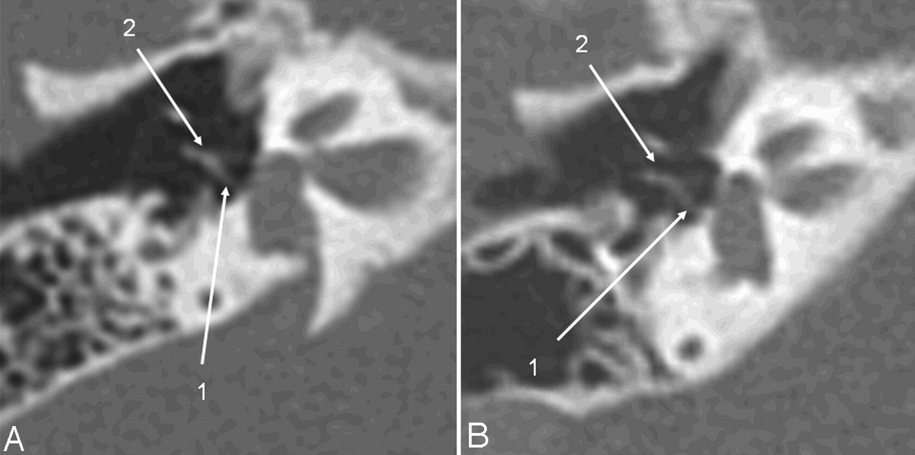
  • Fig 2.
    • Download figure
    • Open in new tab
    • Download powerpoint
    Fig 2.

    Axial CT section of the right temporal bone obtained with a CTDIvol of 63 mGy (A) (14-month-old patient; DLP, 223 mGy cm; estimated Deff, 1.4 mSv) and with the low-dose protocol (B) (16-month-old child; CTDIvol, 10.8 mGy; DLP, 46.9 mGy cm; estimated Deff, 0.35 mSv). Critical structures like the modiolus and the thin bony lamella separating the internal auditory canal from the cochlea (1) and the spiral osseous lamina (2) are delineated well despite the higher image noise.

  • Fig 3.
    • Download figure
    • Open in new tab
    • Download powerpoint
    Fig 3.

    The same patients as in Fig 2. Oblique axial image, reformatted in the stapes main plane, a high-dose scan (A) versus a low-dose scan (B). The posterior stapes crus (1) and the incudostapedial articulation (2) are identifiable on both scans.

Tables

  • Figures
    • View popup
    Table 1:

    Synopsis of the 23 anatomic structures reviewed and the respective primary criteria for image quality assessment

    Structure/ConditionReview Criteria
    CochleaNormal contour, 2.5 turns
    Cochlear patencyAbility to discern intracochlear ossifications
    Spiral osseous laminaPresence, integrity
    ModiolusPresence, integrity
    LabyrinthContour, density
    Vestibular aqueductContour, density
    Cochlear aqueductContour, density
    Cochlear nerve canalPresence
    Bony lamella at auditory canal fundusPresence, integrity of bony lamella separating the internal auditory canal from the cochlea
    Internal auditory canalContour
    Facial nerve canal, cochlear segmentContour, course
    Facial nerve canal, tympanic segmentContour, course
    Facial nerve canal, mastoid segmentContour, course
    Middle ear cavityAeration
    MalleusPresence of all parts
    IncusPresence of all parts
    StapesPresence of all parts
    Round windowPresence, aperture
    Round window nicheBorders, aeration
    Oval windowPresence, borders, footplate position
    Internal carotid artery canalBorders, osseous wall dehiscence
    Jugular foramenBorders, osseous wall dehiscence
    MastoidBony borders, aeration
    • View popup
    Table 2:

    Mean effective doses (millisievert) for 3 age groups when scanned with local low- and high-dose protocols in comparison with literature-derived calculated effective doses and literature-derived effective doses

    AgeLow-Dose ProtocolHigh-Dose ProtocolSwartz et al 20098aThomas et al 20089bLutz et al 20075b
    CTDIDLPDeffCTDIDLPDeffCTDIkVmAsDeffkVmAsDeffkVmAsDeff
    1 year old8.8400.3632851.8341201500.9–11202002.6–––
    5 year old10.8490.25632881.4451202000.97–1.11202001.7–––
    Adult–––––––1203201.2–1.3–––1201400.3
    • a Indicates literature derived calculated effective doses.

    • b – indicates literature-derived effective doses.

    • When available, CTDI (milligray), DLP (milligray × centimeter), and kilovolt/milliampere-second are shown also for literature-derived protocols.

PreviousNext
Back to top

In this issue

American Journal of Neuroradiology: 32 (8)
American Journal of Neuroradiology
Vol. 32, Issue 8
1 Sep 2011
  • Table of Contents
  • Index by author
Advertisement
Print
Download PDF
Email Article

Thank you for your interest in spreading the word on American Journal of Neuroradiology.

NOTE: We only request your email address so that the person you are recommending the page to knows that you wanted them to see it, and that it is not junk mail. We do not capture any email address.

Enter multiple addresses on separate lines or separate them with commas.
Low-Dose Temporal Bone CT in Infants and Young Children: Effective Dose and Image Quality
(Your Name) has sent you a message from American Journal of Neuroradiology
(Your Name) thought you would like to see the American Journal of Neuroradiology web site.
CAPTCHA
This question is for testing whether or not you are a human visitor and to prevent automated spam submissions.
Cite this article
C.B. Nauer, A. Rieke, C. Zubler, C. Candreia, A. Arnold, P. Senn
Low-Dose Temporal Bone CT in Infants and Young Children: Effective Dose and Image Quality
American Journal of Neuroradiology Sep 2011, 32 (8) 1375-1380; DOI: 10.3174/ajnr.A2524

Citation Manager Formats

  • BibTeX
  • Bookends
  • EasyBib
  • EndNote (tagged)
  • EndNote 8 (xml)
  • Medlars
  • Mendeley
  • Papers
  • RefWorks Tagged
  • Ref Manager
  • RIS
  • Zotero
0 Responses
Respond to this article
Share
Bookmark this article
Low-Dose Temporal Bone CT in Infants and Young Children: Effective Dose and Image Quality
C.B. Nauer, A. Rieke, C. Zubler, C. Candreia, A. Arnold, P. Senn
American Journal of Neuroradiology Sep 2011, 32 (8) 1375-1380; DOI: 10.3174/ajnr.A2524
del.icio.us logo Twitter logo Facebook logo Mendeley logo
  • Tweet Widget
  • Facebook Like
  • Google Plus One
Purchase

Jump to section

  • Article
    • Abstract
    • Abbreviations
    • Materials and Methods
    • Results
    • Discussion
    • Conclusions
    • Acknowledgments
    • Footnotes
    • References
  • Figures & Data
  • Info & Metrics
  • Responses
  • References
  • PDF

Related Articles

  • No related articles found.
  • PubMed
  • Google Scholar

Cited By...

  • Temporal Bone CT: Improved Image Quality and Potential for Decreased Radiation Dose Using an Ultra-High-Resolution Scan Mode with an Iterative Reconstruction Algorithm
  • Crossref
  • Google Scholar

This article has not yet been cited by articles in journals that are participating in Crossref Cited-by Linking.

More in this TOC Section

  • Safety of Intrathecal Gadobutrol in Various Doses
  • Impact of Kidney Function on CNS Gadolinium Deposition in Patients Receiving Repeated Doses of Gadobutrol
  • Contrast-Induced Acute Kidney Injury in Radiologic Management of Acute Ischemic Stroke in the Emergency Setting
Show more Patient Safety

Similar Articles

Advertisement

Indexed Content

  • Current Issue
  • Accepted Manuscripts
  • Article Preview
  • Past Issues
  • Editorials
  • Editor's Choice
  • Fellows' Journal Club
  • Letters to the Editor
  • Video Articles

Cases

  • Case Collection
  • Archive - Case of the Week
  • Archive - Case of the Month
  • Archive - Classic Case

More from AJNR

  • Trainee Corner
  • Imaging Protocols
  • MRI Safety Corner
  • Book Reviews

Multimedia

  • AJNR Podcasts
  • AJNR Scantastics

Resources

  • Turnaround Time
  • Submit a Manuscript
  • Submit a Video Article
  • Submit an eLetter to the Editor/Response
  • Manuscript Submission Guidelines
  • Statistical Tips
  • Fast Publishing of Accepted Manuscripts
  • Graphical Abstract Preparation
  • Imaging Protocol Submission
  • Evidence-Based Medicine Level Guide
  • Publishing Checklists
  • Author Policies
  • Become a Reviewer/Academy of Reviewers
  • News and Updates

About Us

  • About AJNR
  • Editorial Board
  • Editorial Board Alumni
  • Alerts
  • Permissions
  • Not an AJNR Subscriber? Join Now
  • Advertise with Us
  • Librarian Resources
  • Feedback
  • Terms and Conditions
  • AJNR Editorial Board Alumni

American Society of Neuroradiology

  • Not an ASNR Member? Join Now

© 2025 by the American Society of Neuroradiology All rights, including for text and data mining, AI training, and similar technologies, are reserved.
Print ISSN: 0195-6108 Online ISSN: 1936-959X

Powered by HighWire