Advertisement

Main menu

  • Home
  • Content
    • Current Issue
    • Accepted Manuscripts
    • Article Preview
    • Past Issue Archive
    • Video Articles
    • AJNR Case Collection
    • Case of the Week Archive
    • Case of the Month Archive
    • Classic Case Archive
  • Special Collections
    • AJNR Awards
    • Low-Field MRI
    • Alzheimer Disease
    • ASNR Foundation Special Collection
    • Photon-Counting CT
    • View All
  • Multimedia
    • AJNR Podcasts
    • AJNR SCANtastic
    • Trainee Corner
    • MRI Safety Corner
    • Imaging Protocols
  • For Authors
    • Submit a Manuscript
    • Submit a Video Article
    • Submit an eLetter to the Editor/Response
    • Manuscript Submission Guidelines
    • Statistical Tips
    • Fast Publishing of Accepted Manuscripts
    • Graphical Abstract Preparation
    • Imaging Protocol Submission
    • Author Policies
  • About Us
    • About AJNR
    • Editorial Board
    • Editorial Board Alumni
  • More
    • Become a Reviewer/Academy of Reviewers
    • Subscribers
    • Permissions
    • Alerts
    • Feedback
    • Advertisers
    • ASNR Home

User menu

  • Alerts
  • Log in

Search

  • Advanced search
American Journal of Neuroradiology

American Journal of Neuroradiology

ASHNR American Society of Functional Neuroradiology ASHNR American Society of Pediatric Neuroradiology ASSR
  • Alerts
  • Log in

Advanced Search

  • Home
  • Content
    • Current Issue
    • Accepted Manuscripts
    • Article Preview
    • Past Issue Archive
    • Video Articles
    • AJNR Case Collection
    • Case of the Week Archive
    • Case of the Month Archive
    • Classic Case Archive
  • Special Collections
    • AJNR Awards
    • Low-Field MRI
    • Alzheimer Disease
    • ASNR Foundation Special Collection
    • Photon-Counting CT
    • View All
  • Multimedia
    • AJNR Podcasts
    • AJNR SCANtastic
    • Trainee Corner
    • MRI Safety Corner
    • Imaging Protocols
  • For Authors
    • Submit a Manuscript
    • Submit a Video Article
    • Submit an eLetter to the Editor/Response
    • Manuscript Submission Guidelines
    • Statistical Tips
    • Fast Publishing of Accepted Manuscripts
    • Graphical Abstract Preparation
    • Imaging Protocol Submission
    • Author Policies
  • About Us
    • About AJNR
    • Editorial Board
    • Editorial Board Alumni
  • More
    • Become a Reviewer/Academy of Reviewers
    • Subscribers
    • Permissions
    • Alerts
    • Feedback
    • Advertisers
    • ASNR Home
  • Follow AJNR on Twitter
  • Visit AJNR on Facebook
  • Follow AJNR on Instagram
  • Join AJNR on LinkedIn
  • RSS Feeds

AJNR Awards, New Junior Editors, and more. Read the latest AJNR updates

Review ArticleReview Articles
Open Access

Neuroradiology of Cholesteatomas

K. Baráth, A.M. Huber, P. Stämpfli, Z. Varga and S. Kollias
American Journal of Neuroradiology February 2011, 32 (2) 221-229; DOI: https://doi.org/10.3174/ajnr.A2052
K. Baráth
  • Find this author on Google Scholar
  • Find this author on PubMed
  • Search for this author on this site
A.M. Huber
  • Find this author on Google Scholar
  • Find this author on PubMed
  • Search for this author on this site
P. Stämpfli
  • Find this author on Google Scholar
  • Find this author on PubMed
  • Search for this author on this site
Z. Varga
  • Find this author on Google Scholar
  • Find this author on PubMed
  • Search for this author on this site
S. Kollias
  • Find this author on Google Scholar
  • Find this author on PubMed
  • Search for this author on this site
  • Article
  • Figures & Data
  • Info & Metrics
  • Responses
  • References
  • PDF
Loading

Article Figures & Data

Figures

  • Tables
  • Fig 1.
    • Download figure
    • Open in new tab
    • Download powerpoint
    Fig 1.

    Congenital cholesteatoma. Coronal (A) and axial HRCT (B) scans demonstrate a round well-defined lesion (arrow) anterosuperior in the tympanic cavity, medial to the ossicular chain. Note the missing ossicular erosion. Based on the position of the lesion and the lack of bone erosion along with the clinical aspects, this is probably a congenital type.

  • Fig 2.
    • Download figure
    • Open in new tab
    • Download powerpoint
    Fig 2.

    Cholesteatoma of the petrous apex. Contrast-enhanced HRCT scans with a soft-tissue window (A) and a bone window (B) show an oval well-delineated, nonenhancing lesion (white arrows) with erosion of the posterior wall of the pyramidal segment of the internal carotid artery (thin black arrow) and the anterior wall of the jugular bulb (thick black arrow). C, DWI demonstrates diffusion restriction in the lesion (white arrow), supporting the diagnosis of a cholesteatoma.

  • Fig 3.
    • Download figure
    • Open in new tab
    • Download powerpoint
    Fig 3.

    EAC cholesteatoma (arrow). Note the typical localization at the inferior wall of the EAC and the small bone fragments along the lesion.

  • Fig 4.
    • Download figure
    • Open in new tab
    • Download powerpoint
    Fig 4.

    HRCT scan, coronal view. A, Pars flaccida cholesteatoma (arrow) filling the Prussak space. Notice the erosion of the scutum (dashed arrow). B, Prussak space is bordered by the pars flaccida of the TM (arrow) lateral, neck of the malleus (thick white arrow) medial, the short process of the malleus (white arrowhead) inferior, and lateral malleal ligament (dashed arrow) superior.
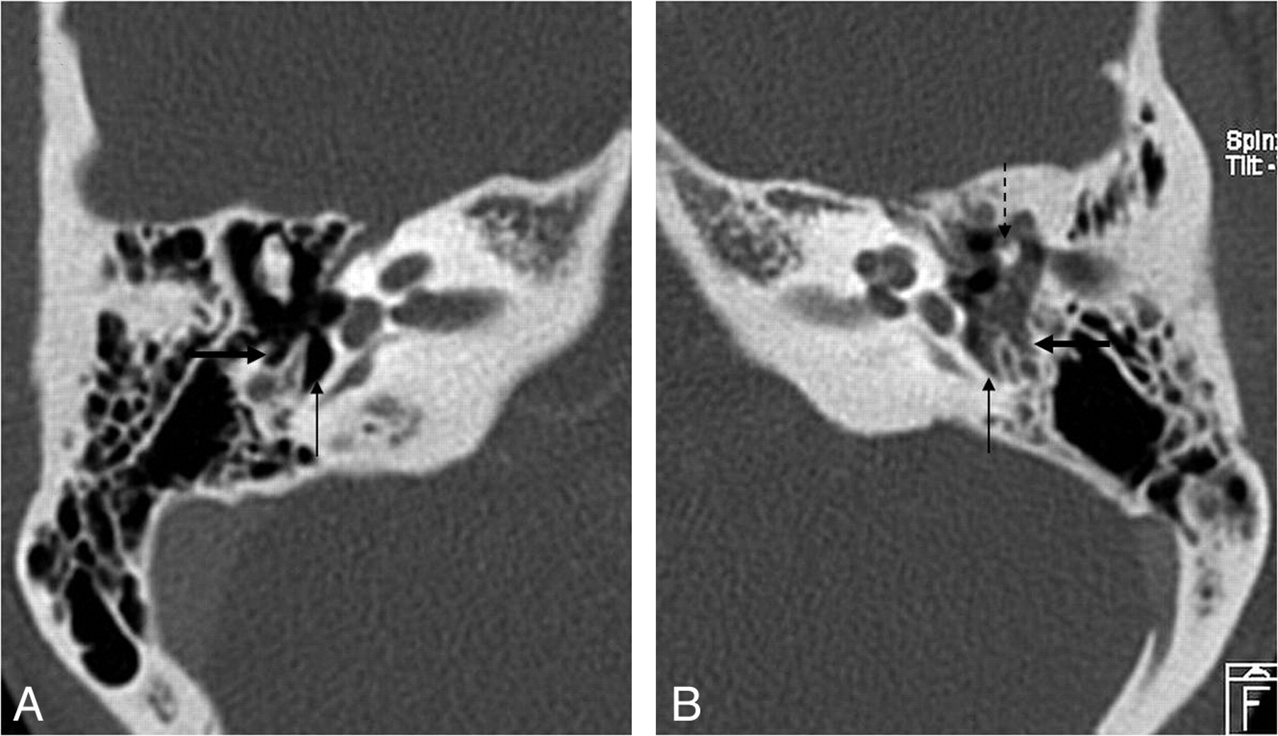
  • Fig 5.
    • Download figure
    • Open in new tab
    • Download powerpoint
    Fig 5.

    Pars tensa cholesteatoma. A, Coronal HRCT scan at the level of the cochlea shows the soft-tissue mass (black arrow) at the pars tensa of the retracted TM (white arrow). B, At the level of the vestibulum, the obliteration of the oval (dashed black arrow) and round (dashed white arrow) window niche is seen. Note the small bony fragments in the oval window niche, probably a sign of erosion of the stapes and the inferior wall of the tympanic segment of the facial canal.
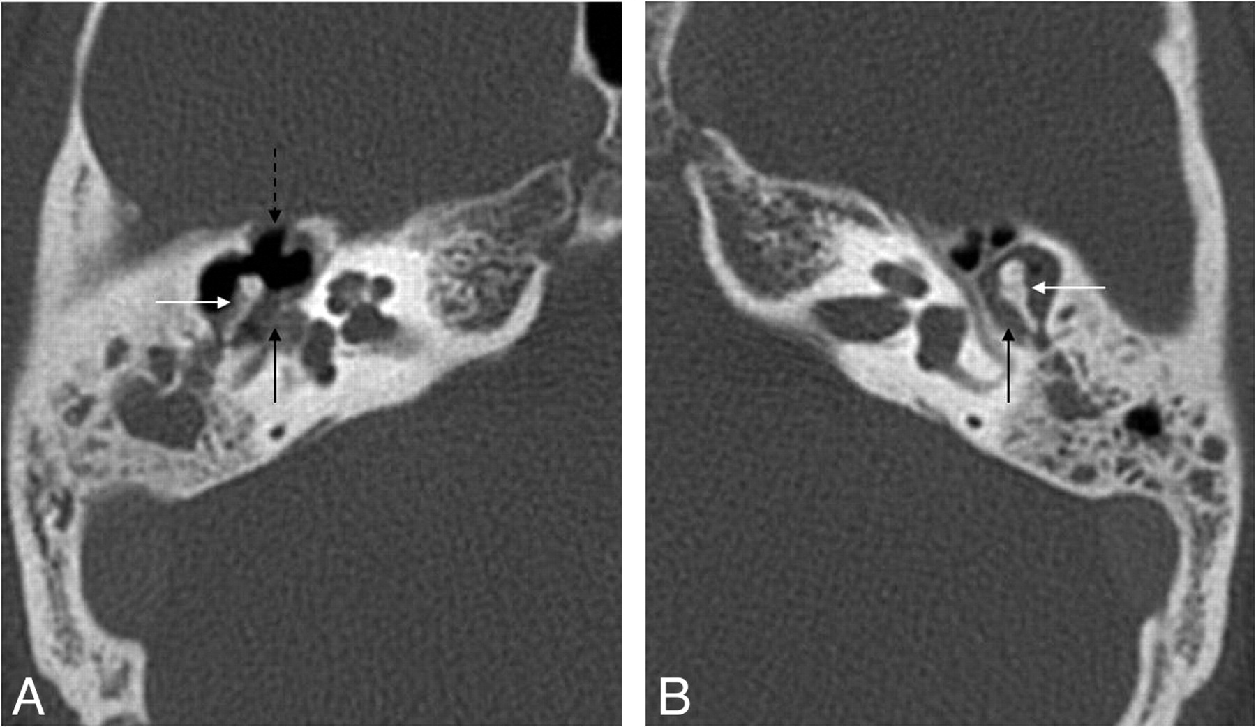
  • Fig 6.
    • Download figure
    • Open in new tab
    • Download powerpoint
    Fig 6.

    Pars tensa cholesteatoma. HRCT scans demonstrate the normal (air-filled) (A) and the obliterated (B) sinus tympani (arrow) and facial recess (thick arrow) due to a pars tensa (sinus) cholesteatoma. Note the ossicular erosion (dashed arrow) on B.
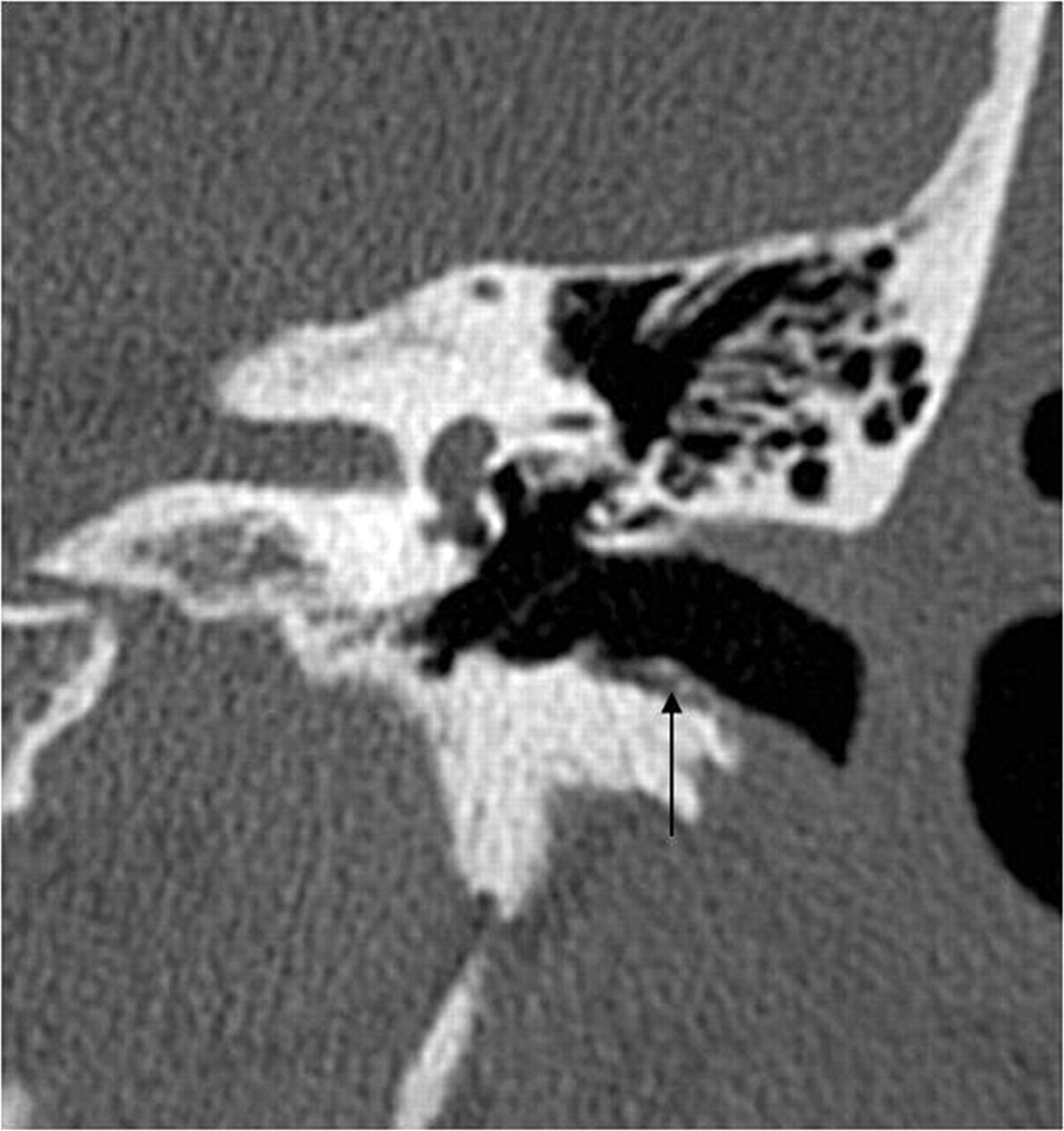
  • Fig 7.
    • Download figure
    • Open in new tab
    • Download powerpoint
    Fig 7.

    Mural cholesteatoma. Axial (A) and coronal (B) HRCT scans show the shell of the cholesteatoma in the epitympanum (black arrow) and the automastoidectomy cavity (thick arrow) without a history of surgery. Note the complete erosion of the ossicles, the fistula of the horizontal semicircular canal (dashed black arrow), and the wall erosion of the tympanic segment of the facial nerve canal (white arrow).

  • Fig 8.
    • Download figure
    • Open in new tab
    • Download powerpoint
    Fig 8.

    Intraoperative images show a typical pearly appearance of a cholesteatoma (arrow, A), in the aditus ad antrum, next to the posterior wall of the EAC (dashed arrow) and a more irregular cholesteatoma (thick arrow, B) accompanied by granulation tissue.
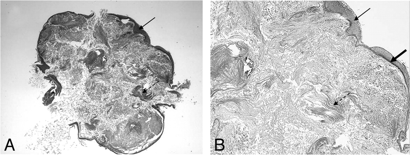
  • Fig 9.
    • Download figure
    • Open in new tab
    • Download powerpoint
    Fig 9.

    Histologic appearance of a cholesteatoma. Hematoxylin-eosin stain. Low-power view (original magnification ×25) (A) and high-power view (original magnification ×400) (B) demonstrate a cystic lesion covered by a strongly keratinizing stratified squamous epithelium (arrows). Within the cyst, there is abundant formation of desquamated keratin lamellas (dashed arrows). Note the prominent strongly hyperchromatic basal layer of the epidermis (thick arrow).

  • Fig 10.
    • Download figure
    • Open in new tab
    • Download powerpoint
    Fig 10.

    Patient with cholesteatoma on the right and chronic otitis media without cholesteatoma on the left. A, Axial HRCT scan shows the mass lesion (black arrow) in the tympanic cavity with ossicle erosion (white arrow) and erosion of the anterior wall of the epitympanum (dashed arrow). B, Axial HRCT scan demonstrates a mass lesion (black arrow) in the epitympanum, but no bony erosion (white arrow).

  • Fig 11.
    • Download figure
    • Open in new tab
    • Download powerpoint
    Fig 11.

    Recurrent cholesteatoma after surgery. A, Coronal HRCT scan shows the obliterated mastoidectomy cavity (white arrow). B, Coronal FIESTA image distinguishes the slightly hyperintense (to brain) cholesteatoma (thick white arrow on B, C, and D) from the strongly hyperintense granulation tissue (dashed arrow on B and C). C, Coronal contrast-enhanced MR image differentiates as well the nonenhancing cholesteatoma from the strongly enhancing granulation tissue. D, Coronal DWI with the intensive intralesional diffusion restriction supports the diagnosis. Note the susceptibility artifacts on the EPI-DWI (black arrow) on the right.

Tables

  • Figures
    • View popup
    Table 1:

    Classification of all cholesteatomas based on pathogenesis

    Initial LocationClinical HistoryStatus of TM (if middle ear)
    Congenital (2%)aAnywhere in the temporal boneNo historyIntact
    AcquiredMiddle earRecurrent ear disease
        Primary (80%)aApparently intact
        Secondary (18%)aPerforated
    • a The percentage refers to the distribution of cholesteatomas in the middle ear.

    • View popup
    Table 2:

    Classification of middle ear cholesteatomas based on location in relation to the TM

    Initial LocationPathogenesis
    Pars flaccida (attic)Epitympanum,Congenital
        lateral to ossiclesPrimary acquired
    Secondary acquired
    Pars tensa (sinus)Mesotympanum,Congenital
        medial to ossiclesSecondary acquired
PreviousNext
Back to top

In this issue

American Journal of Neuroradiology: 32 (2)
American Journal of Neuroradiology
Vol. 32, Issue 2
1 Feb 2011
  • Table of Contents
  • Index by author
Advertisement
Print
Download PDF
Email Article

Thank you for your interest in spreading the word on American Journal of Neuroradiology.

NOTE: We only request your email address so that the person you are recommending the page to knows that you wanted them to see it, and that it is not junk mail. We do not capture any email address.

Enter multiple addresses on separate lines or separate them with commas.
Neuroradiology of Cholesteatomas
(Your Name) has sent you a message from American Journal of Neuroradiology
(Your Name) thought you would like to see the American Journal of Neuroradiology web site.
CAPTCHA
This question is for testing whether or not you are a human visitor and to prevent automated spam submissions.
Cite this article
K. Baráth, A.M. Huber, P. Stämpfli, Z. Varga, S. Kollias
Neuroradiology of Cholesteatomas
American Journal of Neuroradiology Feb 2011, 32 (2) 221-229; DOI: 10.3174/ajnr.A2052

Citation Manager Formats

  • BibTeX
  • Bookends
  • EasyBib
  • EndNote (tagged)
  • EndNote 8 (xml)
  • Medlars
  • Mendeley
  • Papers
  • RefWorks Tagged
  • Ref Manager
  • RIS
  • Zotero
0 Responses
Respond to this article
Share
Bookmark this article
Neuroradiology of Cholesteatomas
K. Baráth, A.M. Huber, P. Stämpfli, Z. Varga, S. Kollias
American Journal of Neuroradiology Feb 2011, 32 (2) 221-229; DOI: 10.3174/ajnr.A2052
del.icio.us logo Twitter logo Facebook logo Mendeley logo
  • Tweet Widget
  • Facebook Like
  • Google Plus One
Purchase

Jump to section

  • Article
    • Abstract
    • Abbreviations
    • Definition
    • History and Etymology
    • Epidemiology
    • Classification
    • Cross-Sectional Imaging
    • Differential Diagnosis
    • References
  • Figures & Data
  • Info & Metrics
  • Responses
  • References
  • PDF

Related Articles

  • No related articles found.
  • PubMed
  • Google Scholar

Cited By...

  • Improved Assessment of Middle Ear Recurrent Cholesteatomas Using a Fusion of Conventional CT and Non-EPI-DWI MRI
  • Mystery Case: Cholesterol granuloma of the petrous apex in Gradenigo syndrome
  • Imaging Findings in Auto-Atticotomy
  • Crossref
  • Google Scholar

This article has not yet been cited by articles in journals that are participating in Crossref Cited-by Linking.

More in this TOC Section

  • An Atlas of Neonatal Neurovascular Imaging Anatomy as Depicted with Microvascular Imaging: The Intracranial Arteries
  • An Atlas of Neonatal Neurovascular Imaging Anatomy as Depicted with Microvascular Imaging: The Intracranial Veins
  • Clinical Translation of Hyperpolarized 13C Metabolic Probes for Glioma Imaging
Show more Review Articles

Similar Articles

Advertisement

Indexed Content

  • Current Issue
  • Accepted Manuscripts
  • Article Preview
  • Past Issues
  • Editorials
  • Editor's Choice
  • Fellows' Journal Club
  • Letters to the Editor
  • Video Articles

Cases

  • Case Collection
  • Archive - Case of the Week
  • Archive - Case of the Month
  • Archive - Classic Case

More from AJNR

  • Trainee Corner
  • Imaging Protocols
  • MRI Safety Corner
  • Book Reviews

Multimedia

  • AJNR Podcasts
  • AJNR Scantastics

Resources

  • Turnaround Time
  • Submit a Manuscript
  • Submit a Video Article
  • Submit an eLetter to the Editor/Response
  • Manuscript Submission Guidelines
  • Statistical Tips
  • Fast Publishing of Accepted Manuscripts
  • Graphical Abstract Preparation
  • Imaging Protocol Submission
  • Evidence-Based Medicine Level Guide
  • Publishing Checklists
  • Author Policies
  • Become a Reviewer/Academy of Reviewers
  • News and Updates

About Us

  • About AJNR
  • Editorial Board
  • Editorial Board Alumni
  • Alerts
  • Permissions
  • Not an AJNR Subscriber? Join Now
  • Advertise with Us
  • Librarian Resources
  • Feedback
  • Terms and Conditions
  • AJNR Editorial Board Alumni

American Society of Neuroradiology

  • Not an ASNR Member? Join Now

© 2025 by the American Society of Neuroradiology All rights, including for text and data mining, AI training, and similar technologies, are reserved.
Print ISSN: 0195-6108 Online ISSN: 1936-959X

Powered by HighWire