Advertisement

Main menu

  • Home
  • Content
    • Current Issue
    • Accepted Manuscripts
    • Article Preview
    • Past Issue Archive
    • Video Articles
    • AJNR Case Collection
    • Case of the Week Archive
    • Case of the Month Archive
    • Classic Case Archive
  • Special Collections
    • AJNR Awards
    • Low-Field MRI
    • Alzheimer Disease
    • ASNR Foundation Special Collection
    • Photon-Counting CT
    • View All
  • Multimedia
    • AJNR Podcasts
    • AJNR SCANtastic
    • Trainee Corner
    • MRI Safety Corner
    • Imaging Protocols
  • For Authors
    • Submit a Manuscript
    • Submit a Video Article
    • Submit an eLetter to the Editor/Response
    • Manuscript Submission Guidelines
    • Statistical Tips
    • Fast Publishing of Accepted Manuscripts
    • Graphical Abstract Preparation
    • Imaging Protocol Submission
    • Author Policies
  • About Us
    • About AJNR
    • Editorial Board
    • Editorial Board Alumni
  • More
    • Become a Reviewer/Academy of Reviewers
    • Subscribers
    • Permissions
    • Alerts
    • Feedback
    • Advertisers
    • ASNR Home

User menu

  • Alerts
  • Log in

Search

  • Advanced search
American Journal of Neuroradiology

American Journal of Neuroradiology

ASHNR American Society of Functional Neuroradiology ASHNR American Society of Pediatric Neuroradiology ASSR
  • Alerts
  • Log in

Advanced Search

  • Home
  • Content
    • Current Issue
    • Accepted Manuscripts
    • Article Preview
    • Past Issue Archive
    • Video Articles
    • AJNR Case Collection
    • Case of the Week Archive
    • Case of the Month Archive
    • Classic Case Archive
  • Special Collections
    • AJNR Awards
    • Low-Field MRI
    • Alzheimer Disease
    • ASNR Foundation Special Collection
    • Photon-Counting CT
    • View All
  • Multimedia
    • AJNR Podcasts
    • AJNR SCANtastic
    • Trainee Corner
    • MRI Safety Corner
    • Imaging Protocols
  • For Authors
    • Submit a Manuscript
    • Submit a Video Article
    • Submit an eLetter to the Editor/Response
    • Manuscript Submission Guidelines
    • Statistical Tips
    • Fast Publishing of Accepted Manuscripts
    • Graphical Abstract Preparation
    • Imaging Protocol Submission
    • Author Policies
  • About Us
    • About AJNR
    • Editorial Board
    • Editorial Board Alumni
  • More
    • Become a Reviewer/Academy of Reviewers
    • Subscribers
    • Permissions
    • Alerts
    • Feedback
    • Advertisers
    • ASNR Home
  • Follow AJNR on Twitter
  • Visit AJNR on Facebook
  • Follow AJNR on Instagram
  • Join AJNR on LinkedIn
  • RSS Feeds

AJNR Awards, New Junior Editors, and more. Read the latest AJNR updates

Research ArticlePediatric Neuroimaging

High-Resolution In Utero 3D MR Imaging of Inner Ear Microstructures in Fetal Sheep

J.-H. Buhk, M. Frisch, J. Yamamura, J. Graessner, G. Adam and U. Wedegärtner
American Journal of Neuroradiology December 2011, 32 (11) 2043-2046; DOI: https://doi.org/10.3174/ajnr.A2736
J.-H. Buhk
aFrom the Department of Diagnostic and Interventional Radiology (J.-H.B., M.F., J.Y., G.A., U.W.), University Medical Center Hamburg Eppendorf, Hamburg, Germany
  • Find this author on Google Scholar
  • Find this author on PubMed
  • Search for this author on this site
M. Frisch
aFrom the Department of Diagnostic and Interventional Radiology (J.-H.B., M.F., J.Y., G.A., U.W.), University Medical Center Hamburg Eppendorf, Hamburg, Germany
  • Find this author on Google Scholar
  • Find this author on PubMed
  • Search for this author on this site
J. Yamamura
aFrom the Department of Diagnostic and Interventional Radiology (J.-H.B., M.F., J.Y., G.A., U.W.), University Medical Center Hamburg Eppendorf, Hamburg, Germany
  • Find this author on Google Scholar
  • Find this author on PubMed
  • Search for this author on this site
J. Graessner
bSiemens Healthcare (J.G.), Hamburg, Germany.
  • Find this author on Google Scholar
  • Find this author on PubMed
  • Search for this author on this site
G. Adam
aFrom the Department of Diagnostic and Interventional Radiology (J.-H.B., M.F., J.Y., G.A., U.W.), University Medical Center Hamburg Eppendorf, Hamburg, Germany
  • Find this author on Google Scholar
  • Find this author on PubMed
  • Search for this author on this site
U. Wedegärtner
aFrom the Department of Diagnostic and Interventional Radiology (J.-H.B., M.F., J.Y., G.A., U.W.), University Medical Center Hamburg Eppendorf, Hamburg, Germany
  • Find this author on Google Scholar
  • Find this author on PubMed
  • Search for this author on this site
  • Article
  • Figures & Data
  • Info & Metrics
  • Responses
  • References
  • PDF
Loading

Article Figures & Data

Figures

  • Tables
  • Fig 1.
    • Download figure
    • Open in new tab
    • Download powerpoint
    Fig 1.

    Coronal view (thin-section MIP; section thickness, 0.5 mm) shows the normally developed cochlea (open arrows) and the tympanic cavity (closed arrows), which are both (normal findings in utero) fluid-filled in the reference examination (A, 3D CISS, stillborn lamb) as well as in vivo (B, 3D True FISP).

  • Fig 2.
    • Download figure
    • Open in new tab
    • Download powerpoint
    Fig 2.

    This set of axial views of the reference examination (A, 3D CISS, stillborn lamb) and in vivo (B, 3D True FISP; both thin-section MIPs; section thickness, 0.7 mm) displays the IAM with the seventh and eighth cranial nerve (arrows) and a cross-section through the normally developed cochlear modiolus, which is also well-depicted.
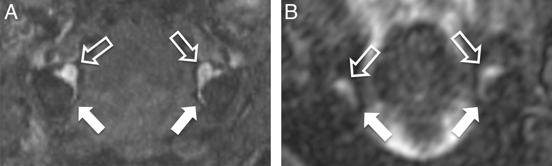
  • Fig 3.
    • Download figure
    • Open in new tab
    • Download powerpoint
    Fig 3.

    The axial views of the reference examination (A, 3D CISS, stillborn lamb) and in vivo (B, 3D True FISP; both thin-section MIPs; section thickness, 2.0 mm) display the utriculus (open arrows) and the medial section of the posterior semicircular canal (closed arrows). The utricular ostium of the lateral semicircular canal can be seen as well in both images.

Tables

  • Figures
  • Scores regarding the identification of the anatomic structures and of the assessment of the artifacts loada

    AnimalCochleaUtriculusLateral CanalAnterior CanalPosterior CanalIAMbArtifacts
    A441243Moderate
    B441144Unremarkable
    C552145Unremarkable
    D331113Severe
    E441133Moderate
    F551144Moderate
    Ref.553353Unremarkable
    • Note:—Ref. indicates reference; 1, not visible; 2, visible, not diagnostic; 3, moderate but sufficient; 4, good quality; 5, excellent quality.

    • ↵a First, the depictability of the lateral and anterior semicircular canals is also moderate in the reference examination and, therefore, likely to be a normal anatomic finding at that stage of development. Second, severe artifacts only occurred in animal D due to breech presentation of the fetus.

    • b Scoring includes visibility of cranial nerves VII and VIII.

PreviousNext
Back to top

In this issue

American Journal of Neuroradiology: 32 (11)
American Journal of Neuroradiology
Vol. 32, Issue 11
1 Dec 2011
  • Table of Contents
  • Index by author
Advertisement
Print
Download PDF
Email Article

Thank you for your interest in spreading the word on American Journal of Neuroradiology.

NOTE: We only request your email address so that the person you are recommending the page to knows that you wanted them to see it, and that it is not junk mail. We do not capture any email address.

Enter multiple addresses on separate lines or separate them with commas.
High-Resolution In Utero 3D MR Imaging of Inner Ear Microstructures in Fetal Sheep
(Your Name) has sent you a message from American Journal of Neuroradiology
(Your Name) thought you would like to see the American Journal of Neuroradiology web site.
CAPTCHA
This question is for testing whether or not you are a human visitor and to prevent automated spam submissions.
Cite this article
J.-H. Buhk, M. Frisch, J. Yamamura, J. Graessner, G. Adam, U. Wedegärtner
High-Resolution In Utero 3D MR Imaging of Inner Ear Microstructures in Fetal Sheep
American Journal of Neuroradiology Dec 2011, 32 (11) 2043-2046; DOI: 10.3174/ajnr.A2736

Citation Manager Formats

  • BibTeX
  • Bookends
  • EasyBib
  • EndNote (tagged)
  • EndNote 8 (xml)
  • Medlars
  • Mendeley
  • Papers
  • RefWorks Tagged
  • Ref Manager
  • RIS
  • Zotero
0 Responses
Respond to this article
Share
Bookmark this article
High-Resolution In Utero 3D MR Imaging of Inner Ear Microstructures in Fetal Sheep
J.-H. Buhk, M. Frisch, J. Yamamura, J. Graessner, G. Adam, U. Wedegärtner
American Journal of Neuroradiology Dec 2011, 32 (11) 2043-2046; DOI: 10.3174/ajnr.A2736
del.icio.us logo Twitter logo Facebook logo Mendeley logo
  • Tweet Widget
  • Facebook Like
  • Google Plus One
Purchase

Jump to section

  • Article
    • Abstract
    • ABBREVIATIONS
    • Materials and Methods
    • Results
    • Discussion
    • Footnotes
    • References
  • Figures & Data
  • Info & Metrics
  • Responses
  • References
  • PDF

Related Articles

  • No related articles found.
  • PubMed
  • Google Scholar

Cited By...

  • No citing articles found.
  • Crossref
  • Google Scholar

This article has not yet been cited by articles in journals that are participating in Crossref Cited-by Linking.

More in this TOC Section

  • Frontal Paraventricular Cysts
  • Sodium MRI in Pediatric Brain Tumors
  • FRACTURE MR in Congenital Vertebral Anomalies
Show more Pediatric Neuroimaging

Similar Articles

Advertisement

Indexed Content

  • Current Issue
  • Accepted Manuscripts
  • Article Preview
  • Past Issues
  • Editorials
  • Editor's Choice
  • Fellows' Journal Club
  • Letters to the Editor
  • Video Articles

Cases

  • Case Collection
  • Archive - Case of the Week
  • Archive - Case of the Month
  • Archive - Classic Case

More from AJNR

  • Trainee Corner
  • Imaging Protocols
  • MRI Safety Corner
  • Book Reviews

Multimedia

  • AJNR Podcasts
  • AJNR Scantastics

Resources

  • Turnaround Time
  • Submit a Manuscript
  • Submit a Video Article
  • Submit an eLetter to the Editor/Response
  • Manuscript Submission Guidelines
  • Statistical Tips
  • Fast Publishing of Accepted Manuscripts
  • Graphical Abstract Preparation
  • Imaging Protocol Submission
  • Evidence-Based Medicine Level Guide
  • Publishing Checklists
  • Author Policies
  • Become a Reviewer/Academy of Reviewers
  • News and Updates

About Us

  • About AJNR
  • Editorial Board
  • Editorial Board Alumni
  • Alerts
  • Permissions
  • Not an AJNR Subscriber? Join Now
  • Advertise with Us
  • Librarian Resources
  • Feedback
  • Terms and Conditions
  • AJNR Editorial Board Alumni

American Society of Neuroradiology

  • Not an ASNR Member? Join Now

© 2025 by the American Society of Neuroradiology All rights, including for text and data mining, AI training, and similar technologies, are reserved.
Print ISSN: 0195-6108 Online ISSN: 1936-959X

Powered by HighWire