Advertisement

Main menu

  • Home
  • Content
    • Current Issue
    • Accepted Manuscripts
    • Article Preview
    • Past Issue Archive
    • Video Articles
    • AJNR Case Collection
    • Case of the Week Archive
    • Case of the Month Archive
    • Classic Case Archive
  • Special Collections
    • AJNR Awards
    • Low-Field MRI
    • Alzheimer Disease
    • ASNR Foundation Special Collection
    • Photon-Counting CT
    • View All
  • Multimedia
    • AJNR Podcasts
    • AJNR SCANtastic
    • Trainee Corner
    • MRI Safety Corner
    • Imaging Protocols
  • For Authors
    • Submit a Manuscript
    • Submit a Video Article
    • Submit an eLetter to the Editor/Response
    • Manuscript Submission Guidelines
    • Statistical Tips
    • Fast Publishing of Accepted Manuscripts
    • Graphical Abstract Preparation
    • Imaging Protocol Submission
    • Author Policies
  • About Us
    • About AJNR
    • Editorial Board
    • Editorial Board Alumni
  • More
    • Become a Reviewer/Academy of Reviewers
    • Subscribers
    • Permissions
    • Alerts
    • Feedback
    • Advertisers
    • ASNR Home

User menu

  • Alerts
  • Log in

Search

  • Advanced search
American Journal of Neuroradiology

American Journal of Neuroradiology

ASHNR American Society of Functional Neuroradiology ASHNR American Society of Pediatric Neuroradiology ASSR
  • Alerts
  • Log in

Advanced Search

  • Home
  • Content
    • Current Issue
    • Accepted Manuscripts
    • Article Preview
    • Past Issue Archive
    • Video Articles
    • AJNR Case Collection
    • Case of the Week Archive
    • Case of the Month Archive
    • Classic Case Archive
  • Special Collections
    • AJNR Awards
    • Low-Field MRI
    • Alzheimer Disease
    • ASNR Foundation Special Collection
    • Photon-Counting CT
    • View All
  • Multimedia
    • AJNR Podcasts
    • AJNR SCANtastic
    • Trainee Corner
    • MRI Safety Corner
    • Imaging Protocols
  • For Authors
    • Submit a Manuscript
    • Submit a Video Article
    • Submit an eLetter to the Editor/Response
    • Manuscript Submission Guidelines
    • Statistical Tips
    • Fast Publishing of Accepted Manuscripts
    • Graphical Abstract Preparation
    • Imaging Protocol Submission
    • Author Policies
  • About Us
    • About AJNR
    • Editorial Board
    • Editorial Board Alumni
  • More
    • Become a Reviewer/Academy of Reviewers
    • Subscribers
    • Permissions
    • Alerts
    • Feedback
    • Advertisers
    • ASNR Home
  • Follow AJNR on Twitter
  • Visit AJNR on Facebook
  • Follow AJNR on Instagram
  • Join AJNR on LinkedIn
  • RSS Feeds

AJNR Awards, New Junior Editors, and more. Read the latest AJNR updates

Research ArticlePediatric Neuroimaging
Open Access

Brain Perfusion in Asphyxiated Newborns Treated with Therapeutic Hypothermia

P. Wintermark, A. Hansen, M.C. Gregas, J. Soul, M. Labrecque, R.L. Robertson and S.K. Warfield
American Journal of Neuroradiology December 2011, 32 (11) 2023-2029; DOI: https://doi.org/10.3174/ajnr.A2708
P. Wintermark
aFrom the Division of Newborn Medicine (P.W., A.H., M.L.)
bDepartment of Radiology (P.W., R.L.R., S.K.W.)
eDivision of Newborn Medicine (P.W.), Montreal Children's McGill Hospital, Quebec, Ontario, Canada.
  • Find this author on Google Scholar
  • Find this author on PubMed
  • Search for this author on this site
A. Hansen
aFrom the Division of Newborn Medicine (P.W., A.H., M.L.)
  • Find this author on Google Scholar
  • Find this author on PubMed
  • Search for this author on this site
M.C. Gregas
cClinical Research Program (M.C.G.)
dDepartment of Neurology (M.C.G., J.S.), Children's Hospital Boston, Boston, Massachusetts
  • Find this author on Google Scholar
  • Find this author on PubMed
  • Search for this author on this site
J. Soul
dDepartment of Neurology (M.C.G., J.S.), Children's Hospital Boston, Boston, Massachusetts
  • Find this author on Google Scholar
  • Find this author on PubMed
  • Search for this author on this site
M. Labrecque
aFrom the Division of Newborn Medicine (P.W., A.H., M.L.)
  • Find this author on Google Scholar
  • Find this author on PubMed
  • Search for this author on this site
R.L. Robertson
bDepartment of Radiology (P.W., R.L.R., S.K.W.)
  • Find this author on Google Scholar
  • Find this author on PubMed
  • Search for this author on this site
S.K. Warfield
bDepartment of Radiology (P.W., R.L.R., S.K.W.)
  • Find this author on Google Scholar
  • Find this author on PubMed
  • Search for this author on this site
  • Article
  • Figures & Data
  • Supplemental
  • Info & Metrics
  • Responses
  • References
  • PDF
Loading

Article Figures & Data

Figures

  • Fig 1.
    • Download figure
    • Open in new tab
    • Download powerpoint
    Fig 1.

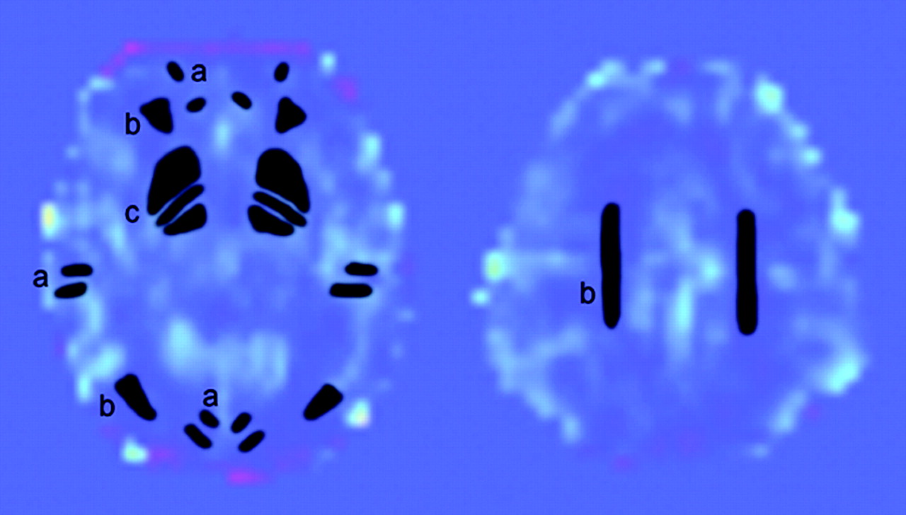
    Axial PASL images showing the manually drawn regions of interest. A total of 9 regions of interest were placed in 3 types of tissue. a, CGM (within frontal, parietal, and occipital cortices). b and c, WM (within the frontal and posterior WM and the centrum semiovale) (b) and BG (within the lentiform nucleus and the posterior limb of internal capsule and the thalamus) (c).15,17,24 Measurements were obtained in the right and left sides of the cerebrum in these tissue regions. Regions of interest were always drawn on the ASL data by 1 observer in similar brain areas for all the MR images by looking at the same time at the ASL data and the corresponding T2-weighted imaging.

  • Fig 2.
    • Download figure
    • Open in new tab
    • Download powerpoint
    Fig 2.

    Brain MR imaging in a control neonate; comparison between the perfusion map and images obtained from conventional sequences. A, CBF map obtained by perfusion imaging by ASL shows higher brain perfusion in GM and in BG compared with WM. B, ADC map. C, DWI. D, T2-weighted imaging.

  • Fig 3.
    • Download figure
    • Open in new tab
    • Download powerpoint
    Fig 3.

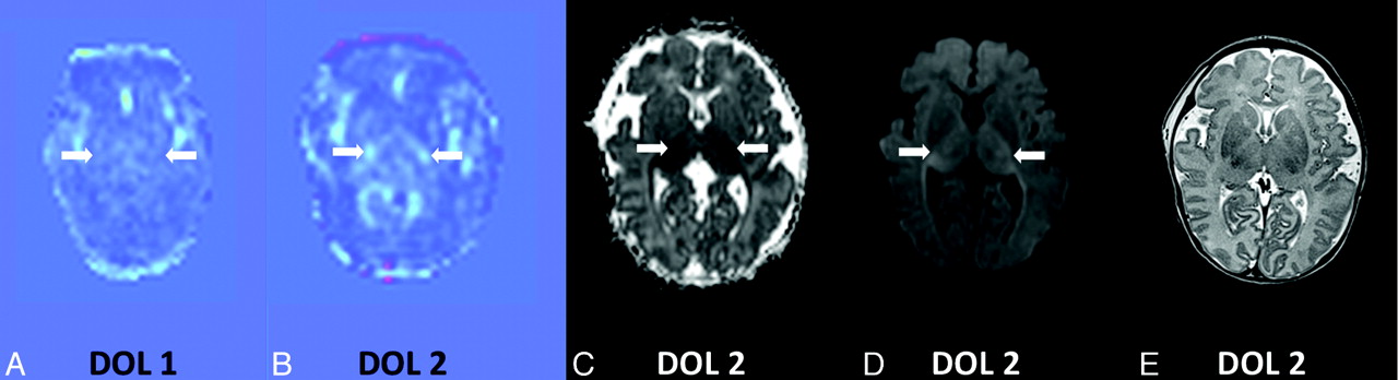
    Brain MR imaging on DOL 2 in patient 5 while he was treated with induced hypothermia; comparison between the perfusion map and images obtained from conventional sequences. A and B, CBF maps obtained by perfusion imaging by ASL show decreased brain perfusion on DOL 1 (A) followed by increased brain perfusion on DOL 2 (B) in the GM, WM, and BGs bilaterally, compared with the control patient. C and D, ADC map (C) and DWI (D) on DOL 2 display multifocal areas of restricted diffusion within bilateral BGs and both cerebral hemispheres involving GM and WM. E, Changes on T2-weighted imaging on DOL 2 are subtle.

  • Fig 4.
    • Download figure
    • Open in new tab
    • Download powerpoint
    Fig 4.

    Brain MR imaging on DOL 2 in patient 8, while he was treated with induced hypothermia; comparison between the perfusion map and images obtained from conventional sequences. A and B, CBF maps obtained by perfusion imaging by ASL show decreased brain perfusion on DOL 1 (A) followed by increased brain perfusion on DOL 2 (B) in bilateral thalami, and relatively lower perfusion in GM and the remaining BG, compared with the control patient. C and D, ADC map (C) and DWI (D) on DOL 2 display restricted diffusion within bilateral thalami. E, Changes on T2-weighted imaging on DOL 2 are subtle.

  • Fig 5.
    • Download figure
    • Open in new tab
    • Download powerpoint
    Fig 5.

    Comparison of means of CBF values in each region of interest between neonates developing brain injury in these regions of interest compared with those who did not, on DOL 1 (A) and on DOL 2–6 (B). Box-and-whisker plots (median, minimum, and maximum, in milliliters/100 g/minute) representation. The group of neonates developing brain injury is noted as the “INJURY” group, and the group of neonates who did not as “NO injury” group. The different regions of interest consist of the following: 1) CGM, including parietal CGM and remaining CGM with frontal and occipital cortical regions; 2) WM, including frontal and posterior WM and the centrum semiovale; 3) BG, including the thalamus and remaining BG with the lentiform nucleus and posterior limb of internal capsule. Dashed lines are drawn horizontally through the mean values of CBF in the CGM, WM, and BG of control neonates, to facilitate comparisons. Of note, CBF values of patient 12 are not included in the graph A, because this subject was the only asphyxiated neonate not treated with induced hypothermia; he presented with hyperperfusion on DOL 1. Patient 9 with BG injury in whom CBF was still decreased in the BG on DOL 2 is not included in the graph B because he was the only patient with persistent relative hypoperfusion on DOL 2.

PreviousNext
Back to top

In this issue

American Journal of Neuroradiology: 32 (11)
American Journal of Neuroradiology
Vol. 32, Issue 11
1 Dec 2011
  • Table of Contents
  • Index by author
Advertisement
Print
Download PDF
Email Article

Thank you for your interest in spreading the word on American Journal of Neuroradiology.

NOTE: We only request your email address so that the person you are recommending the page to knows that you wanted them to see it, and that it is not junk mail. We do not capture any email address.

Enter multiple addresses on separate lines or separate them with commas.
Brain Perfusion in Asphyxiated Newborns Treated with Therapeutic Hypothermia
(Your Name) has sent you a message from American Journal of Neuroradiology
(Your Name) thought you would like to see the American Journal of Neuroradiology web site.
CAPTCHA
This question is for testing whether or not you are a human visitor and to prevent automated spam submissions.
Cite this article
P. Wintermark, A. Hansen, M.C. Gregas, J. Soul, M. Labrecque, R.L. Robertson, S.K. Warfield
Brain Perfusion in Asphyxiated Newborns Treated with Therapeutic Hypothermia
American Journal of Neuroradiology Dec 2011, 32 (11) 2023-2029; DOI: 10.3174/ajnr.A2708

Citation Manager Formats

  • BibTeX
  • Bookends
  • EasyBib
  • EndNote (tagged)
  • EndNote 8 (xml)
  • Medlars
  • Mendeley
  • Papers
  • RefWorks Tagged
  • Ref Manager
  • RIS
  • Zotero
0 Responses
Respond to this article
Share
Bookmark this article
Brain Perfusion in Asphyxiated Newborns Treated with Therapeutic Hypothermia
P. Wintermark, A. Hansen, M.C. Gregas, J. Soul, M. Labrecque, R.L. Robertson, S.K. Warfield
American Journal of Neuroradiology Dec 2011, 32 (11) 2023-2029; DOI: 10.3174/ajnr.A2708
del.icio.us logo Twitter logo Facebook logo Mendeley logo
  • Tweet Widget
  • Facebook Like
  • Google Plus One
Purchase

Jump to section

  • Article
    • Abstract
    • ABBREVIATIONS
    • Materials and Methods
    • Results
    • Discussion
    • Conclusions
    • Acknowledgments
    • Footnotes
    • References
  • Figures & Data
  • Supplemental
  • Info & Metrics
  • Responses
  • References
  • PDF

Related Articles

  • PubMed
  • Google Scholar

Cited By...

  • Long-term consequences of neonatal encephalopathy in the hypothermia era: protocol for a follow-up cohort study at 9 years of age
  • Anoxic Brain Injury Detection with the Normalized Diffusion to ASL Perfusion Ratio: Implications for Blood-Brain Barrier Injury and Permeability
  • Cerebral Perfusion Is Perturbed by Preterm Birth and Brain Injury
  • Brain Temperature Is Increased During the First Days of Life in Asphyxiated Newborns: Developing Brain Injury Despite Hypothermia Treatment
  • Brain Perfusion Imaging in Neonates: An Overview
  • Medial Occipital Lobe Hyperperfusion Identified by Arterial Spin-Labeling: A Poor Prognostic Sign in Patients with Hypoxic-Ischemic Encephalopathy
  • Acquisition Guidelines and Quality Assessment Tools for Analyzing Neonatal Diffusion Tensor MRI Data
  • Brain Perfusion in Encephalopathic Newborns after Therapeutic Hypothermia
  • Crossref
  • Google Scholar

This article has not yet been cited by articles in journals that are participating in Crossref Cited-by Linking.

More in this TOC Section

  • Frontal Paraventricular Cysts
  • Sodium MRI in Pediatric Brain Tumors
  • FRACTURE MR in Congenital Vertebral Anomalies
Show more Pediatric Neuroimaging

Similar Articles

Advertisement

Indexed Content

  • Current Issue
  • Accepted Manuscripts
  • Article Preview
  • Past Issues
  • Editorials
  • Editor's Choice
  • Fellows' Journal Club
  • Letters to the Editor
  • Video Articles

Cases

  • Case Collection
  • Archive - Case of the Week
  • Archive - Case of the Month
  • Archive - Classic Case

More from AJNR

  • Trainee Corner
  • Imaging Protocols
  • MRI Safety Corner
  • Book Reviews

Multimedia

  • AJNR Podcasts
  • AJNR Scantastics

Resources

  • Turnaround Time
  • Submit a Manuscript
  • Submit a Video Article
  • Submit an eLetter to the Editor/Response
  • Manuscript Submission Guidelines
  • Statistical Tips
  • Fast Publishing of Accepted Manuscripts
  • Graphical Abstract Preparation
  • Imaging Protocol Submission
  • Evidence-Based Medicine Level Guide
  • Publishing Checklists
  • Author Policies
  • Become a Reviewer/Academy of Reviewers
  • News and Updates

About Us

  • About AJNR
  • Editorial Board
  • Editorial Board Alumni
  • Alerts
  • Permissions
  • Not an AJNR Subscriber? Join Now
  • Advertise with Us
  • Librarian Resources
  • Feedback
  • Terms and Conditions
  • AJNR Editorial Board Alumni

American Society of Neuroradiology

  • Not an ASNR Member? Join Now

© 2025 by the American Society of Neuroradiology All rights, including for text and data mining, AI training, and similar technologies, are reserved.
Print ISSN: 0195-6108 Online ISSN: 1936-959X

Powered by HighWire