Advertisement

Main menu

  • Home
  • Content
    • Current Issue
    • Accepted Manuscripts
    • Article Preview
    • Past Issue Archive
    • Video Articles
    • AJNR Case Collection
    • Case of the Week Archive
    • Case of the Month Archive
    • Classic Case Archive
  • Special Collections
    • AJNR Awards
    • Low-Field MRI
    • Alzheimer Disease
    • ASNR Foundation Special Collection
    • Photon-Counting CT
    • View All
  • Multimedia
    • AJNR Podcasts
    • AJNR SCANtastic
    • Trainee Corner
    • MRI Safety Corner
    • Imaging Protocols
  • For Authors
    • Submit a Manuscript
    • Submit a Video Article
    • Submit an eLetter to the Editor/Response
    • Manuscript Submission Guidelines
    • Statistical Tips
    • Fast Publishing of Accepted Manuscripts
    • Graphical Abstract Preparation
    • Imaging Protocol Submission
    • Author Policies
  • About Us
    • About AJNR
    • Editorial Board
    • Editorial Board Alumni
  • More
    • Become a Reviewer/Academy of Reviewers
    • Subscribers
    • Permissions
    • Alerts
    • Feedback
    • Advertisers
    • ASNR Home

User menu

  • Alerts
  • Log in

Search

  • Advanced search
American Journal of Neuroradiology

American Journal of Neuroradiology

ASHNR American Society of Functional Neuroradiology ASHNR American Society of Pediatric Neuroradiology ASSR
  • Alerts
  • Log in

Advanced Search

  • Home
  • Content
    • Current Issue
    • Accepted Manuscripts
    • Article Preview
    • Past Issue Archive
    • Video Articles
    • AJNR Case Collection
    • Case of the Week Archive
    • Case of the Month Archive
    • Classic Case Archive
  • Special Collections
    • AJNR Awards
    • Low-Field MRI
    • Alzheimer Disease
    • ASNR Foundation Special Collection
    • Photon-Counting CT
    • View All
  • Multimedia
    • AJNR Podcasts
    • AJNR SCANtastic
    • Trainee Corner
    • MRI Safety Corner
    • Imaging Protocols
  • For Authors
    • Submit a Manuscript
    • Submit a Video Article
    • Submit an eLetter to the Editor/Response
    • Manuscript Submission Guidelines
    • Statistical Tips
    • Fast Publishing of Accepted Manuscripts
    • Graphical Abstract Preparation
    • Imaging Protocol Submission
    • Author Policies
  • About Us
    • About AJNR
    • Editorial Board
    • Editorial Board Alumni
  • More
    • Become a Reviewer/Academy of Reviewers
    • Subscribers
    • Permissions
    • Alerts
    • Feedback
    • Advertisers
    • ASNR Home
  • Follow AJNR on Twitter
  • Visit AJNR on Facebook
  • Follow AJNR on Instagram
  • Join AJNR on LinkedIn
  • RSS Feeds

AJNR Awards, New Junior Editors, and more. Read the latest AJNR updates

Getting new auth cookie, if you see this message a lot, tell someone!
Review ArticleReview Articles
Open Access

Pseudoprogression and Pseudoresponse: Imaging Challenges in the Assessment of Posttreatment Glioma

L.C. Hygino da Cruz, I. Rodriguez, R.C. Domingues, E.L. Gasparetto and A.G. Sorensen
American Journal of Neuroradiology December 2011, 32 (11) 1978-1985; DOI: https://doi.org/10.3174/ajnr.A2397
L.C. Hygino da Cruz Jr
aFrom the Department of Radiology (L.C.H.C.), Clinics IRM, Clinica de Diagnóstico por Imagen and Multi-Imagem, Federal University of Rio de Janeiro, Rio de Janeiro, Brazil
  • Find this author on Google Scholar
  • Find this author on PubMed
  • Search for this author on this site
I. Rodriguez
bImagen Médica Avanzada (I.R.), Madrid, Spain
  • Find this author on Google Scholar
  • Find this author on PubMed
  • Search for this author on this site
R.C. Domingues
cClinics Clinica de Diagnóstico por Imagen and Multi-Imagem (R.C.D.), Rio de Janeiro, Brazil
  • Find this author on Google Scholar
  • Find this author on PubMed
  • Search for this author on this site
E.L. Gasparetto
dDepartment of Radiology (E.L.G.), Clinics Clinica de Diagnóstico por Imagen and Multi-Imagem, Federal University of Rio de Janeiro, Rio de Janeiro, Brazil
  • Find this author on Google Scholar
  • Find this author on PubMed
  • Search for this author on this site
A.G. Sorensen
eDepartment of Radiology, (A.G.S.), Athinoula A. Martinos Center for Biomedical Imaging, Massachusetts General Hospital and Harvard Medical School, Charlestown, Massachusetts.
  • Find this author on Google Scholar
  • Find this author on PubMed
  • Search for this author on this site
  • Article
  • Figures & Data
  • Info & Metrics
  • Responses
  • References
  • PDF
Loading

Article Figures & Data

Figures

  • Tables
  • Fig 1.
    • Download figure
    • Open in new tab
    • Download powerpoint
    Fig 1.

    Pseudoprogression. A 59-year-old man with GBM. An MR image obtained 1 month after RT-TMZ demonstrates an expansion of the right temporal lesion. Reductions in both the enhancing portion and the surrounding abnormal hyperintense area in the T2-weighted imaging were seen in the follow-up MR imaging examinations
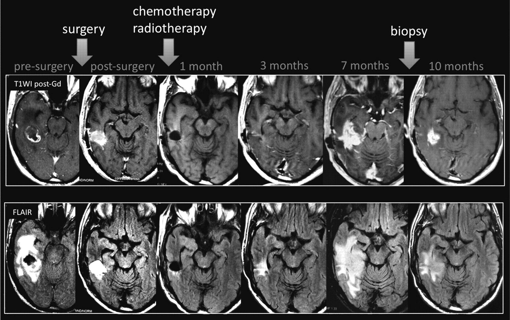
  • Fig 2.
    • Download figure
    • Open in new tab
    • Download powerpoint
    Fig 2.

    Pseudoprogression. A 63-year-old man with GBM. A follow-up MR imaging examination performed 7 months after RT-TMZ demonstrates increased lesion size. The histopathology samples (not shown) demonstrated a mixed tissue with treatment-related changes, associated with a few areas of viable tumor cells.
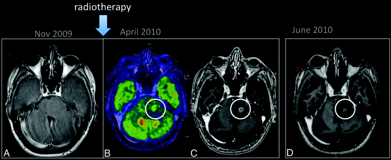
  • Fig 3.
    • Download figure
    • Open in new tab
    • Download powerpoint
    Fig 3.

    Pseudoprogression. A 25-year-old man with a low-grade glioma in the left aspect of the pons (A, arrow) was treated with only RT. PET-MR imaging (B) showed hypermetabolism in the enhancing portion of the lesion (C). An MR imaging examination performed 1 month later (D) shows a reduction in the enhancing portion of lesion.

  • Fig 4.
    • Download figure
    • Open in new tab
    • Download powerpoint
    Fig 4.

    Pseudoresponse is characterized by a marked decreased in the enhancing portion of the lesion some months after initiation of treatment. However, in some such cases, the FLAIR sequence shows a clear expansion of the lesion.

  • Fig 5.
    • Download figure
    • Open in new tab
    • Download powerpoint
    Fig 5.

    Pseudoresponse. A 47-year-old man with GBM. A reduction of the enhancing portion of the lesion is observed 1 day after initiation of cediranib treatment. Four weeks later, besides a continuing reduction in the enhancing portion, an expansion is observed in the FLAIR images. Expansions in both the enhancing area and abnormal hyperintense areas consistent with tumor progression were observed subsequently.

Tables

  • Figures
  • Incidence of pseudoprogression

    PublicationNo. of PatientsResponse CriteriaaCriteria for Early ProgressionbNo. of Early ProgressionPseudo-Progression (% of Early Progression)Pseudo-Progression (% of Patients)
    Brandes et al11103Enhancement increase for earlier progression than Macdonald criteria4 Weeks50/10332/5032/103
    J Clin Oncol 200820(48.5%)(64%)(31%)
    Taal et al85Macdonald Criteria4 Weeks36/8515/31a15/85
    Cancer 200810(42%)(48%)(17.6%)
    Clarke et al80Increased contrast enhancementNot specified33/808/25a8/80
    (abstract) J Clin Oncol 200817(41%)(32%)(10%)
    Gerstner et al45Macdonald Criteria17–28 Days24/4513/2413/45
    J Neurooncol 200918(53%)(54%)(29%)
    Jefferies et al15Not specified6 Months9/153/93/15
    abstract Clin Oncol 200719(60%)(33%)(20%)
    Chaski et al54Increased contrast enhancement6 Months25/543/253/54
    Surg Neurol 200914(46%)(12%)(5.5%)
    Sanghera et al104RECIST8 Weeks27/1047/22a7/104
    Can J Neurol Sci 201016(26%)(32%)(7%)
    Mangla et al36Macdonald Criteria4 Weeks19/367/197/36
    Radiology 201015(53%)(37%)(20%)
    • ↵a —All were defined in the text.

    • ↵b Defined as increase of the enhancing portion of the lesion in the follow-up MR exam.

PreviousNext
Back to top

In this issue

American Journal of Neuroradiology: 32 (11)
American Journal of Neuroradiology
Vol. 32, Issue 11
1 Dec 2011
  • Table of Contents
  • Index by author
Advertisement
Print
Download PDF
Email Article

Thank you for your interest in spreading the word on American Journal of Neuroradiology.

NOTE: We only request your email address so that the person you are recommending the page to knows that you wanted them to see it, and that it is not junk mail. We do not capture any email address.

Enter multiple addresses on separate lines or separate them with commas.
Pseudoprogression and Pseudoresponse: Imaging Challenges in the Assessment of Posttreatment Glioma
(Your Name) has sent you a message from American Journal of Neuroradiology
(Your Name) thought you would like to see the American Journal of Neuroradiology web site.
CAPTCHA
This question is for testing whether or not you are a human visitor and to prevent automated spam submissions.
Cite this article
L.C. Hygino da Cruz, I. Rodriguez, R.C. Domingues, E.L. Gasparetto, A.G. Sorensen
Pseudoprogression and Pseudoresponse: Imaging Challenges in the Assessment of Posttreatment Glioma
American Journal of Neuroradiology Dec 2011, 32 (11) 1978-1985; DOI: 10.3174/ajnr.A2397

Citation Manager Formats

  • BibTeX
  • Bookends
  • EasyBib
  • EndNote (tagged)
  • EndNote 8 (xml)
  • Medlars
  • Mendeley
  • Papers
  • RefWorks Tagged
  • Ref Manager
  • RIS
  • Zotero
0 Responses
Respond to this article
Share
Bookmark this article
Pseudoprogression and Pseudoresponse: Imaging Challenges in the Assessment of Posttreatment Glioma
L.C. Hygino da Cruz, I. Rodriguez, R.C. Domingues, E.L. Gasparetto, A.G. Sorensen
American Journal of Neuroradiology Dec 2011, 32 (11) 1978-1985; DOI: 10.3174/ajnr.A2397
del.icio.us logo Twitter logo Facebook logo Mendeley logo
  • Tweet Widget
  • Facebook Like
  • Google Plus One
Purchase

Jump to section

  • Article
    • Abstract
    • ABBREVIATIONS
    • Macdonald Criteria
    • Patterns of Early Enhancement in Posttreatment MR Imaging Follow-Up
    • Radiation Injury
    • Pseudoprogression
    • O6-Methylguanine DNA MGMT Promoter
    • Incidence
    • Pathophysiology
    • Diagnosis
    • Advanced MR Imaging Techniques
    • PET
    • Clinical Management
    • Conclusions
    • Acknowledgments
    • References
  • Figures & Data
  • Info & Metrics
  • Responses
  • References
  • PDF

Related Articles

  • No related articles found.
  • PubMed
  • Google Scholar

Cited By...

  • Patient-derived glioblastoma organoids as real-time avatars for assessing responses to clinical CAR-T cell therapy
  • Identification of a Single-Dose, Low-Flip-Angle-Based CBV Threshold for Fractional Tumor Burden Mapping in Recurrent Glioblastoma
  • Diagnostic Accuracy of MR Spectroscopic Imaging and 18F-FET PET for Identifying Glioma: A Biopsy-Controlled Hybrid PET/MRI Study
  • Distinguishing Progression from Pseudoprogression in Glioblastoma Using 18F-Fluciclovine PET
  • Amino Acid PET in Neurooncology
  • Amino Acid PET in Neurooncology
  • Application of 7T MRS to High-Grade Gliomas
  • Glioblastoma states are defined by cohabitating cellular populations with progression-, imaging- and sex-distinct patterns
  • In vivo imaging of nanoparticle-labeled CAR T cells
  • Pan-cancer imaging of TERT expression using deuterium magnetic resonance spectroscopy-based assessment of pyruvate metabolism
  • PET/MRI Improves Management of Children with Cancer
  • Diagnosis of Pseudoprogression Following Lomustine-Temozolomide Chemoradiation in Newly Diagnosed Glioblastoma Patients Using FET-PET
  • Spatiotemporal Heterogeneity in Multiparametric Physiologic MRI Is Associated with Patient Outcomes in IDH-Wildtype Glioblastoma
  • 18F-FET PET Imaging in Differentiating Glioma Progression from Treatment-Related Changes: A Single-Center Experience
  • A comprehensive proteomic SWATH-MS workflow for profiling blood extracellular vesicles: a new avenue for glioma tumour surveillance
  • Dynamic Contrast-Enhanced MRI in Patients with Brain Metastases Undergoing Laser Interstitial Thermal Therapy: A Pilot Study
  • Non-Contrast-Enhancing Tumor: A New Frontier in Glioblastoma Research
  • Radiogenomics-based Risk Prediction of Glioblastoma Multiforme with Clinical Relevance
  • Synthesizing a Contrast-Enhancement Map in Patients with High-Grade Gliomas Based on a Postcontrast MR Imaging Quantification Only
  • Radiomics in Brain Tumor: Image Assessment, Quantitative Feature Descriptors, and Machine-Learning Approaches
  • Diagnostic Accuracy of Centrally Restricted Diffusion in the Differentiation of Treatment-Related Necrosis from Tumor Recurrence in High-Grade Gliomas
  • Detection of immune responses after immunotherapy in glioblastoma using PET and MRI
  • Multiparametric Evaluation in Differentiating Glioma Recurrence from Treatment-Induced Necrosis Using Simultaneous 18F-FDG-PET/MRI: A Single-Institution Retrospective Study
  • Preradiotherapy MR Imaging: A Prospective Pilot Study of the Usefulness of Performing an MR Examination Shortly before Radiation Therapy in Patients with Glioblastoma
  • Early Biomarkers from Conventional and Delayed-Contrast MRI to Predict the Response to Bevacizumab in Recurrent High-Grade Gliomas
  • Temozolomide induces radiologic pseudoprogression and tumor cell vanishing in oligodendroglioma
  • Late Pseudoprogression in Glioblastoma: Diagnostic Value of Dynamic O-(2-[18F]fluoroethyl)-L-Tyrosine PET
  • Differentiating Tumor Progression from Pseudoprogression in Patients with Glioblastomas Using Diffusion Tensor Imaging and Dynamic Susceptibility Contrast MRI
  • Parametric Response Mapping of Apparent Diffusion Coefficient as an Imaging Biomarker to Distinguish Pseudoprogression from True Tumor Progression in Peptide-Based Vaccine Therapy for Pediatric Diffuse Intrinsic Pontine Glioma
  • Repeatability of Standardized and Normalized Relative CBV in Patients with Newly Diagnosed Glioblastoma
  • Radiation Necrosis in Pediatric Patients with Brain Tumors Treated with Proton Radiotherapy
  • Diffusion and Perfusion MRI to Differentiate Treatment-Related Changes Including Pseudoprogression from Recurrent Tumors in High-Grade Gliomas with Histopathologic Evidence
  • Differentiation of Tumor Progression from Pseudoprogression in Patients with Posttreatment Glioblastoma Using Multiparametric Histogram Analysis
  • In vivo chemical exchange saturation transfer imaging allows early detection of a therapeutic response in glioblastoma
  • Longitudinal Restriction Spectrum Imaging Is Resistant to Pseudoresponse in Patients with High-Grade Gliomas Treated with Bevacizumab
  • Response Classification Based on a Minimal Model of Glioblastoma Growth Is Prognostic for Clinical Outcomes and Distinguishes Progression from Pseudoprogression
  • Imaging Changes in Very Young Children with Brain Tumors Treated with Proton Therapy and Chemotherapy
  • Multimodal Magnetic Resonance Imaging and 18F-L-Dihydroxyphenylalanine Positron Emission Tomography in Early Characterization of Pseudoresponse and Nonenhancing Tumor Progression in a Pediatric Patient With Malignant Transformation of Ganglioglioma Treated With Bevacizumab
  • Metabolic Response of Glioblastoma to Superselective Intra-Arterial Cerebral Infusion of Bevacizumab: A Proton MR Spectroscopic Imaging Study
  • Late and Prolonged Pseudoprogression in Glioblastoma After Treatment With Lomustine and Temozolomide
  • Crossref
  • Google Scholar

This article has not yet been cited by articles in journals that are participating in Crossref Cited-by Linking.

More in this TOC Section

  • An Atlas of Neonatal Neurovascular Imaging Anatomy as Depicted with Microvascular Imaging: The Intracranial Arteries
  • An Atlas of Neonatal Neurovascular Imaging Anatomy as Depicted with Microvascular Imaging: The Intracranial Veins
  • Clinical Translation of Hyperpolarized 13C Metabolic Probes for Glioma Imaging
Show more Review Articles

Similar Articles

Advertisement

Indexed Content

  • Current Issue
  • Accepted Manuscripts
  • Article Preview
  • Past Issues
  • Editorials
  • Editor's Choice
  • Fellows' Journal Club
  • Letters to the Editor
  • Video Articles

Cases

  • Case Collection
  • Archive - Case of the Week
  • Archive - Case of the Month
  • Archive - Classic Case

More from AJNR

  • Trainee Corner
  • Imaging Protocols
  • MRI Safety Corner
  • Book Reviews

Multimedia

  • AJNR Podcasts
  • AJNR Scantastics

Resources

  • Turnaround Time
  • Submit a Manuscript
  • Submit a Video Article
  • Submit an eLetter to the Editor/Response
  • Manuscript Submission Guidelines
  • Statistical Tips
  • Fast Publishing of Accepted Manuscripts
  • Graphical Abstract Preparation
  • Imaging Protocol Submission
  • Evidence-Based Medicine Level Guide
  • Publishing Checklists
  • Author Policies
  • Become a Reviewer/Academy of Reviewers
  • News and Updates

About Us

  • About AJNR
  • Editorial Board
  • Editorial Board Alumni
  • Alerts
  • Permissions
  • Not an AJNR Subscriber? Join Now
  • Advertise with Us
  • Librarian Resources
  • Feedback
  • Terms and Conditions
  • AJNR Editorial Board Alumni

American Society of Neuroradiology

  • Not an ASNR Member? Join Now

© 2025 by the American Society of Neuroradiology All rights, including for text and data mining, AI training, and similar technologies, are reserved.
Print ISSN: 0195-6108 Online ISSN: 1936-959X

Powered by HighWire