Advertisement

Main menu

  • Home
  • Content
    • Current Issue
    • Accepted Manuscripts
    • Article Preview
    • Past Issue Archive
    • Video Articles
    • AJNR Case Collection
    • Case of the Week Archive
    • Case of the Month Archive
    • Classic Case Archive
  • Special Collections
    • AJNR Awards
    • Low-Field MRI
    • Alzheimer Disease
    • ASNR Foundation Special Collection
    • Photon-Counting CT
    • View All
  • Multimedia
    • AJNR Podcasts
    • AJNR SCANtastic
    • Trainee Corner
    • MRI Safety Corner
    • Imaging Protocols
  • For Authors
    • Submit a Manuscript
    • Submit a Video Article
    • Submit an eLetter to the Editor/Response
    • Manuscript Submission Guidelines
    • Statistical Tips
    • Fast Publishing of Accepted Manuscripts
    • Graphical Abstract Preparation
    • Imaging Protocol Submission
    • Author Policies
  • About Us
    • About AJNR
    • Editorial Board
    • Editorial Board Alumni
  • More
    • Become a Reviewer/Academy of Reviewers
    • Subscribers
    • Permissions
    • Alerts
    • Feedback
    • Advertisers
    • ASNR Home

User menu

  • Alerts
  • Log in

Search

  • Advanced search
American Journal of Neuroradiology

American Journal of Neuroradiology

ASHNR American Society of Functional Neuroradiology ASHNR American Society of Pediatric Neuroradiology ASSR
  • Alerts
  • Log in

Advanced Search

  • Home
  • Content
    • Current Issue
    • Accepted Manuscripts
    • Article Preview
    • Past Issue Archive
    • Video Articles
    • AJNR Case Collection
    • Case of the Week Archive
    • Case of the Month Archive
    • Classic Case Archive
  • Special Collections
    • AJNR Awards
    • Low-Field MRI
    • Alzheimer Disease
    • ASNR Foundation Special Collection
    • Photon-Counting CT
    • View All
  • Multimedia
    • AJNR Podcasts
    • AJNR SCANtastic
    • Trainee Corner
    • MRI Safety Corner
    • Imaging Protocols
  • For Authors
    • Submit a Manuscript
    • Submit a Video Article
    • Submit an eLetter to the Editor/Response
    • Manuscript Submission Guidelines
    • Statistical Tips
    • Fast Publishing of Accepted Manuscripts
    • Graphical Abstract Preparation
    • Imaging Protocol Submission
    • Author Policies
  • About Us
    • About AJNR
    • Editorial Board
    • Editorial Board Alumni
  • More
    • Become a Reviewer/Academy of Reviewers
    • Subscribers
    • Permissions
    • Alerts
    • Feedback
    • Advertisers
    • ASNR Home
  • Follow AJNR on Twitter
  • Visit AJNR on Facebook
  • Follow AJNR on Instagram
  • Join AJNR on LinkedIn
  • RSS Feeds

AJNR Awards, New Junior Editors, and more. Read the latest AJNR updates

Research ArticlePediatric NeuroimagingF

Posterior Circulation and High Prevalence of Ischemic Stroke among Young Pediatric Patients with Moyamoya Disease: Evidence of Angiography-Based Differences by Age at Diagnosis

S. Mugikura, S. Higano, R. Shirane, M. Fujimura, Y. Shimanuki and S. Takahashi
American Journal of Neuroradiology January 2011, 32 (1) 192-198; DOI: https://doi.org/10.3174/ajnr.A2216
S. Mugikura
  • Find this author on Google Scholar
  • Find this author on PubMed
  • Search for this author on this site
S. Higano
  • Find this author on Google Scholar
  • Find this author on PubMed
  • Search for this author on this site
R. Shirane
  • Find this author on Google Scholar
  • Find this author on PubMed
  • Search for this author on this site
M. Fujimura
  • Find this author on Google Scholar
  • Find this author on PubMed
  • Search for this author on this site
Y. Shimanuki
  • Find this author on Google Scholar
  • Find this author on PubMed
  • Search for this author on this site
S. Takahashi
  • Find this author on Google Scholar
  • Find this author on PubMed
  • Search for this author on this site
  • Article
  • Figures & Data
  • Info & Metrics
  • Responses
  • References
  • PDF
Loading

Article Figures & Data

Figures

  • Fig 1.
    • Download figure
    • Open in new tab
    • Download powerpoint
    Fig 1.

    The relationship between the 4 diagnostic age groups and clinical manifestations (A), number of infarcted zones (B), distribution of corticosubcortical infarcted zones (C), ICA staging (D), PCA staging (E), and number of transdural collaterals (F). Asterisks indicate significant differences. Double asterisks indicate a significant difference and a positive relationship to diagnostic age among the 3 oldest diagnostic age groups.
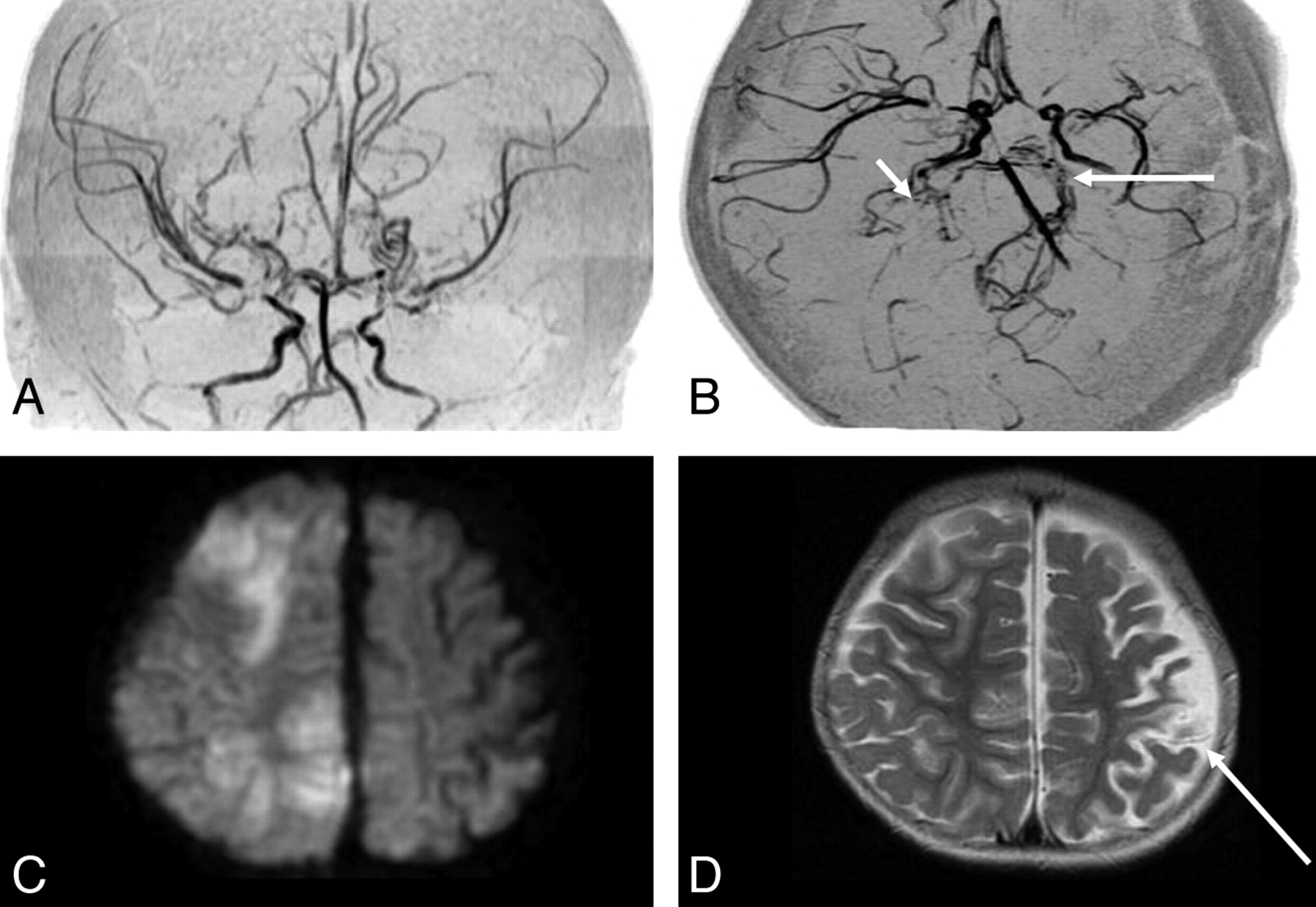
  • Fig 2.
    • Download figure
    • Open in new tab
    • Download powerpoint
    Fig 2.

    A 15-month-old girl (diagnostic age group younger than 4 years of age) initially presented with CS with right upper motor paresis a month before and subsequently presented with contralateral hemiparesis CS. Moyamoya disease was diagnosed. Her clinical manifestation was CS. A and B, Coronal and axial time-of-flight MR angiograms show steno-occlusive bilateral lesions in the terminal part of the ICA and in the proximal parts of the ACA and MCA cerebral arteries (ICA stage II, bilaterally). The right PCA is almost completely occluded (short arrow), with well-developed dilated perforators around it (stage III); the proximal part of the left PCA is stenotic (long arrow, PCA stage II). Note that in this patient, the bilateral steno-occlusive changes involved the PCAs even in the hemispheres with less advanced ICA lesions (ICA stage II, bilaterally). C, Diffusion-weighted MR image shows hyperintense regions of recent infarction in the anterior and posterior watersheds and the boundary zone between the regions of the ACA and PCA in the right. Five infarcted zones are seen on the right. D, T2-weighted MR image additionally shows an old cortical infarction in the posterior MCA territory (arrow), seen on the left.

  • Fig 3.
    • Download figure
    • Open in new tab
    • Download powerpoint
    Fig 3.

    A 7-year-old girl (diagnostic age group, 4–7 years) who had recurrent transient left hemiparesis for 5 years was diagnosed with Moyamoya disease, which manifested clinically as TIA. A and B, Coronal and axial time-of-flight MR angiograms show advanced steno-occlusive changes at or around the terminal part of the right ICA, with poorly visualized ACA and MCA branches (ICA stage III, right). Moderate steno-occlusive changes at or around the terminal part of the left ICA with relatively good visualization of the ACA (small arrows) and MCA (large arrows) cortical branches (ICA stage II) are seen on the left. No steno-occlusive lesions are seen bilaterally in the posterior cerebral artery (PCA stage 1, bilaterally). C, Anteroposterior view of the vertebral angiogram shows no steno-occlusive lesions bilaterally in the PCA and well-developed leptomeningial collateral circulation to the anterior circulation. D, Left lateral external carotid angiogram shows dilated anterior branches of the middle meningeal artery providing transdural collaterals to the contralateral frontal region (large arrows), with the medial branches of the maxillary artery providing transdural collaterals to the right anterior basal region (small arrow). Two transdural collaterals can be seen on the right, and none are seen on the left (anteroposterior view of the left external carotid angiogram, not shown). E, Axial T2-weighted MR image shows no infarction bilaterally (ie, the number of infarcted regions is zero on either side). Note that in this patient, the steno-occlusive changes do not involve the PCA, even in the right hemisphere, where ICA lesions are advanced (stage III).
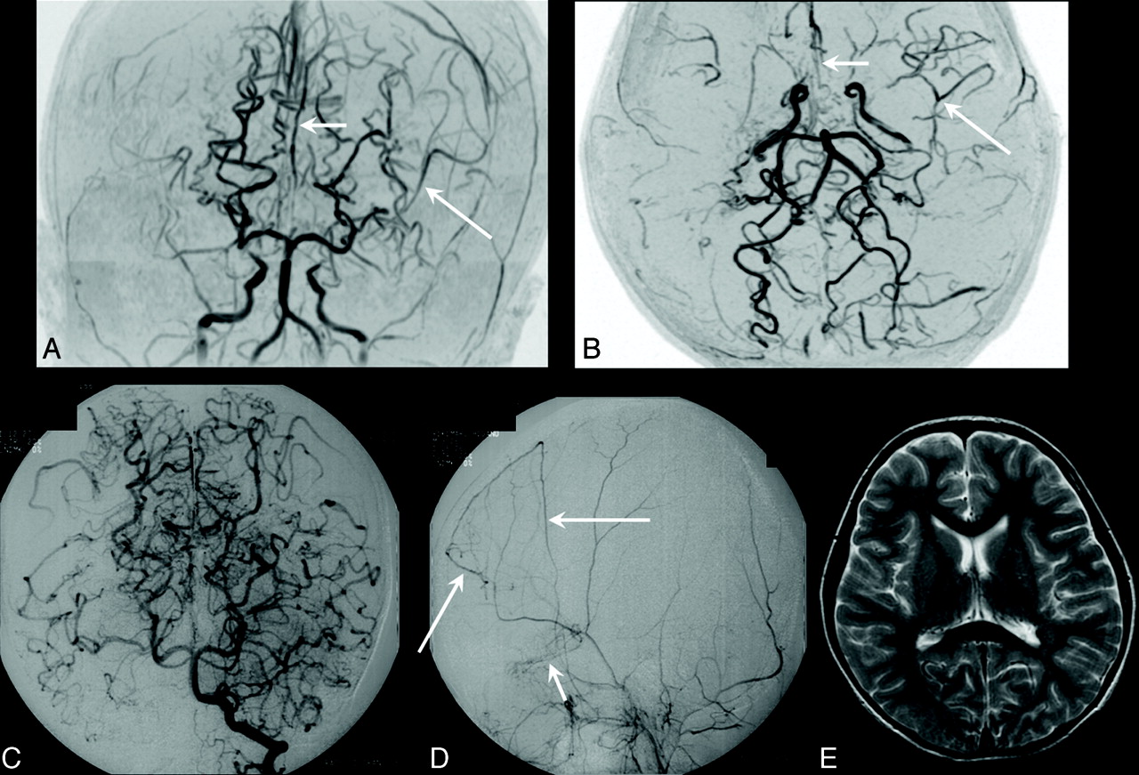
  • Fig 4.
    • Download figure
    • Open in new tab
    • Download powerpoint
    Fig 4.

    A 12-year-old boy (diagnostic age group, 12–15 years) with right and left transient motor paresis. His clinical manifestation was TIA. A, Axial time-of-flight MR angiogram shows advanced steno-occlusive changes bilaterally at or around the terminal parts of the ICAs, with no apparent ACA and MCA branches (ICA stage IV, bilaterally). Advanced steno-occlusive lesions bilaterally in the PCAs with well-developed Moyamoya vessels from the PCA are seen (PCA stage 3, bilaterally). B, Anteroposterior view of the vertebral angiogram shows advanced steno-occlusive lesions bilaterally in the PCAs (PCA stage III, bilaterally). C and D, Lateral view of the arterial (C) and capillary (D) phases of the right external carotid angiograms shows that the dilated posterior branch of the middle meningeal artery (large arrows) and the meningeal branch of the occipital artery (arrowhead) provide transdural collaterals mainly to the right posterior part of the cerebral hemispheres. The frontal branch of the superficial temporal artery (small arrow) and medial branches of the internal maxillary artery provide transdural collaterals to the frontobasal region. Three transdural collaterals are seen on the right. The right cerebral hemisphere is largely supplied through the transdural collaterals (D). Three transdural collaterals were seen on the left (left external carotid angiogram, not shown). E, Axial T2-weighted MR image shows an old small infarction in the left anterior MCA territory. No infarcted regions are seen on the right, but 1 is seen on the left. Note that this patient has severe steno-occlusive PCA changes that parallel the advanced ICA lesions. This progressive state should have provoked the development of transdural collaterals, which might have prevented a large infarction and CS.

  • Fig 5.
    • Download figure
    • Open in new tab
    • Download powerpoint
    Fig 5.

    The relationship between the 4 diagnostic age groups and PCA stage in the 117 hemispheres with less advanced steno-occlusive ICA lesions (ICA stage I or II). The asterisk indicates a significant difference between diagnostic age groups younger than 4 years and 4–7 years of age.

PreviousNext
Back to top

In this issue

American Journal of Neuroradiology: 32 (1)
American Journal of Neuroradiology
Vol. 32, Issue 1
1 Jan 2011
  • Table of Contents
  • Index by author
Advertisement
Print
Download PDF
Email Article

Thank you for your interest in spreading the word on American Journal of Neuroradiology.

NOTE: We only request your email address so that the person you are recommending the page to knows that you wanted them to see it, and that it is not junk mail. We do not capture any email address.

Enter multiple addresses on separate lines or separate them with commas.
Posterior Circulation and High Prevalence of Ischemic Stroke among Young Pediatric Patients with Moyamoya Disease: Evidence of Angiography-Based Differences by Age at Diagnosis
(Your Name) has sent you a message from American Journal of Neuroradiology
(Your Name) thought you would like to see the American Journal of Neuroradiology web site.
CAPTCHA
This question is for testing whether or not you are a human visitor and to prevent automated spam submissions.
Cite this article
S. Mugikura, S. Higano, R. Shirane, M. Fujimura, Y. Shimanuki, S. Takahashi
Posterior Circulation and High Prevalence of Ischemic Stroke among Young Pediatric Patients with Moyamoya Disease: Evidence of Angiography-Based Differences by Age at Diagnosis
American Journal of Neuroradiology Jan 2011, 32 (1) 192-198; DOI: 10.3174/ajnr.A2216

Citation Manager Formats

  • BibTeX
  • Bookends
  • EasyBib
  • EndNote (tagged)
  • EndNote 8 (xml)
  • Medlars
  • Mendeley
  • Papers
  • RefWorks Tagged
  • Ref Manager
  • RIS
  • Zotero
0 Responses
Respond to this article
Share
Bookmark this article
Posterior Circulation and High Prevalence of Ischemic Stroke among Young Pediatric Patients with Moyamoya Disease: Evidence of Angiography-Based Differences by Age at Diagnosis
S. Mugikura, S. Higano, R. Shirane, M. Fujimura, Y. Shimanuki, S. Takahashi
American Journal of Neuroradiology Jan 2011, 32 (1) 192-198; DOI: 10.3174/ajnr.A2216
del.icio.us logo Twitter logo Facebook logo Mendeley logo
  • Tweet Widget
  • Facebook Like
  • Google Plus One
Purchase

Jump to section

  • Article
    • Abstract
    • Abbreviations
    • Materials and Methods
    • Results
    • Discussion
    • Conclusions
    • Acknowledgments
    • Footnotes
    • References
  • Figures & Data
  • Info & Metrics
  • Responses
  • References
  • PDF

Related Articles

  • No related articles found.
  • PubMed
  • Google Scholar

Cited By...

  • Predictive role of heterozygous p.R4810K of RNF213 in the phenotype of Chinese moyamoya disease
  • Clinical and Angiographic Features and Stroke Types in Adult Moyamoya Disease
  • Homozygous c.14576G>A variant of RNF213 predicts early-onset and severe form of moyamoya disease
  • Crossref
  • Google Scholar

This article has not yet been cited by articles in journals that are participating in Crossref Cited-by Linking.

More in this TOC Section

  • Frontal Paraventricular Cysts
  • Sodium MRI in Pediatric Brain Tumors
  • FRACTURE MR in Congenital Vertebral Anomalies
Show more Pediatric Neuroimaging

Similar Articles

Advertisement

Indexed Content

  • Current Issue
  • Accepted Manuscripts
  • Article Preview
  • Past Issues
  • Editorials
  • Editor's Choice
  • Fellows' Journal Club
  • Letters to the Editor
  • Video Articles

Cases

  • Case Collection
  • Archive - Case of the Week
  • Archive - Case of the Month
  • Archive - Classic Case

More from AJNR

  • Trainee Corner
  • Imaging Protocols
  • MRI Safety Corner
  • Book Reviews

Multimedia

  • AJNR Podcasts
  • AJNR Scantastics

Resources

  • Turnaround Time
  • Submit a Manuscript
  • Submit a Video Article
  • Submit an eLetter to the Editor/Response
  • Manuscript Submission Guidelines
  • Statistical Tips
  • Fast Publishing of Accepted Manuscripts
  • Graphical Abstract Preparation
  • Imaging Protocol Submission
  • Evidence-Based Medicine Level Guide
  • Publishing Checklists
  • Author Policies
  • Become a Reviewer/Academy of Reviewers
  • News and Updates

About Us

  • About AJNR
  • Editorial Board
  • Editorial Board Alumni
  • Alerts
  • Permissions
  • Not an AJNR Subscriber? Join Now
  • Advertise with Us
  • Librarian Resources
  • Feedback
  • Terms and Conditions
  • AJNR Editorial Board Alumni

American Society of Neuroradiology

  • Not an ASNR Member? Join Now

© 2025 by the American Society of Neuroradiology All rights, including for text and data mining, AI training, and similar technologies, are reserved.
Print ISSN: 0195-6108 Online ISSN: 1936-959X

Powered by HighWire