
Article Figures & Data
Figures
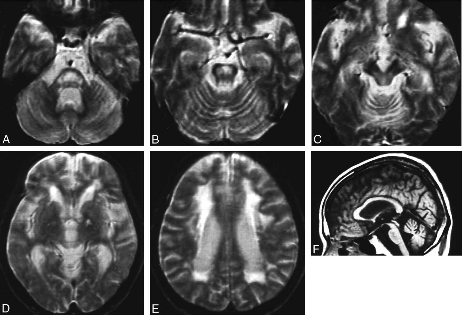
- Fig 1.
MR images of patient 1 (with adult-onset DRPLA). A–E, The T2-weighted axial images obtained at 60 years of age show high-signal-intensity lesions in the middle and upper pons, midbrain tegmentum, and cerebral white matter, in addition to a left pallidal high-signal-intensity spot due to an old lacunar infarction (D). F, A T1-weighted midsagittal image shows atrophy of the brain stem and cerebellum.
- Fig 2.
MR images of patient 2 (with juvenile-onset DRPLA). A–C, The T2-weighted axial images obtained at 35 years of age show marked atrophy of the pons, cerebellum, and cerebral hemispheres. High-signal-intensity lesions are observed only in the periventricular white matter at this point in time (C). D–F, On T2-weighted MR images obtained at 39 years of age, marked progression of atrophic changes is demonstrated in the cerebellum, brain stem, and cerebral hemispheres. Additionally, high-signal-intensity lesions in the cerebral white matter are clearly observed (F).
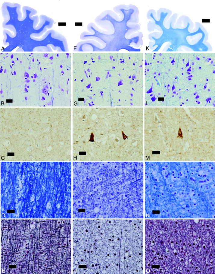
- Fig 3.
F, Patient 1 shows no evident atrophy of the cerebral cortex and white matter. G, The cortical neurons are slightly atrophic. H, Accumulation of phosphorylated neurofilaments is noted in several cortical neurons in the fifth layer. I and J, The cerebral white matter shows marked loss of myelinated fibers. K and L, The cerebral white matter of patient 2 is severely atrophic, and the width of the cerebral cortex is generally reduced. The cortical neurons are shrunken, and the neuropile is markedly atrophic in every layer (L). M, Some cortical neurons especially in the fifth layer show accumulation of phosphorylated neurofilaments. N and O, The cerebral white matter shows moderate loss of myelinated fibers. A–E, Control. F–J, Patient 1. K–O, Patient 2. A, B, D, F, G, I, K, L, N, KB staining; C, H, and M, phosphorylated neurofilament immunohistochemistry. E, J, and O, Bodian preparation.
- Fig 4.
D–F, Patient 1 shows moderate atrophy of the pons and marked reduction of myelinated fibers, particularly in the base (D–F) compared with the control (A–C). G–I, The pons of patient 2 is markedly atrophic (G), the volume of the neuropile is severely reduced (H), and the attenuation of myelinated fibers in the base is preserved (I). A–C, Control. D–F, Patient 1. G–I, Patient 2. A, B, D, E, G, H, KB staining. C, F, and I, Bodian preparation.


