Advertisement

Main menu

  • Home
  • Content
    • Current Issue
    • Accepted Manuscripts
    • Article Preview
    • Past Issue Archive
    • Video Articles
    • AJNR Case Collection
    • Case of the Week Archive
    • Case of the Month Archive
    • Classic Case Archive
  • Special Collections
    • AJNR Awards
    • Low-Field MRI
    • Alzheimer Disease
    • ASNR Foundation Special Collection
    • Photon-Counting CT
    • View All
  • Multimedia
    • AJNR Podcasts
    • AJNR SCANtastic
    • Trainee Corner
    • MRI Safety Corner
    • Imaging Protocols
  • For Authors
    • Submit a Manuscript
    • Submit a Video Article
    • Submit an eLetter to the Editor/Response
    • Manuscript Submission Guidelines
    • Statistical Tips
    • Fast Publishing of Accepted Manuscripts
    • Graphical Abstract Preparation
    • Imaging Protocol Submission
    • Author Policies
  • About Us
    • About AJNR
    • Editorial Board
    • Editorial Board Alumni
  • More
    • Become a Reviewer/Academy of Reviewers
    • Subscribers
    • Permissions
    • Alerts
    • Feedback
    • Advertisers
    • ASNR Home

User menu

  • Alerts
  • Log in

Search

  • Advanced search
American Journal of Neuroradiology

American Journal of Neuroradiology

ASHNR American Society of Functional Neuroradiology ASHNR American Society of Pediatric Neuroradiology ASSR
  • Alerts
  • Log in

Advanced Search

  • Home
  • Content
    • Current Issue
    • Accepted Manuscripts
    • Article Preview
    • Past Issue Archive
    • Video Articles
    • AJNR Case Collection
    • Case of the Week Archive
    • Case of the Month Archive
    • Classic Case Archive
  • Special Collections
    • AJNR Awards
    • Low-Field MRI
    • Alzheimer Disease
    • ASNR Foundation Special Collection
    • Photon-Counting CT
    • View All
  • Multimedia
    • AJNR Podcasts
    • AJNR SCANtastic
    • Trainee Corner
    • MRI Safety Corner
    • Imaging Protocols
  • For Authors
    • Submit a Manuscript
    • Submit a Video Article
    • Submit an eLetter to the Editor/Response
    • Manuscript Submission Guidelines
    • Statistical Tips
    • Fast Publishing of Accepted Manuscripts
    • Graphical Abstract Preparation
    • Imaging Protocol Submission
    • Author Policies
  • About Us
    • About AJNR
    • Editorial Board
    • Editorial Board Alumni
  • More
    • Become a Reviewer/Academy of Reviewers
    • Subscribers
    • Permissions
    • Alerts
    • Feedback
    • Advertisers
    • ASNR Home
  • Follow AJNR on Twitter
  • Visit AJNR on Facebook
  • Follow AJNR on Instagram
  • Join AJNR on LinkedIn
  • RSS Feeds

AJNR Awards, New Junior Editors, and more. Read the latest AJNR updates

Research ArticleBrain

Added Value of High-Resolution MR Imaging in the Diagnosis of Vertebral Artery Dissection

O. Naggara, F. Louillet, E. Touzé, D. Roy, X. Leclerc, J.-L. Mas, J.-P. Pruvo, J.-F. Meder and C. Oppenheim
American Journal of Neuroradiology October 2010, 31 (9) 1707-1712; DOI: https://doi.org/10.3174/ajnr.A2165
O. Naggara
  • Find this author on Google Scholar
  • Find this author on PubMed
  • Search for this author on this site
F. Louillet
  • Find this author on Google Scholar
  • Find this author on PubMed
  • Search for this author on this site
E. Touzé
  • Find this author on Google Scholar
  • Find this author on PubMed
  • Search for this author on this site
D. Roy
  • Find this author on Google Scholar
  • Find this author on PubMed
  • Search for this author on this site
X. Leclerc
  • Find this author on Google Scholar
  • Find this author on PubMed
  • Search for this author on this site
J.-L. Mas
  • Find this author on Google Scholar
  • Find this author on PubMed
  • Search for this author on this site
J.-P. Pruvo
  • Find this author on Google Scholar
  • Find this author on PubMed
  • Search for this author on this site
J.-F. Meder
  • Find this author on Google Scholar
  • Find this author on PubMed
  • Search for this author on this site
C. Oppenheim
  • Find this author on Google Scholar
  • Find this author on PubMed
  • Search for this author on this site
  • Article
  • Figures & Data
  • Info & Metrics
  • Responses
  • References
  • PDF
Loading

Article Figures & Data

Figures

  • Tables
  • Fig 1.
    • Download figure
    • Open in new tab
    • Download powerpoint
    Fig 1.

    Distinction between mural hematoma and perivertebral hypertrophic venous plexus on HR-MR imaging. Axial high-resolution fat-suppressed PDWI (A and E), fat-suppressed T2WI (B and F), T1WI (C and G), and TOF sequences (D and H). In case 1 (top row), crescentic high signal intensity of the vertebral wall, brighter than the signal intensity of the sternocleidomastoid muscle on all sequences (A−D), is associated with a low signal intensity of the intimal layer between the lumen and the crescentic hyperintense mural thickening on TOF (D, arrow), corresponding to a mural hematoma. In case 2 (bottom row), crescentic high signal intensity of the vertebral wall, brighter than muscle signal intensity on TOF, PDWI, and T2WI (E, F, and H), is isointense to the muscle on T1WI (G), without magnetic susceptibility artifacts (H), leading to the diagnosis of inflow enhancement of a hypertrophic venous structure.
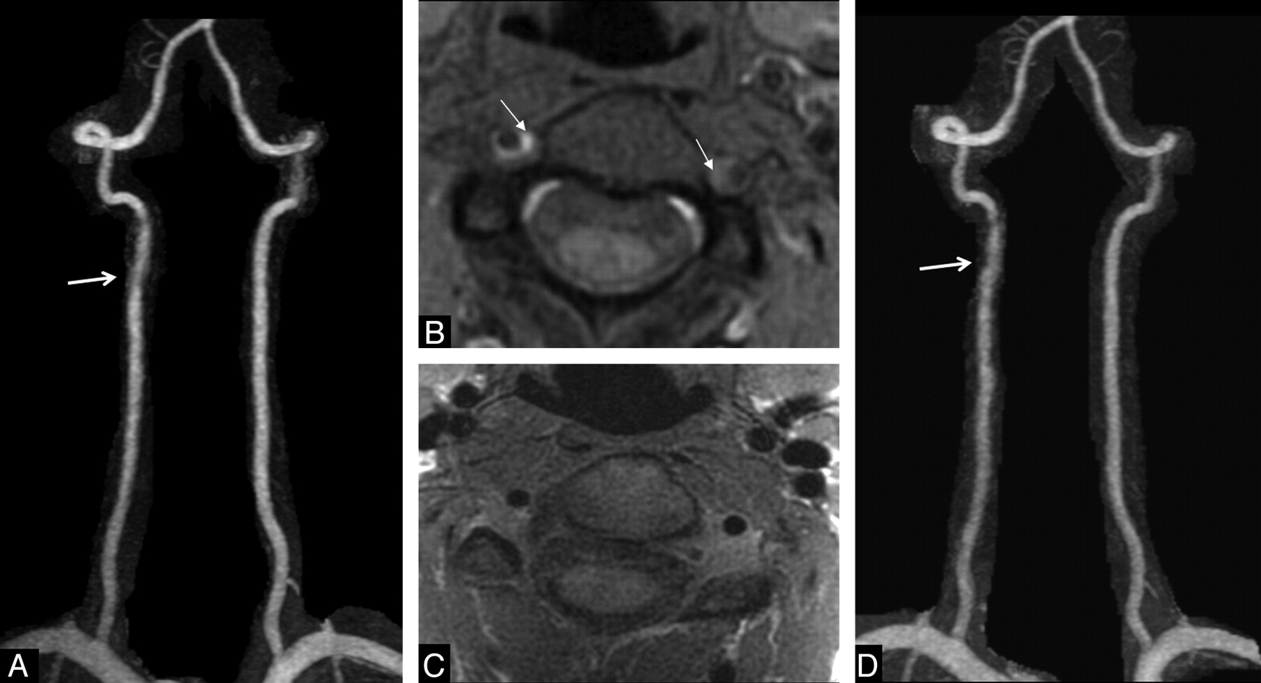
  • Fig 2.
    • Download figure
    • Open in new tab
    • Download powerpoint
    Fig 2.

    Example of discordance between the standard approach and HR-MR imaging. Suspicion of traumatic right acute VA dissection in a 25-year-old woman with sudden neck pain and dizziness, without stroke. DUS findings were consistent with a right vertebral mural hematoma (not shown). A, On CE-MRA, note irregularity of the right V2 segment (arrow). B, On axial fat-suppressed cervical T1WI, note hyperintense crescentic thickening of the right VA, associated with a slight crescentic hyperintensity of the left VA (arrows). C, On axial high-resolution T1WI, obtained 24 hours later, the crescentic hyperintense mural thickening is no longer present, leading to the diagnosis of inflow enhancement of the hypertrophic venous structure. D, On 6-month follow-up CE-MRA, the lumen of the right VA remains unchanged (arrow).
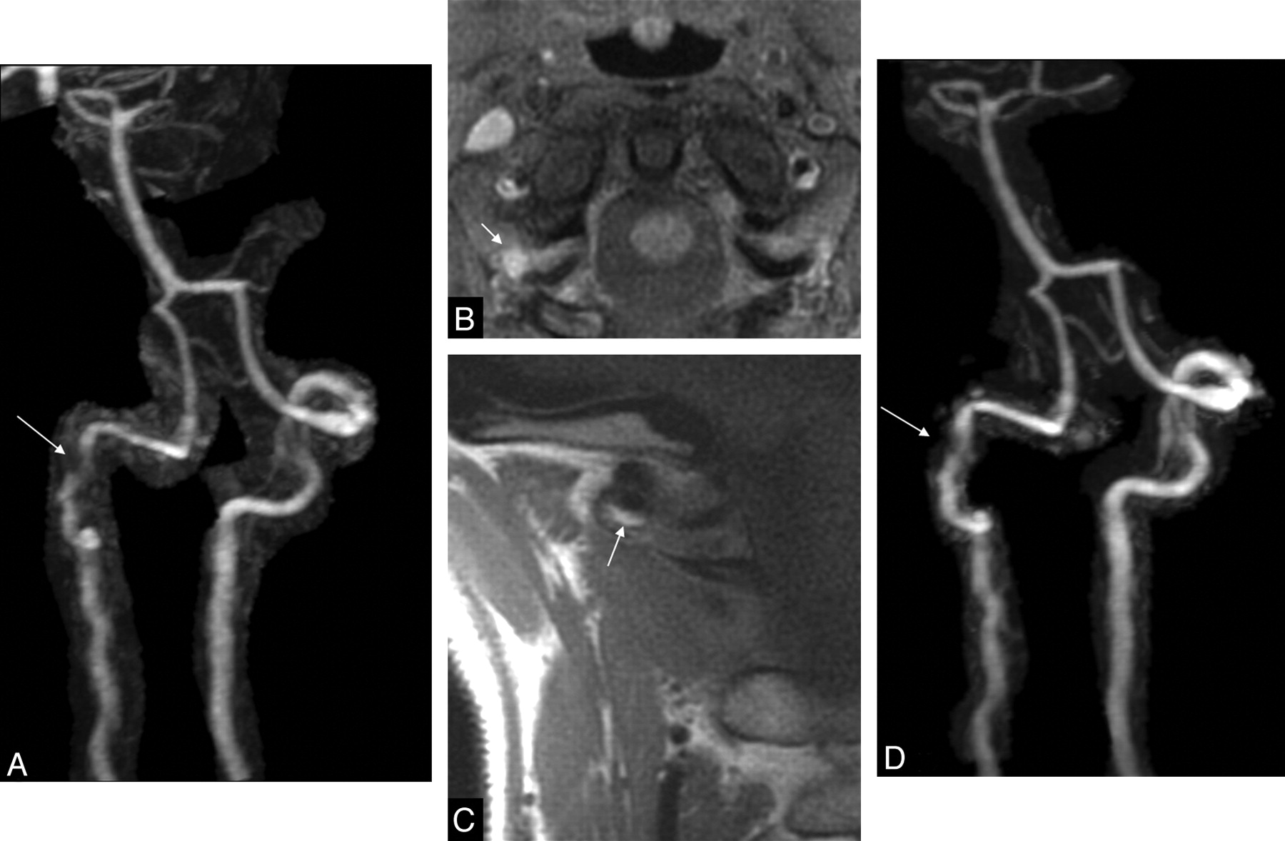
  • Fig 3.
    • Download figure
    • Open in new tab
    • Download powerpoint
    Fig 3.

    Example of discordance between the standard approach and HR-MR imaging. Suspicion of spontaneous acute dissection in a 40-year-old woman with left neck pain and laterobulbar acute stroke. A, On CE-MRA, note occlusion of the right VA and stenosis (arrow) of the left V3 segment. B, On axial fat-suppressed cervical T1WI, note inconclusive crescentic hyperintense mural thickening of the left VA (arrow) and a hyperintense occluded lumen of the right V3 segment (double arrow). DUS did not demonstrate any mural hematoma (not shown). C, On axial high-resolution fat-suppressed T2WI, obtained 24 hours later, note crescentic hyperintense mural thickening (arrow). D, On 5-month follow-up CE-MRA, note clear improvement of the left V3 stenosis. This favors the diagnosis of VAD.
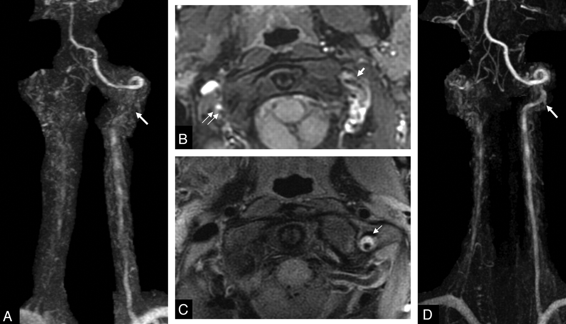
  • Fig 4.
    • Download figure
    • Open in new tab
    • Download powerpoint
    Fig 4.

    Example of discordance between the standard approach and HR-MR imaging. Suspicion of spontaneous acute dissection in a 47-year-old woman with postchiropractic right neck pain and transient dizziness, without stroke. Findings of DUS were inconclusive (not shown). B, On standard axial fat-suppressed cervical T1WI, note nonspecific hyperintense crescentic mural thickening of the right V3 segment (arrows). A and D, On CE-MRA, note stenosis (arrow, A) of the right V3 segment, which was partially resolved on 1-year follow-up CE-MRA (D, arrow). C, Coronal high-resolution T1WI, obtained 24 hours after CE-MRA, demonstrates a clear hyperintense crescentic mural thickening of the right V3 segment (arrow). This supports the diagnosis of VAD.

Tables

  • Figures
    • View popup
    Table 1:

    Demographic and clinical data

    Demographic Data
    Patients35
    Age (yr, mean, range)41.5 ± 8.5 (25–56)
    Male14 (40%)
    Migraine14
    Diabetes1
    Smoking19
    Hypercholesterolemia4
    Elevated blood pressure2
    Clinical presentation
        Trauma15
        Vertebral manipulation3
        Headache22
        Neck pain33
        Dizziness21
        Horner syndrome3
        Diplopia7
        Cerebellar signs13
        NIHSS score (mean, range)1.1 ± 1.9 (0–9)
    Imaging
        Stroke on DWI14
        Onset-to-HR-MR imaging delay (day, mean, range)8.6 ± 6.0 (3–16)
        Clinical follow-up (month, mean, range)9.6 ± 3.6 (6–32)
        Imaging follow-up (month, mean, range)7.4 ± 6.3 (4–32)
    • View popup
    Table 2:

    Comparison of neurologists' and HR-MR classifications

    Neurologists' ClassificationsTotal
    VADNo VAD
    HR-MR imaging classification
        VAD14418
        No VAD41317
    Total181735
PreviousNext
Back to top

In this issue

American Journal of Neuroradiology: 31 (9)
American Journal of Neuroradiology
Vol. 31, Issue 9
1 Oct 2010
  • Table of Contents
  • Index by author
Advertisement
Print
Download PDF
Email Article

Thank you for your interest in spreading the word on American Journal of Neuroradiology.

NOTE: We only request your email address so that the person you are recommending the page to knows that you wanted them to see it, and that it is not junk mail. We do not capture any email address.

Enter multiple addresses on separate lines or separate them with commas.
Added Value of High-Resolution MR Imaging in the Diagnosis of Vertebral Artery Dissection
(Your Name) has sent you a message from American Journal of Neuroradiology
(Your Name) thought you would like to see the American Journal of Neuroradiology web site.
CAPTCHA
This question is for testing whether or not you are a human visitor and to prevent automated spam submissions.
Cite this article
O. Naggara, F. Louillet, E. Touzé, D. Roy, X. Leclerc, J.-L. Mas, J.-P. Pruvo, J.-F. Meder, C. Oppenheim
Added Value of High-Resolution MR Imaging in the Diagnosis of Vertebral Artery Dissection
American Journal of Neuroradiology Oct 2010, 31 (9) 1707-1712; DOI: 10.3174/ajnr.A2165

Citation Manager Formats

  • BibTeX
  • Bookends
  • EasyBib
  • EndNote (tagged)
  • EndNote 8 (xml)
  • Medlars
  • Mendeley
  • Papers
  • RefWorks Tagged
  • Ref Manager
  • RIS
  • Zotero
0 Responses
Respond to this article
Share
Bookmark this article
Added Value of High-Resolution MR Imaging in the Diagnosis of Vertebral Artery Dissection
O. Naggara, F. Louillet, E. Touzé, D. Roy, X. Leclerc, J.-L. Mas, J.-P. Pruvo, J.-F. Meder, C. Oppenheim
American Journal of Neuroradiology Oct 2010, 31 (9) 1707-1712; DOI: 10.3174/ajnr.A2165
del.icio.us logo Twitter logo Facebook logo Mendeley logo
  • Tweet Widget
  • Facebook Like
  • Google Plus One
Purchase

Jump to section

  • Article
    • Abstract
    • Abbreviations
    • Materials and Methods
    • Results
    • Discussion
    • Conclusions
    • References
  • Figures & Data
  • Info & Metrics
  • Responses
  • References
  • PDF

Related Articles

  • No related articles found.
  • PubMed
  • Google Scholar

Cited By...

  • Assessment of Apparent Internal Carotid Tandem Occlusion on High-Resolution Vessel Wall Imaging: Comparison with Digital Subtraction Angiography
  • 3D T1-weighted black blood sequence at 3.0 Tesla for the diagnosis of cervical artery dissection
  • Vertebral artery dissection in evolution found during chiropractic examination
  • Vertebral artery hypoplasia and vertebral artery dissection: A hospital-based cohort study
  • Cervical Arterial Dissections and Association With Cervical Manipulative Therapy: A Statement for Healthcare Professionals From the American Heart Association/American Stroke Association
  • 3D Fast Spin-Echo T1 Black-Blood Imaging for the Diagnosis of Cervical Artery Dissection
  • Mechanism of Ischemic Infarct in Spontaneous Cervical Artery Dissection
  • Vertebral Artery Dissection: Looking for the Ideal Study Protocol
  • Reply:
  • Crossref
  • Google Scholar

This article has not yet been cited by articles in journals that are participating in Crossref Cited-by Linking.

More in this TOC Section

  • Predictors of Reperfusion in Patients with Acute Ischemic Stroke
  • Enhanced Axonal Metabolism during Early Natalizumab Treatment in Relapsing-Remitting Multiple Sclerosis
  • Progression of Microstructural Damage in Spinocerebellar Ataxia Type 2: A Longitudinal DTI Study
Show more Brain

Similar Articles

Advertisement

Indexed Content

  • Current Issue
  • Accepted Manuscripts
  • Article Preview
  • Past Issues
  • Editorials
  • Editor's Choice
  • Fellows' Journal Club
  • Letters to the Editor
  • Video Articles

Cases

  • Case Collection
  • Archive - Case of the Week
  • Archive - Case of the Month
  • Archive - Classic Case

More from AJNR

  • Trainee Corner
  • Imaging Protocols
  • MRI Safety Corner
  • Book Reviews

Multimedia

  • AJNR Podcasts
  • AJNR Scantastics

Resources

  • Turnaround Time
  • Submit a Manuscript
  • Submit a Video Article
  • Submit an eLetter to the Editor/Response
  • Manuscript Submission Guidelines
  • Statistical Tips
  • Fast Publishing of Accepted Manuscripts
  • Graphical Abstract Preparation
  • Imaging Protocol Submission
  • Evidence-Based Medicine Level Guide
  • Publishing Checklists
  • Author Policies
  • Become a Reviewer/Academy of Reviewers
  • News and Updates

About Us

  • About AJNR
  • Editorial Board
  • Editorial Board Alumni
  • Alerts
  • Permissions
  • Not an AJNR Subscriber? Join Now
  • Advertise with Us
  • Librarian Resources
  • Feedback
  • Terms and Conditions
  • AJNR Editorial Board Alumni

American Society of Neuroradiology

  • Not an ASNR Member? Join Now

© 2025 by the American Society of Neuroradiology All rights, including for text and data mining, AI training, and similar technologies, are reserved.
Print ISSN: 0195-6108 Online ISSN: 1936-959X

Powered by HighWire