
Article Figures & Data
Figures
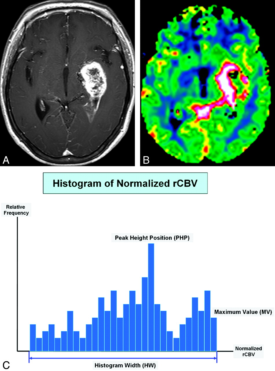
- Fig 1.
The schema of histogram distribution in a patient with GBM. A and B, Axial postcontrast T1-weighted image (A) and the normalized CBV map (B) show typical findings of GBM. C, Three histogram parameters are defined as HW, PHP, and MV.
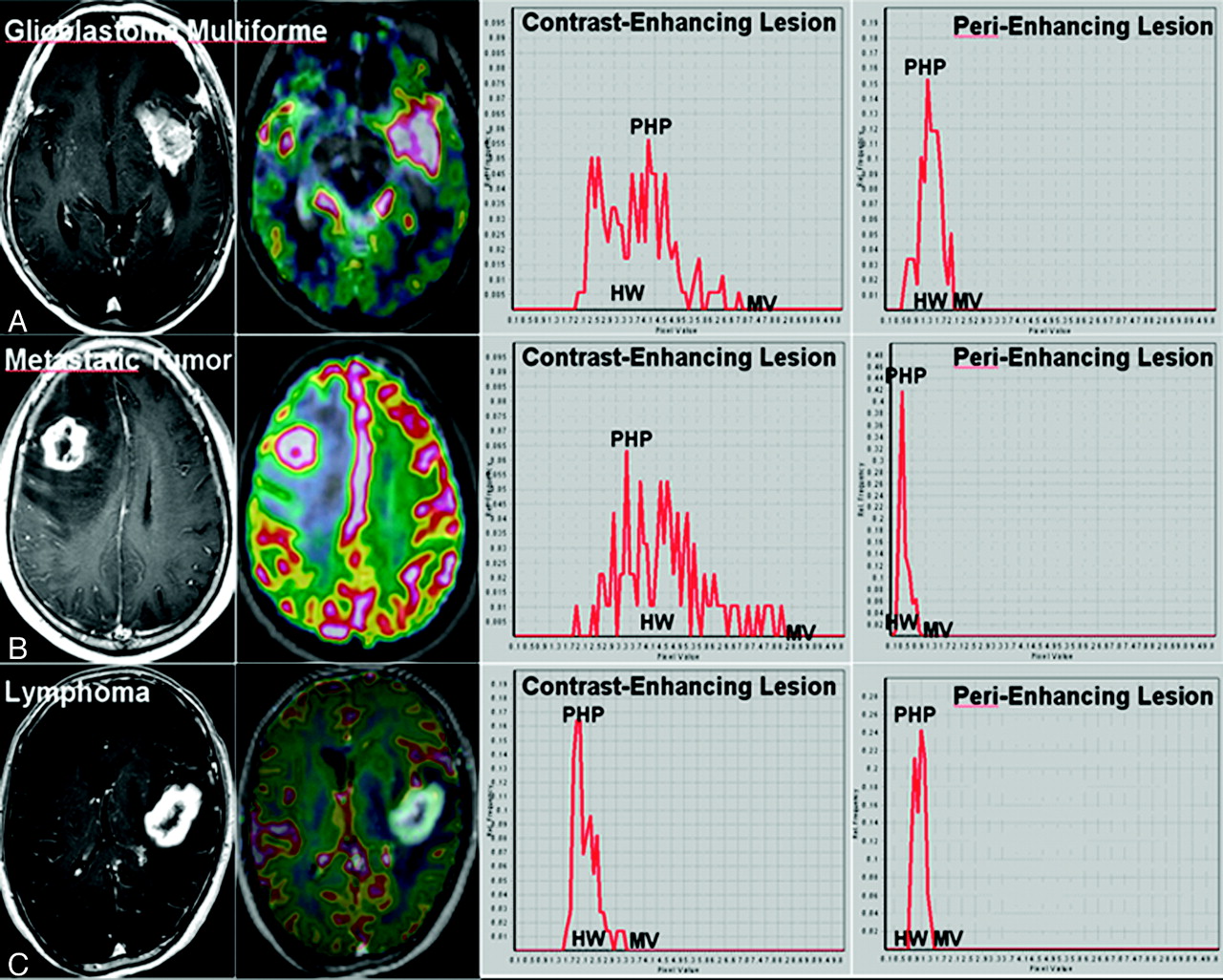
- Fig 2.
MR images and dynamic susceptibility contrast-enhanced imaging histograms in patients with GBM (A), metastatic tumor (B), and lymphoma (C).
- Fig 3.
Box-and-whisker plots of 6 histogram parameters for 3 pathologic groups of solitary enhancing brain lesions. A, HW in contrast-enhancing lesions. B, PHP in contrast-enhancing lesions. C, MV in contrast-enhancing lesions. D, HW in perienhancing lesions. E, PHP in perienhancing lesions. F, MV in perienhancing lesions.
- Fig 4.
Comparison of ROC curves of 6 histogram parameters in the contrast-enhancing lesions and perienhancing lesions for differentiating GBM and SMT (A), GBM and lymphoma (B), and SMT and lymphoma (C).


Tables
- Table 1:
Mean ± SD of histogram parameters for glioblastomas, metastatic tumors, and lymphomas
Parameters Glioblastoma Metastatic Tumor Lymphoma HWCEL 6.01 ± 1.15 6.36 ± 1.50 1.93 ± 0.79 PHPCEL 4.79 ± 1.31 3.32 ± 1.10 2.08 ± 0.54 MVCEL 7.43 ± 1.28 8.04 ± 1.20 2.83 ± 0.83 HWPEL 1.60 ± 0.22 0.65 ± 0.20 0.71 ± 0.42 PHPPEL 1.42 ± 0.28 0.46 ± 0.23 0.91 ± 0.37 MVPEL 1.90 ± 0.26 0.80 ± 0.21 1.27 ± 0.34 - Table 2:
P values for the differential diagnosis among glioblastomas, metastatic tumors, and lymphomas using each histogram parameter
Parameters GBM versus SMT GBM versus Lymphoma SMT versus Lymphoma HWCEL .502 <.0001 <.0001 PHPCEL .031 .007 .019 MVCEL .177 <.0001 <.0001 HWPEL <.0001 <.0001 .263 PHPPEL .005 .011 .093 MVPEL <.0001 <.0001 .038 - Table 3:
Optimum threshold, sensitivity, and specificity of each histogram parameter for distinguishing glioblastoma from SMT
GBM versus SMT PHPCEL HWPEL PHPPEL MVPEL Threshold 3.5 1.2 1.1 1.4 Sensitivity 89.2% 96.4% 89.2% 100.0% Specificity 72.7% 100.0% 95.4% 100.0% - Table 4:
Optimum threshold, sensitivity, and specificity of each histogram parameter for distinguishing glioblastoma from lymphoma
GBM versus Lymphoma HWCEL PHPCEL MVCEL HWPEL PHPPEL MVPEL Threshold 2.7 2.7 3.9 1.3 0.9 1.2 Sensitivity 100.0% 96.4% 100.0% 85.7% 95.4% 92.2% Specificity 100.0% 100.0% 100.0% 100.0% 91.7% 100.0% - Table 5:
Optimum threshold, sensitivity, and specificity of each histogram parameter for distinguishing SMT from lymphoma
SMT versus Lymphoma HWCEL PHPCEL MVCEL MVPEL Threshold 2.5 2.4 3.9 0.8 Sensitivity 100.0% 85.7% 100.0% 83.3% Specificity 92.7% 83.3% 100.0% 67.7%
In this issue


Jump to section
Related Articles
Cited By...
- Discrimination between Glioblastoma and Solitary Brain Metastasis: Comparison of Inflow-Based Vascular-Space-Occupancy and Dynamic Susceptibility Contrast MR Imaging
- Tumor-related Perfusion Changes in White Matter Adjacent to Brain Tumors: Pharmacodynamic Analysis of Dynamic 3T Magnetic Resonance Imaging
- Differentiation of Primary Central Nervous System Lymphomas and Glioblastomas: Comparisons of Diagnostic Performance of Dynamic Susceptibility Contrast-Enhanced Perfusion MR Imaging without and with Contrast-Leakage Correction
- Differentiation between Brain Glioblastoma Multiforme and Solitary Metastasis: Qualitative and Quantitative Analysis Based on Routine MR Imaging