Advertisement

Main menu

  • Home
  • Content
    • Current Issue
    • Accepted Manuscripts
    • Article Preview
    • Past Issue Archive
    • Video Articles
    • AJNR Case Collection
    • Case of the Week Archive
    • Case of the Month Archive
    • Classic Case Archive
  • Special Collections
    • AJNR Awards
    • Low-Field MRI
    • Alzheimer Disease
    • ASNR Foundation Special Collection
    • Photon-Counting CT
    • View All
  • Multimedia
    • AJNR Podcasts
    • AJNR SCANtastic
    • Trainee Corner
    • MRI Safety Corner
    • Imaging Protocols
  • For Authors
    • Submit a Manuscript
    • Submit a Video Article
    • Submit an eLetter to the Editor/Response
    • Manuscript Submission Guidelines
    • Statistical Tips
    • Fast Publishing of Accepted Manuscripts
    • Graphical Abstract Preparation
    • Imaging Protocol Submission
    • Author Policies
  • About Us
    • About AJNR
    • Editorial Board
    • Editorial Board Alumni
  • More
    • Become a Reviewer/Academy of Reviewers
    • Subscribers
    • Permissions
    • Alerts
    • Feedback
    • Advertisers
    • ASNR Home

User menu

  • Alerts
  • Log in

Search

  • Advanced search
American Journal of Neuroradiology

American Journal of Neuroradiology

ASHNR American Society of Functional Neuroradiology ASHNR American Society of Pediatric Neuroradiology ASSR
  • Alerts
  • Log in

Advanced Search

  • Home
  • Content
    • Current Issue
    • Accepted Manuscripts
    • Article Preview
    • Past Issue Archive
    • Video Articles
    • AJNR Case Collection
    • Case of the Week Archive
    • Case of the Month Archive
    • Classic Case Archive
  • Special Collections
    • AJNR Awards
    • Low-Field MRI
    • Alzheimer Disease
    • ASNR Foundation Special Collection
    • Photon-Counting CT
    • View All
  • Multimedia
    • AJNR Podcasts
    • AJNR SCANtastic
    • Trainee Corner
    • MRI Safety Corner
    • Imaging Protocols
  • For Authors
    • Submit a Manuscript
    • Submit a Video Article
    • Submit an eLetter to the Editor/Response
    • Manuscript Submission Guidelines
    • Statistical Tips
    • Fast Publishing of Accepted Manuscripts
    • Graphical Abstract Preparation
    • Imaging Protocol Submission
    • Author Policies
  • About Us
    • About AJNR
    • Editorial Board
    • Editorial Board Alumni
  • More
    • Become a Reviewer/Academy of Reviewers
    • Subscribers
    • Permissions
    • Alerts
    • Feedback
    • Advertisers
    • ASNR Home
  • Follow AJNR on Twitter
  • Visit AJNR on Facebook
  • Follow AJNR on Instagram
  • Join AJNR on LinkedIn
  • RSS Feeds

AJNR Awards, New Junior Editors, and more. Read the latest AJNR updates

Getting new auth cookie, if you see this message a lot, tell someone!
Research ArticleBrain

Histologically Confirmed Hippocampal Structural Features Revealed by 3T MR Imaging: Potential to Increase Diagnostic Specificity of Mesial Temporal Sclerosis

K.L. Howe, D. Dimitri, C. Heyn, T.-R. Kiehl, D. Mikulis and T. Valiante
American Journal of Neuroradiology October 2010, 31 (9) 1682-1689; DOI: https://doi.org/10.3174/ajnr.A2154
K.L. Howe
  • Find this author on Google Scholar
  • Find this author on PubMed
  • Search for this author on this site
D. Dimitri
  • Find this author on Google Scholar
  • Find this author on PubMed
  • Search for this author on this site
C. Heyn
  • Find this author on Google Scholar
  • Find this author on PubMed
  • Search for this author on this site
T.-R. Kiehl
  • Find this author on Google Scholar
  • Find this author on PubMed
  • Search for this author on this site
D. Mikulis
  • Find this author on Google Scholar
  • Find this author on PubMed
  • Search for this author on this site
T. Valiante
  • Find this author on Google Scholar
  • Find this author on PubMed
  • Search for this author on this site
  • Article
  • Figures & Data
  • Info & Metrics
  • Responses
  • References
  • PDF
Loading

Article Figures & Data

Figures

  • Tables
  • Fig 1.
    • Download figure
    • Open in new tab
    • Download powerpoint
    Fig 1.

    Right hippocampus in a coronal section showing anatomic detail in 3T MR imaging (A) versus 1.5T MR imaging (B). Small arrows indicate the hypoattenuated region later determined to represent the stratum lacunosum (a) and lateral margin (b), both of which are significantly more distinguishable on 3T MR imaging. C, Right hippocampus shows ultrastructural detail determined on 3T MR imaging (T2 FSTIR). Coronal view of the right hippocampus shows distances that can be measured by using improved neuroanatomic detail. Arrowheads point to a consistently observed band seen on coronal sections in 3T MR imaging.

  • Fig 2.
    • Download figure
    • Open in new tab
    • Download powerpoint
    Fig 2.

    Sample images from patient 4 depicting a coronal section showing left-sided hippocampal sclerosis (A) with enlarged images of the right (B) and left (C) sides. Note the yellow markers used to depict the superior, inferior, and lateral measurements (sample only, actual images with measurements from Advantage Workstation could not be exported). D, The enlarged image depicts the hippocampus in a sagittal section with the head of the hippocampus indicated by the dashed line, used to determine the appropriate coronal image from which measurements could be taken. E, Corresponding coronal T2 FLAIR image shows increased signal intensity on the left side, a radiographic marker for a sclerotic process suggestive of gliosis.

  • Fig 3.
    • Download figure
    • Open in new tab
    • Download powerpoint
    Fig 3.

    Cumulative hippocampal measurements of superior, inferior, and lateral margins comparing the MTS side with the contralateral internal control. Values are presented in millimeters (mean ± SD, P < .001; n = 20).
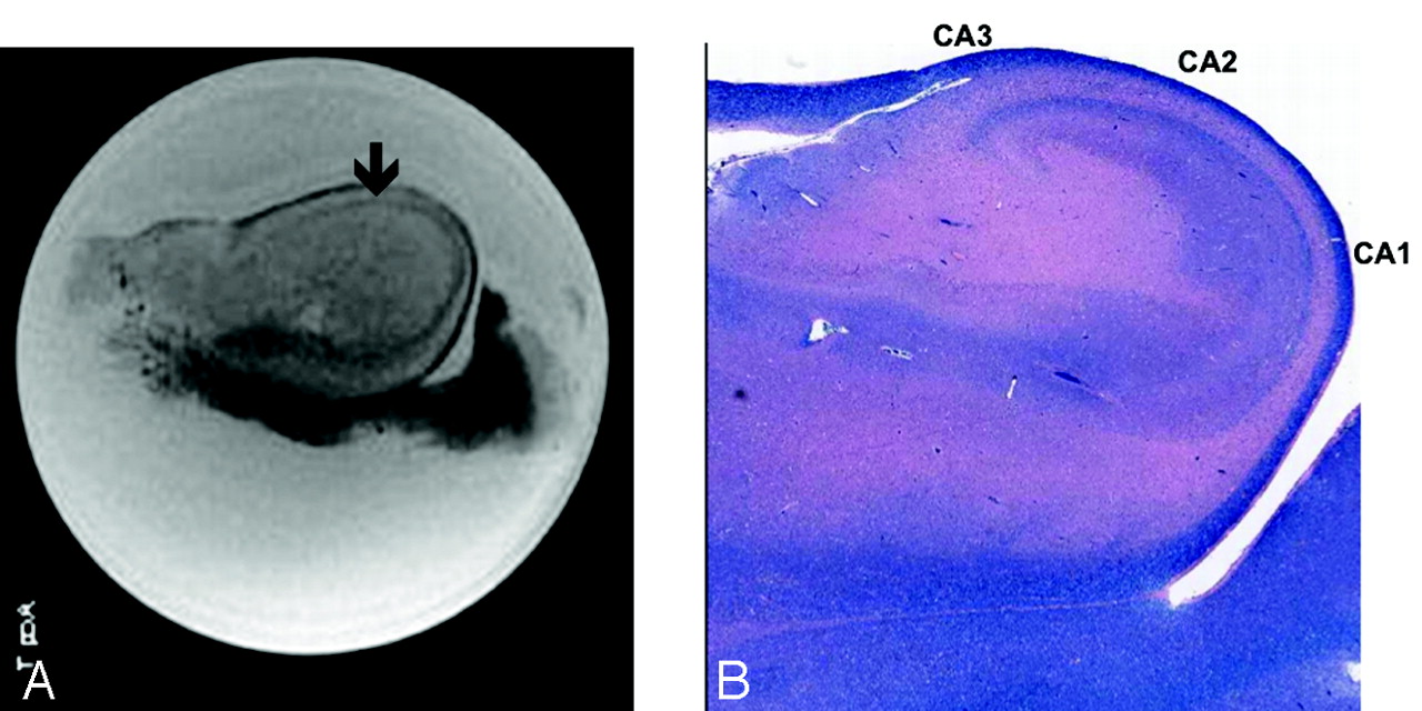
  • Fig 4.
    • Download figure
    • Open in new tab
    • Download powerpoint
    Fig 4.

    A, Postoperative ex vivo MR image of the hippocampal specimen in a coronal section. B, Scanned image of corresponding histologic slide (× 20 magnification) with CA1, CA2, and CA3 regions marked accordingly. Overlay of both images clarifies structural MR imaging information, such as the dark band that was ultimately identified as the stratum lacunosum (arrow).

  • Fig 5.
    • Download figure
    • Open in new tab
    • Download powerpoint
    Fig 5.

    A, Comparison of layer thickness within the CA1 between the control and 2 cases of MTS. Note the marked reduction in thickness, particularly in the stratum pyramidale and radiatum in cases 4 and 5. B, Thickness measurement of the distance between the stratum oriens and stratum lacunosum in all 3 CA regions. Again note the marked size reduction in cases 4 and 5 compared with all 3 controls (C1, C2, C3). C, Specific thickness measurements of the stratum pyramidale in regions CA1, -2, and -3 with marked size reduction apparent in the CA1 region of cases 4 and 5.
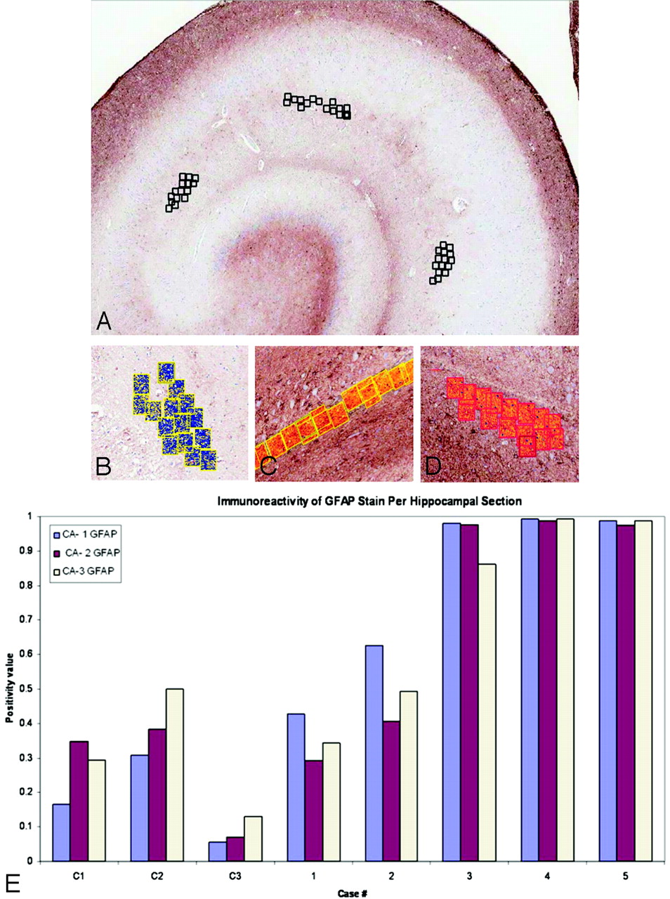
  • Fig 6.
    • Download figure
    • Open in new tab
    • Download powerpoint
    Fig 6.

    A, Methodology for Positive Pixel Count Tool. Placement of pixel count boxes in 3 sections of the hippocampus: CA1, -2, and -3. B−D, Positive Pixel Count Tool: immunoreactivity is converted to blue (LFB-HE) or red and orange (GFAP stain). E, Analysis of immunoreactivity of the GFAP stain within each CA region of the hippocampal specimens. Note marked increase in GFAP staining in all MTS cases compared with control cases.

Tables

  • Figures
  • Patient demographics (ex-vivo and histopathologic analysis)

    Patient No.Age (yr)SexSeizure History (yr)Radiographic FeatureEEGNeuropsychiatric AssessmentConcordance (Yes/No)Side of OperationPostoperative Seizure History
    124F7 R diffuse temporal dysplasiaR anterior temporal lobeNondominant dysfunctionYesR1 Year seizure-free
    234F19R MTSR anterior temporal lobeNondominant dysfunctionYesR1 Year seizure-free
    344F44R MTSR mesial temporal lobeNonverbal memory dysfunctionYesR1 Year seizure-free
    451F5L MTSL anterior temporal lobeDecreased verbal memoryYesL1 Year seizure recurrence
    542F31L MTSL temporal lobeL mesial temporal dysfunctionYesL1 Year seizure-free
PreviousNext
Back to top

In this issue

American Journal of Neuroradiology: 31 (9)
American Journal of Neuroradiology
Vol. 31, Issue 9
1 Oct 2010
  • Table of Contents
  • Index by author
Advertisement
Print
Download PDF
Email Article

Thank you for your interest in spreading the word on American Journal of Neuroradiology.

NOTE: We only request your email address so that the person you are recommending the page to knows that you wanted them to see it, and that it is not junk mail. We do not capture any email address.

Enter multiple addresses on separate lines or separate them with commas.
Histologically Confirmed Hippocampal Structural Features Revealed by 3T MR Imaging: Potential to Increase Diagnostic Specificity of Mesial Temporal Sclerosis
(Your Name) has sent you a message from American Journal of Neuroradiology
(Your Name) thought you would like to see the American Journal of Neuroradiology web site.
CAPTCHA
This question is for testing whether or not you are a human visitor and to prevent automated spam submissions.
Cite this article
K.L. Howe, D. Dimitri, C. Heyn, T.-R. Kiehl, D. Mikulis, T. Valiante
Histologically Confirmed Hippocampal Structural Features Revealed by 3T MR Imaging: Potential to Increase Diagnostic Specificity of Mesial Temporal Sclerosis
American Journal of Neuroradiology Oct 2010, 31 (9) 1682-1689; DOI: 10.3174/ajnr.A2154

Citation Manager Formats

  • BibTeX
  • Bookends
  • EasyBib
  • EndNote (tagged)
  • EndNote 8 (xml)
  • Medlars
  • Mendeley
  • Papers
  • RefWorks Tagged
  • Ref Manager
  • RIS
  • Zotero
0 Responses
Respond to this article
Share
Bookmark this article
Histologically Confirmed Hippocampal Structural Features Revealed by 3T MR Imaging: Potential to Increase Diagnostic Specificity of Mesial Temporal Sclerosis
K.L. Howe, D. Dimitri, C. Heyn, T.-R. Kiehl, D. Mikulis, T. Valiante
American Journal of Neuroradiology Oct 2010, 31 (9) 1682-1689; DOI: 10.3174/ajnr.A2154
del.icio.us logo Twitter logo Facebook logo Mendeley logo
  • Tweet Widget
  • Facebook Like
  • Google Plus One
Purchase

Jump to section

  • Article
    • Abstract
    • Abbreviations
    • Materials and Methods
    • Results
    • Discussion
    • Conclusions
    • Acknowledgments
    • References
  • Figures & Data
  • Info & Metrics
  • Responses
  • References
  • PDF

Related Articles

  • No related articles found.
  • PubMed
  • Google Scholar

Cited By...

  • Improved Detection of Subtle Mesial Temporal Sclerosis: Validation of a Commercially Available Software for Automated Segmentation of Hippocampal Volume
  • 3T MRI Quantification of Hippocampal Volume and Signal in Mesial Temporal Lobe Epilepsy Improves Detection of Hippocampal Sclerosis
  • 3.0 T MRI of 2000 consecutive patients with localisation-related epilepsy
  • Crossref
  • Google Scholar

This article has not yet been cited by articles in journals that are participating in Crossref Cited-by Linking.

More in this TOC Section

  • Usefulness of Quantitative Susceptibility Mapping for the Diagnosis of Parkinson Disease
  • White Matter Alterations in the Brains of Patients with Active, Remitted, and Cured Cushing Syndrome: A DTI Study
  • Qualitative and Quantitative Analysis of MR Imaging Findings in Patients with Middle Cerebral Artery Stroke Implanted with Mesenchymal Stem Cells
Show more Brain

Similar Articles

Advertisement

Indexed Content

  • Current Issue
  • Accepted Manuscripts
  • Article Preview
  • Past Issues
  • Editorials
  • Editor's Choice
  • Fellows' Journal Club
  • Letters to the Editor
  • Video Articles

Cases

  • Case Collection
  • Archive - Case of the Week
  • Archive - Case of the Month
  • Archive - Classic Case

More from AJNR

  • Trainee Corner
  • Imaging Protocols
  • MRI Safety Corner
  • Book Reviews

Multimedia

  • AJNR Podcasts
  • AJNR Scantastics

Resources

  • Turnaround Time
  • Submit a Manuscript
  • Submit a Video Article
  • Submit an eLetter to the Editor/Response
  • Manuscript Submission Guidelines
  • Statistical Tips
  • Fast Publishing of Accepted Manuscripts
  • Graphical Abstract Preparation
  • Imaging Protocol Submission
  • Evidence-Based Medicine Level Guide
  • Publishing Checklists
  • Author Policies
  • Become a Reviewer/Academy of Reviewers
  • News and Updates

About Us

  • About AJNR
  • Editorial Board
  • Editorial Board Alumni
  • Alerts
  • Permissions
  • Not an AJNR Subscriber? Join Now
  • Advertise with Us
  • Librarian Resources
  • Feedback
  • Terms and Conditions
  • AJNR Editorial Board Alumni

American Society of Neuroradiology

  • Not an ASNR Member? Join Now

© 2025 by the American Society of Neuroradiology All rights, including for text and data mining, AI training, and similar technologies, are reserved.
Print ISSN: 0195-6108 Online ISSN: 1936-959X

Powered by HighWire