
Article Figures & Data
Figures
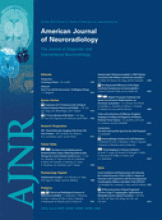
- Fig 1.
A 45-year-old woman without a history of hypertension and with intact coagulation presented with acute onset of headache and visual changes. A and B, High-probability NCCT scan demonstrates an acute right occipital ICH with calcifications along its posteroinferior margin (arrowhead, B; SICH score, 6). There was associated subdural hemorrhage overlying the right temporal lobe but no associated IVH or SAH. C, CTA source image demonstrates a tangle of abnormal vessels along the posteroinferior aspect of the ICH (arrowhead) with associated calcifications (arrow), consistent with an AVM. D, CTA MIP image in the axial plane redemonstrates the right occipital AVM (arrowhead) with arterial supply from branches of the right posterior cerebral artery and drainage to the right transverse sinus.
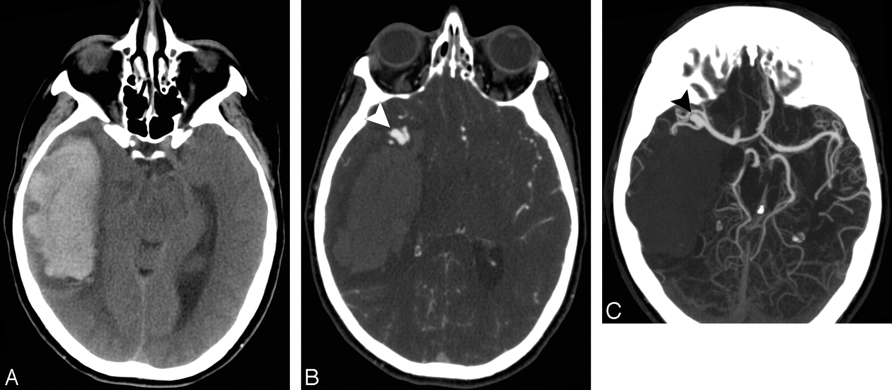
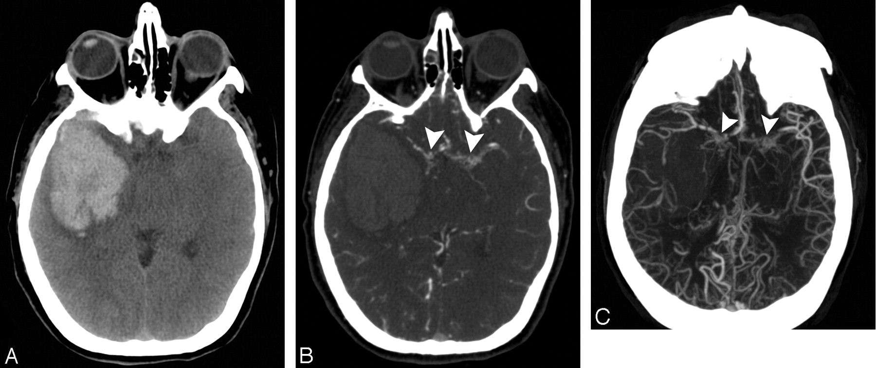
- Fig 2.
A 50-year-old woman with a history of hypertension and intact coagulation presented with acute onset of left-sided weakness. A, Indeterminate NCCT scan demonstrates an acute right temporal ICH without associated IVH or SAH (SICH score, 3). B, CTA source image demonstrates an 11-mm outpouching arising from the right MCA bifurcation (arrowhead), consistent with an aneurysm. C, CTA MIP image in the axial plane redemonstrates the right MCA bifurcation aneurysm (arrowhead).
- Fig 3.
A 60-year-old woman without a history of hypertension and with intact coagulation presented with increasing headache during the past several days. A and B, High-probability NCCT scan demonstrates an acute right mesiotemporal ICH with subtle associated hyperattenuation within the distal right vein of Labbe (arrow, B) and right transverse sinus (arrowhead, B; SICH score,5). C, Coronal NCCT scan reformation improves depiction of the hyperattenuation within the distal right vein of Labbe (arrow) and right transverse sinus (arrowhead). D, CT venogram source image obtained immediately after the CTA demonstrates nonopacification of the right transverse and sigmoid sinuses (arrowheads), consistent with DVST. E, CT venogram MIP image after calvarial segmentation redemonstrates the right transverse and sigmoid sinus thrombosis (arrowheads).
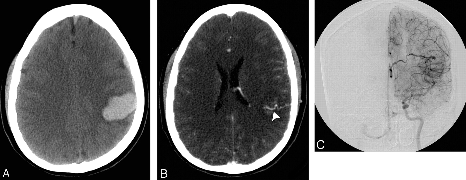
- Fig 4.
A 44-year-old woman without history of hypertension and with intact coagulation presented with headache. A, Indeterminate NCCT scan demonstrates a left parietal ICH (SICH score, 5). There was associated subdural hemorrhage overlying the left frontal lobe but no associated IVH or SAH. B, CTA source image demonstrates an abnormal vessel along the inferior aspect of the ICH in the left parietal lobe, consistent with an AVF (arrowhead). C, Frontal left internal carotid artery catheter angiogram confirms the presence of a left parietal AVF with deep venous drainage into the left internal cerebral vein.
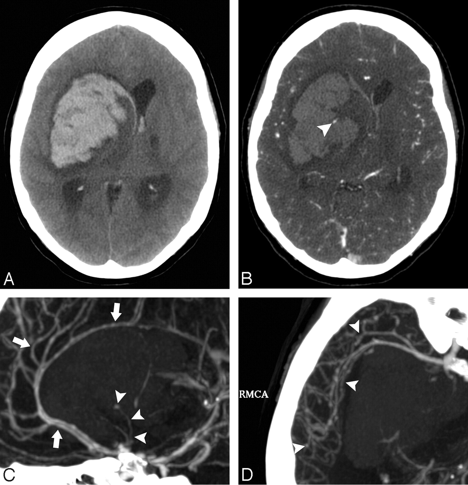
- Fig 5.
A 50-year-old woman without a history of hypertension and with intact coagulation presented with acute onset of unresponsiveness. A, Low-probability NCCT scan demonstrates an acute right basal ganglia ICH with associated IVH (SICH score, 3). B, CTA source image demonstrates a 3-mm outpouching arising from a lenticulostriate branch of the right middle cerebral artery (arrowhead), consistent with an aneurysm. C, CTA MIP image in the sagittal plane redemonstrates the right lenticulostriate aneurysm (arrowheads) as well as a diffuse luminal irregularity in the visualized anterior cerebral artery branches (arrows). D, CTA MIP image in the axial plane demonstrates diffuse luminal irregularity in the right middle cerebral artery branches (arrowheads). These findings are consistent with vasculitis with secondary pseudoaneurysm formation and rupture. The patient was ultimately found to have Lyme disease affecting the central nervous system.
- Fig 6.
A 42-year-old woman without a history of hypertension and with intact coagulation presented with severe headache. A, Indeterminate NCCT scan demonstrates an acute right temporal ICH without associated IVH or SAH (SICH score, 5). B, CTA source image demonstrates occlusion of the supraclinoid segments of the internal carotid arteries and proximal M1 segments of the middle cerebral arteries bilaterally, with numerous associated lenticulostriate collateral vessels (arrowheads), consistent with Moyamoya phenomenon. C, CTA MIP image in the axial plane redemonstrates the findings of Moyamoya phenomenon (arrowheads).


Tables
Etiology Retrospective Cohort (n = 91) Prospective Cohort (n = 29) All Patients (n = 120) No. % No. % No. % AVM 40 44 15 51.7 55 45.8 Aneurysm 21a 23 5 17.2 26a 21.7 DVST 17 18.7 3 10.3 20 16.7 AVF 8 8.8 3 10.3 11 9.2 Vasculopathy 3 3.3 1b 3.5 4b 3.3 Moyamoya 2 2.2 2 7 4 3.3 -
a Includes 3 pseudoaneurysms.
-
b Includes a patient in whom vasculitis led to pseudoaneurysm formation and rupture.
-
Parameter Points NCCT categorizationa High probability 2 Indeterminate 1 Low probability 0 Age group 18–45 years 2 46–70 years 1 ≥71 years 0 Sex Female 1 Male 0 Neither known HTN nor impaired coagulationb Yes 1 No 0 -
Note:—The SICH score is calculated by adding the total number of points for a given patient.
-
a High-probability NCCT: an examination with either 1) enlarged vessels or calcifications along the margins of the ICH or 2) hyperattenuation within a dural venous sinus or cortical vein along the presumed venous drainage path of the ICH. Low-probability NCCT: an examination in which neither 1) nor 2) is present and the ICH is located in the basal ganglia, thalamus, or brain stem. Indeterminate NCCT: an examination that does not meet criteria for a high- or low-probability NCCT.
-
b Impaired coagulation defined as admission INR >3, aPTT >80 seconds, platelet count <50,000, or daily antiplatelet therapy.
-
Score Retrospective-Derivation Cohort (n = 623) Prospective-Validation Cohort (n = 222) All Patients (n = 845) No. (%) % Positive CTAs No. (%) % Positive CTAs No. (%) % Positive CTAs 0 37 (5.9) 0 15 (6.8) 0 52 (6.1) 0 1 145 (23.3) 1.4 67 (30.2) 1.5 212 (25.1) 1.4 2 209 (33.5) 5.3 68 (30.6) 4.4 277 (32.8) 5.1 3 138 (22.2) 18.1 40 (18.0) 20 178 (21.1) 18.5 4 61 (9.8) 39.3 21 (9.5) 38.1 82 (9.7) 39 5 28 (4.5) 85.7 10 (4.5) 80 38 (4.5) 84.2 6 5 (0.8) 100 1 (0.4) 100 6 (0.7) 100 AUC (95% CI) 0.86 (0.83–0.89) 0.87 (0.82–0.91) 0.87 (0.84–0.89) MOP >2 >2 >2 Sensitivity 85.7 86.2 85.8 Specificity 71.1 75.6 72.3 P value <.0001 <.0001 <.0001
In this issue


Jump to section
Related Articles
Cited By...
- Management of Intracerebral Hemorrhage: JACC Focus Seminar
- Predicting the presence of macrovascular causes in non-traumatic intracerebral haemorrhage: the DIAGRAM prediction score
- Diagnostic yield and accuracy of CT angiography, MR angiography, and digital subtraction angiography for detection of macrovascular causes of intracerebral haemorrhage: prospective, multicentre cohort study
- External Validation of the Secondary Intracerebral Hemorrhage Score in The Netherlands
- Emergency Noninvasive Angiography for Acute Intracerebral Hemorrhage
- Yield of catheter angiography in patients with intracerebral hemorrhage with and without intraventricular extension
- Frequency of Adequate Contrast Opacification of the Major Intracranial Venous Structures with CT Angiography in the Setting of Intracerebral Hemorrhage: Comparison of 16- and 64-Section CT Angiography Techniques
- Scaling Back on Scales with a Scale of Scales