Advertisement

Main menu

  • Home
  • Content
    • Current Issue
    • Accepted Manuscripts
    • Article Preview
    • Past Issue Archive
    • Video Articles
    • AJNR Case Collection
    • Case of the Week Archive
    • Case of the Month Archive
    • Classic Case Archive
  • Special Collections
    • AJNR Awards
    • Low-Field MRI
    • Alzheimer Disease
    • ASNR Foundation Special Collection
    • Photon-Counting CT
    • View All
  • Multimedia
    • AJNR Podcasts
    • AJNR SCANtastic
    • Trainee Corner
    • MRI Safety Corner
    • Imaging Protocols
  • For Authors
    • Submit a Manuscript
    • Submit a Video Article
    • Submit an eLetter to the Editor/Response
    • Manuscript Submission Guidelines
    • Statistical Tips
    • Fast Publishing of Accepted Manuscripts
    • Graphical Abstract Preparation
    • Imaging Protocol Submission
    • Author Policies
  • About Us
    • About AJNR
    • Editorial Board
    • Editorial Board Alumni
  • More
    • Become a Reviewer/Academy of Reviewers
    • Subscribers
    • Permissions
    • Alerts
    • Feedback
    • Advertisers
    • ASNR Home

User menu

  • Alerts
  • Log in

Search

  • Advanced search
American Journal of Neuroradiology

American Journal of Neuroradiology

ASHNR American Society of Functional Neuroradiology ASHNR American Society of Pediatric Neuroradiology ASSR
  • Alerts
  • Log in

Advanced Search

  • Home
  • Content
    • Current Issue
    • Accepted Manuscripts
    • Article Preview
    • Past Issue Archive
    • Video Articles
    • AJNR Case Collection
    • Case of the Week Archive
    • Case of the Month Archive
    • Classic Case Archive
  • Special Collections
    • AJNR Awards
    • Low-Field MRI
    • Alzheimer Disease
    • ASNR Foundation Special Collection
    • Photon-Counting CT
    • View All
  • Multimedia
    • AJNR Podcasts
    • AJNR SCANtastic
    • Trainee Corner
    • MRI Safety Corner
    • Imaging Protocols
  • For Authors
    • Submit a Manuscript
    • Submit a Video Article
    • Submit an eLetter to the Editor/Response
    • Manuscript Submission Guidelines
    • Statistical Tips
    • Fast Publishing of Accepted Manuscripts
    • Graphical Abstract Preparation
    • Imaging Protocol Submission
    • Author Policies
  • About Us
    • About AJNR
    • Editorial Board
    • Editorial Board Alumni
  • More
    • Become a Reviewer/Academy of Reviewers
    • Subscribers
    • Permissions
    • Alerts
    • Feedback
    • Advertisers
    • ASNR Home
  • Follow AJNR on Twitter
  • Visit AJNR on Facebook
  • Follow AJNR on Instagram
  • Join AJNR on LinkedIn
  • RSS Feeds

AJNR Awards, New Junior Editors, and more. Read the latest AJNR updates

Research ArticleSpine Imaging and Spine Image-Guided Interventions
Open Access

Assessment of Craniospinal Pressure-Volume Indices

A. Wåhlin, K. Ambarki, R. Birgander, N. Alperin, J. Malm and A. Eklund
American Journal of Neuroradiology October 2010, 31 (9) 1645-1650; DOI: https://doi.org/10.3174/ajnr.A2166
A. Wåhlin
  • Find this author on Google Scholar
  • Find this author on PubMed
  • Search for this author on this site
K. Ambarki
  • Find this author on Google Scholar
  • Find this author on PubMed
  • Search for this author on this site
R. Birgander
  • Find this author on Google Scholar
  • Find this author on PubMed
  • Search for this author on this site
N. Alperin
  • Find this author on Google Scholar
  • Find this author on PubMed
  • Search for this author on this site
J. Malm
  • Find this author on Google Scholar
  • Find this author on PubMed
  • Search for this author on this site
A. Eklund
  • Find this author on Google Scholar
  • Find this author on PubMed
  • Search for this author on this site
  • Article
  • Figures & Data
  • Info & Metrics
  • Responses
  • References
  • PDF
Loading

Article Figures & Data

Figures

  • Tables
  • Fig 1.
    • Download figure
    • Open in new tab
    • Download powerpoint
    Fig 1.

    A subject recording of ΔICP and ICP from a constant pressure lumbar CSF infusion test. The RPPC is the slope of the linear regression line between ΔICP and ICP. P0 is calculated as the pressure at which this line intersects with ΔICP = 0.

  • Fig 2.
    • Download figure
    • Open in new tab
    • Download powerpoint
    Fig 2.

    An overview of how the infusion and MR imaging modalities are combined to estimate compliance indices.
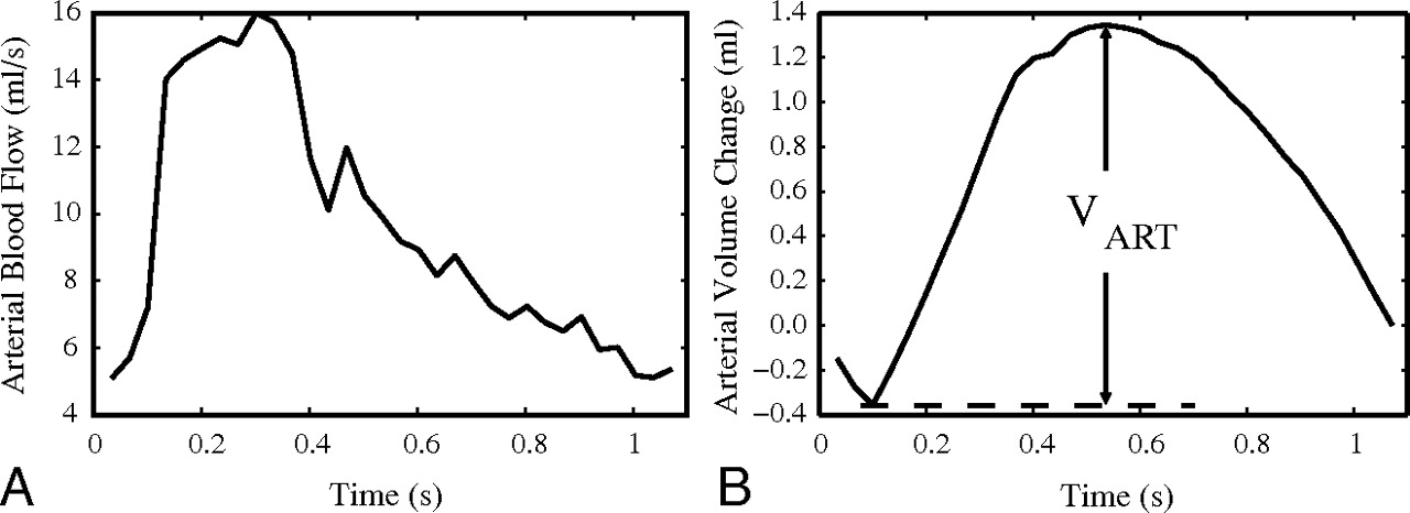
  • Fig 3.
    • Download figure
    • Open in new tab
    • Download powerpoint
    Fig 3.

    A, Total cerebral arterial blood flow (summated flow of the internal carotid and vertebral arteries). B, Cumulative integration of the curve in A with the average flow subtracted yields the arterial volume change during a cardiac cycle. ΔVART was defined as the largest volume difference during a cardiac cycle.
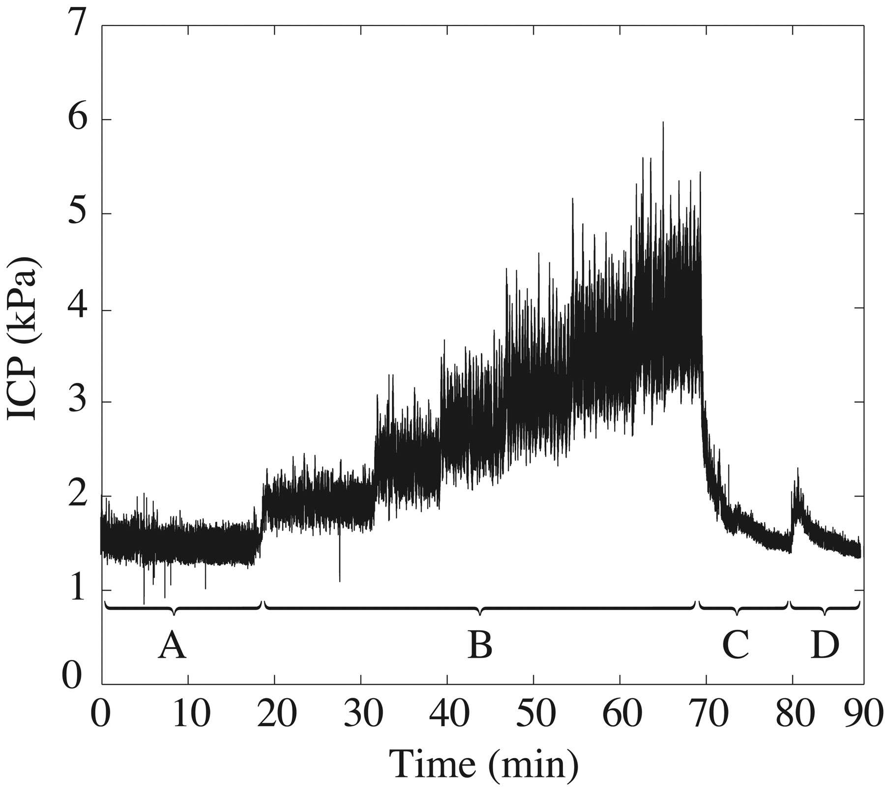
  • Fig 4.
    • Download figure
    • Open in new tab
    • Download powerpoint
    Fig 4.

    An ICP recording from a lumbar CSF infusion test. A, Baseline ICP recording. B, Infusion to predetermined ICP levels. C, Relaxation phase allowing ICP to normalize. D, A bolus test (rapid infusion of a 5.6-mL artificial CSF).

  • Fig 5.
    • Download figure
    • Open in new tab
    • Download powerpoint
    Fig 5.

    The craniospinal PVI calculated from combined MR imaging and infusion (y-xis) and from the bolus test (x-axis). The difference was not statistically different from zero, P = .83 from a paired t test. There was no significant correlation after excluding 2 extreme values (dashed contours) (P = .8778).

Tables

  • Figures
  • Reports of PVICC in various states

    SubjectsPVICC (mL)Method
    Normal pressure hydrocephalus (n = 69)1616.4Repeated bolus injections
    Idiopathic normal pressure hydrocephalus (n = 47)1616.7Repeated bolus injections
    Head injury: ICP, <20 mm Hg (n = 9)1820.8Bolus injection/CSF withdrawal
    Hydrocephalus, ventriculomegaly (n = 27)199.6Constant-rate infusion
    Hydrocephalus, ventriculomegaly (n = 27)1916.5Relaxation preceding infusion
    Mechanically ventilated, pathologic autoregulation (n = 35)3420.0Bolus injection
    Mechanically ventilated, functioning autoregulation (n = 24)3431.6Bolus injection
    Hydrocephalus, ventriculomegaly (n = 46)208.9Constant-rate infusion
    Adult patients without intracranial masses (n = 7)125.9Bolus injection/CSF withdrawal
PreviousNext
Back to top

In this issue

American Journal of Neuroradiology: 31 (9)
American Journal of Neuroradiology
Vol. 31, Issue 9
1 Oct 2010
  • Table of Contents
  • Index by author
Advertisement
Print
Download PDF
Email Article

Thank you for your interest in spreading the word on American Journal of Neuroradiology.

NOTE: We only request your email address so that the person you are recommending the page to knows that you wanted them to see it, and that it is not junk mail. We do not capture any email address.

Enter multiple addresses on separate lines or separate them with commas.
Assessment of Craniospinal Pressure-Volume Indices
(Your Name) has sent you a message from American Journal of Neuroradiology
(Your Name) thought you would like to see the American Journal of Neuroradiology web site.
CAPTCHA
This question is for testing whether or not you are a human visitor and to prevent automated spam submissions.
Cite this article
A. Wåhlin, K. Ambarki, R. Birgander, N. Alperin, J. Malm, A. Eklund
Assessment of Craniospinal Pressure-Volume Indices
American Journal of Neuroradiology Oct 2010, 31 (9) 1645-1650; DOI: 10.3174/ajnr.A2166

Citation Manager Formats

  • BibTeX
  • Bookends
  • EasyBib
  • EndNote (tagged)
  • EndNote 8 (xml)
  • Medlars
  • Mendeley
  • Papers
  • RefWorks Tagged
  • Ref Manager
  • RIS
  • Zotero
0 Responses
Respond to this article
Share
Bookmark this article
Assessment of Craniospinal Pressure-Volume Indices
A. Wåhlin, K. Ambarki, R. Birgander, N. Alperin, J. Malm, A. Eklund
American Journal of Neuroradiology Oct 2010, 31 (9) 1645-1650; DOI: 10.3174/ajnr.A2166
del.icio.us logo Twitter logo Facebook logo Mendeley logo
  • Tweet Widget
  • Facebook Like
  • Google Plus One
Purchase

Jump to section

  • Article
    • Abstract
    • Abbreviations
    • Materials and Methods
    • Results
    • Discussion
    • Conclusions
    • Appendix
    • Footnotes
    • References
  • Figures & Data
  • Info & Metrics
  • Responses
  • References
  • PDF

Related Articles

  • No related articles found.
  • PubMed
  • Google Scholar

Cited By...

  • fMRI-Based CSF Flow Quantification Identifies Cardiac Pulsatility as the Dominant Driver Over Respiratory and Slow Vasomotion Cycles
  • Assessing Cerebral Microvascular Volumetric Pulsatility with High-Resolution 4D CBV MRI at 7T
  • Modeling CSF circulation and the glymphatic system during infusion using subject specific intracranial pressures and brain geometries
  • Human intracranial pulsatility during the cardiac cycle: a computational modelling framework
  • Spinal Compliance Curves: Preliminary Experience with a New Tool for Evaluating Suspected CSF Venous Fistulas on CT Myelography in Patients with Spontaneous Intracranial Hypotension
  • Measuring Pulsatile Flow in Cerebral Arteries Using 4D Phase-Contrast MR Imaging
  • Pulsatility in CSF dynamics: pathophysiology of idiopathic normal pressure hydrocephalus
  • Blood Flow of Ophthalmic Artery in Healthy Individuals Determined by Phase-Contrast Magnetic Resonance Imaging
  • Crossref
  • Google Scholar

This article has not yet been cited by articles in journals that are participating in Crossref Cited-by Linking.

More in this TOC Section

  • Optimization of Photon Counting CT Myelography
  • Characteristics of SIH Type I Culprit Lesions
  • Management Outcomes For VO Spine Biopsy
Show more Spine Imaging and Spine Image-Guided Interventions

Similar Articles

Advertisement

Indexed Content

  • Current Issue
  • Accepted Manuscripts
  • Article Preview
  • Past Issues
  • Editorials
  • Editor's Choice
  • Fellows' Journal Club
  • Letters to the Editor
  • Video Articles

Cases

  • Case Collection
  • Archive - Case of the Week
  • Archive - Case of the Month
  • Archive - Classic Case

More from AJNR

  • Trainee Corner
  • Imaging Protocols
  • MRI Safety Corner
  • Book Reviews

Multimedia

  • AJNR Podcasts
  • AJNR Scantastics

Resources

  • Turnaround Time
  • Submit a Manuscript
  • Submit a Video Article
  • Submit an eLetter to the Editor/Response
  • Manuscript Submission Guidelines
  • Statistical Tips
  • Fast Publishing of Accepted Manuscripts
  • Graphical Abstract Preparation
  • Imaging Protocol Submission
  • Evidence-Based Medicine Level Guide
  • Publishing Checklists
  • Author Policies
  • Become a Reviewer/Academy of Reviewers
  • News and Updates

About Us

  • About AJNR
  • Editorial Board
  • Editorial Board Alumni
  • Alerts
  • Permissions
  • Not an AJNR Subscriber? Join Now
  • Advertise with Us
  • Librarian Resources
  • Feedback
  • Terms and Conditions
  • AJNR Editorial Board Alumni

American Society of Neuroradiology

  • Not an ASNR Member? Join Now

© 2025 by the American Society of Neuroradiology All rights, including for text and data mining, AI training, and similar technologies, are reserved.
Print ISSN: 0195-6108 Online ISSN: 1936-959X

Powered by HighWire