Advertisement

Main menu

  • Home
  • Content
    • Current Issue
    • Accepted Manuscripts
    • Article Preview
    • Past Issue Archive
    • Video Articles
    • AJNR Case Collection
    • Case of the Week Archive
    • Case of the Month Archive
    • Classic Case Archive
  • Special Collections
    • AJNR Awards
    • Low-Field MRI
    • Alzheimer Disease
    • ASNR Foundation Special Collection
    • Photon-Counting CT
    • View All
  • Multimedia
    • AJNR Podcasts
    • AJNR SCANtastic
    • Trainee Corner
    • MRI Safety Corner
    • Imaging Protocols
  • For Authors
    • Submit a Manuscript
    • Submit a Video Article
    • Submit an eLetter to the Editor/Response
    • Manuscript Submission Guidelines
    • Statistical Tips
    • Fast Publishing of Accepted Manuscripts
    • Graphical Abstract Preparation
    • Imaging Protocol Submission
    • Author Policies
  • About Us
    • About AJNR
    • Editorial Board
    • Editorial Board Alumni
  • More
    • Become a Reviewer/Academy of Reviewers
    • Subscribers
    • Permissions
    • Alerts
    • Feedback
    • Advertisers
    • ASNR Home

User menu

  • Alerts
  • Log in

Search

  • Advanced search
American Journal of Neuroradiology

American Journal of Neuroradiology

ASHNR American Society of Functional Neuroradiology ASHNR American Society of Pediatric Neuroradiology ASSR
  • Alerts
  • Log in

Advanced Search

  • Home
  • Content
    • Current Issue
    • Accepted Manuscripts
    • Article Preview
    • Past Issue Archive
    • Video Articles
    • AJNR Case Collection
    • Case of the Week Archive
    • Case of the Month Archive
    • Classic Case Archive
  • Special Collections
    • AJNR Awards
    • Low-Field MRI
    • Alzheimer Disease
    • ASNR Foundation Special Collection
    • Photon-Counting CT
    • View All
  • Multimedia
    • AJNR Podcasts
    • AJNR SCANtastic
    • Trainee Corner
    • MRI Safety Corner
    • Imaging Protocols
  • For Authors
    • Submit a Manuscript
    • Submit a Video Article
    • Submit an eLetter to the Editor/Response
    • Manuscript Submission Guidelines
    • Statistical Tips
    • Fast Publishing of Accepted Manuscripts
    • Graphical Abstract Preparation
    • Imaging Protocol Submission
    • Author Policies
  • About Us
    • About AJNR
    • Editorial Board
    • Editorial Board Alumni
  • More
    • Become a Reviewer/Academy of Reviewers
    • Subscribers
    • Permissions
    • Alerts
    • Feedback
    • Advertisers
    • ASNR Home
  • Follow AJNR on Twitter
  • Visit AJNR on Facebook
  • Follow AJNR on Instagram
  • Join AJNR on LinkedIn
  • RSS Feeds

AJNR Awards, New Junior Editors, and more. Read the latest AJNR updates

Research ArticlePediatric Neuroimaging

Neuroimaging In Cockayne Syndrome

M. Koob, V. Laugel, M. Durand, H. Fothergill, C. Dalloz, F. Sauvanaud, H. Dollfus, I.J. Namer and J.-L. Dietemann
American Journal of Neuroradiology October 2010, 31 (9) 1623-1630; DOI: https://doi.org/10.3174/ajnr.A2135
M. Koob
  • Find this author on Google Scholar
  • Find this author on PubMed
  • Search for this author on this site
V. Laugel
  • Find this author on Google Scholar
  • Find this author on PubMed
  • Search for this author on this site
M. Durand
  • Find this author on Google Scholar
  • Find this author on PubMed
  • Search for this author on this site
H. Fothergill
  • Find this author on Google Scholar
  • Find this author on PubMed
  • Search for this author on this site
C. Dalloz
  • Find this author on Google Scholar
  • Find this author on PubMed
  • Search for this author on this site
F. Sauvanaud
  • Find this author on Google Scholar
  • Find this author on PubMed
  • Search for this author on this site
H. Dollfus
  • Find this author on Google Scholar
  • Find this author on PubMed
  • Search for this author on this site
I.J. Namer
  • Find this author on Google Scholar
  • Find this author on PubMed
  • Search for this author on this site
J.-L. Dietemann
  • Find this author on Google Scholar
  • Find this author on PubMed
  • Search for this author on this site
  • Article
  • Figures & Data
  • Supplemental
  • Info & Metrics
  • Responses
  • References
  • PDF
Loading

Article Figures & Data

Figures

  • Fig 1.
    • Download figure
    • Open in new tab
    • Download powerpoint
    Fig 1.

    Patient 6 at 6 years of age (CS I). A, Axial CT scan at the basal ganglia level shows severe bilateral calcifications in the putamen. Subtle calcifications are seen in the frontal and parietal cortex at the depths of the sulci (arrows). B, Axial CT scan of the posterior fossa shows bilateral slight calcifications in the dentate nuclei (arrows).

  • Fig 2.
    • Download figure
    • Open in new tab
    • Download powerpoint
    Fig 2.

    Patient 14 (CS II) at 2 years of age. A, Axial CT scan at the basal ganglia level shows bilateral calcifications in the cortex at the depths of the sulci in the parietal regions (arrows), in addition to mild bilateral putaminal calcifications (arrowheads). B, Coronal reformatted CT scan shows bilateral calcifications in the cortex at the depths of the sulci in the frontal regions (arrows). C, Axial maximum-intensity-projection reformation of the CT scan (A) shows calcified leptomeningeal vessels, notably in the right insular region (arrow).
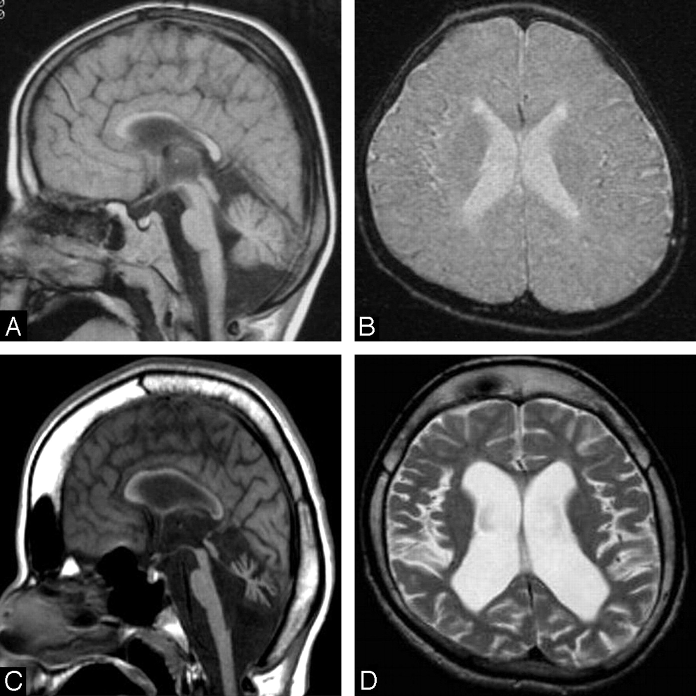
  • Fig 3.
    • Download figure
    • Open in new tab
    • Download powerpoint
    Fig 3.

    Patient 10 (CS I) at 4 (A and B) and 13 (C and D) years of age. A and C, Midline sagittal T1-weighted images demonstrate the progressive development of generalized cerebral and cerebellar atrophy, which is very severe at 13 years of age, with substantial corpus callosum and brain stem thinning. Note the thickened cranial vault and frontal and sphenoid sinus dilation at 13 years of age related to brain atrophy. B and D, Axial T2-weighted images show only a slight ventricular dilation at 4 years of age but considerable atrophy of the cortex as well as the white matter at 13 years of age, with increased ventricular dilation.
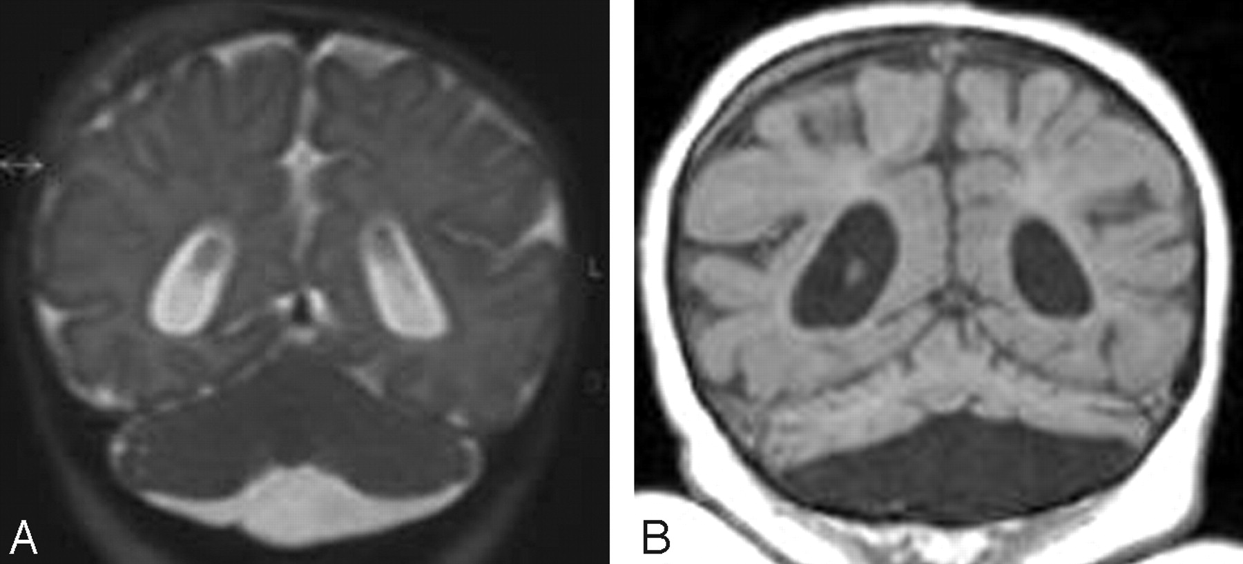
  • Fig 4.
    • Download figure
    • Open in new tab
    • Download powerpoint
    Fig 4.

    Patient 18 (COFS) at 3 months (A) and 2 years (B) of age. A, Coronal T2-weighted image shows cerebellar hypoplasia. B, Coronal reformatted T1-weighted image demonstrates on follow-up that the cerebellum is now extremely atrophic.
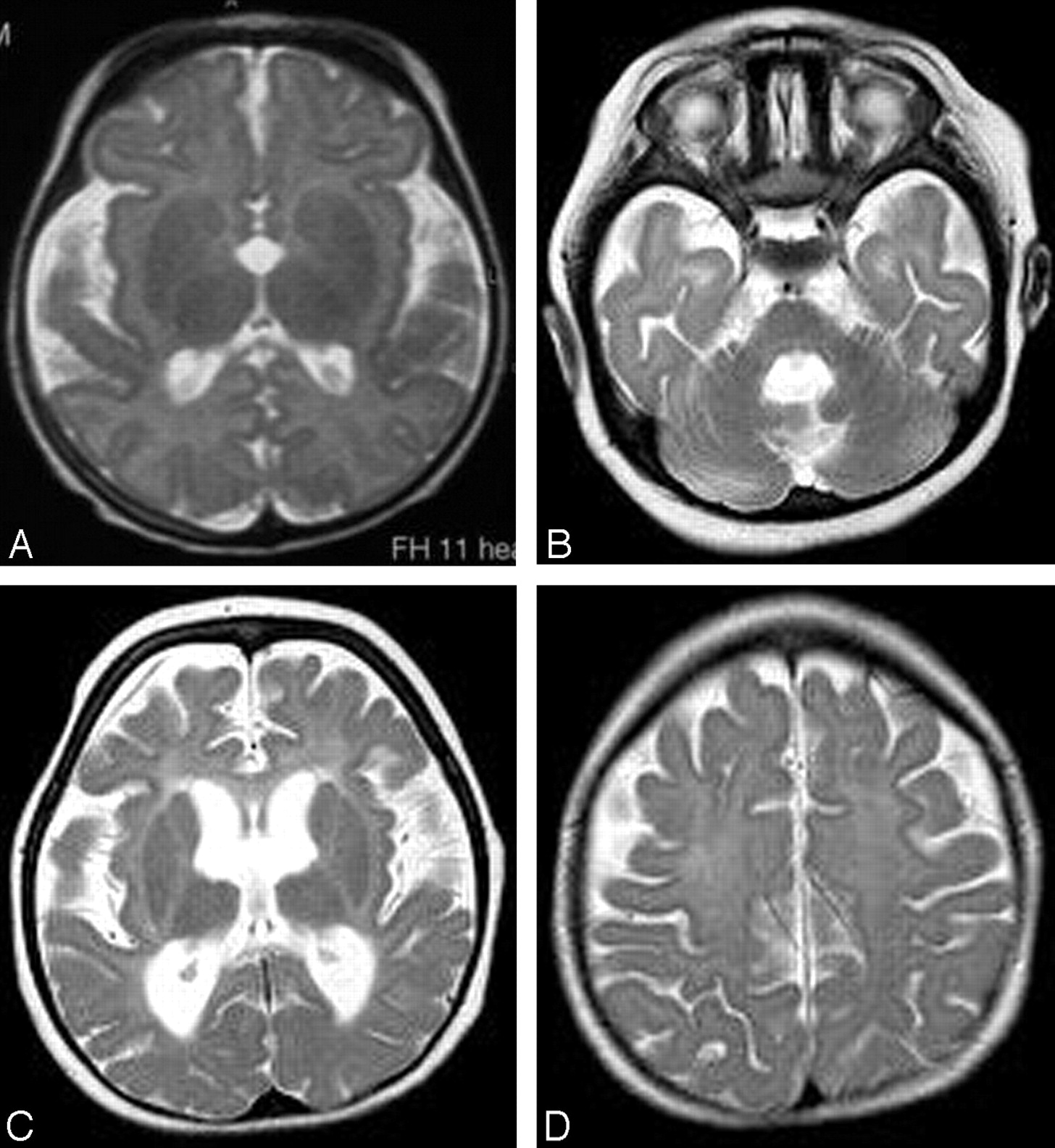
  • Fig 5.
    • Download figure
    • Open in new tab
    • Download powerpoint
    Fig 5.

    Axial T2-weighted images of patient 18 (COFS) at 3 months (A) and 2 years (B–D) of age. A, High signal intensity is present in the posterior limb of the internal capsule, consistent with an early lack of myelin. B–D, At 2 years of age, the myelination in the posterior fossa is normal (B), but no myelin is seen in the supratentorial white matter (C and D), which shows diffuse mild hyperintensity, including in the internal and external capsules (C). This aspect is consistent with hypomyelination. Note also the progressive cerebral atrophy.

  • Fig 6.
    • Download figure
    • Open in new tab
    • Download powerpoint
    Fig 6.

    Axial T2-weighted images of patient 1 (CS III) at 15 years of age. A, The cerebellum and the brain stem are fully myelinated. B, Myelination is also seen in the corpus callosum and the posterior limb of the internal capsule. The T2 hyperintensity lining of the medial part of the right internal capsule is probably the result of its progressive atrophy. C, The semioval center shows a punctuate appearance (arrow), with hypointense foci within the hyperintense white matter, reflecting preserved perivascular myelin. This corresponds to the tigroid leukodystrophy found in CS neuropathology.

PreviousNext
Back to top

In this issue

American Journal of Neuroradiology: 31 (9)
American Journal of Neuroradiology
Vol. 31, Issue 9
1 Oct 2010
  • Table of Contents
  • Index by author
Advertisement
Print
Download PDF
Email Article

Thank you for your interest in spreading the word on American Journal of Neuroradiology.

NOTE: We only request your email address so that the person you are recommending the page to knows that you wanted them to see it, and that it is not junk mail. We do not capture any email address.

Enter multiple addresses on separate lines or separate them with commas.
Neuroimaging In Cockayne Syndrome
(Your Name) has sent you a message from American Journal of Neuroradiology
(Your Name) thought you would like to see the American Journal of Neuroradiology web site.
CAPTCHA
This question is for testing whether or not you are a human visitor and to prevent automated spam submissions.
Cite this article
M. Koob, V. Laugel, M. Durand, H. Fothergill, C. Dalloz, F. Sauvanaud, H. Dollfus, I.J. Namer, J.-L. Dietemann
Neuroimaging In Cockayne Syndrome
American Journal of Neuroradiology Oct 2010, 31 (9) 1623-1630; DOI: 10.3174/ajnr.A2135

Citation Manager Formats

  • BibTeX
  • Bookends
  • EasyBib
  • EndNote (tagged)
  • EndNote 8 (xml)
  • Medlars
  • Mendeley
  • Papers
  • RefWorks Tagged
  • Ref Manager
  • RIS
  • Zotero
0 Responses
Respond to this article
Share
Bookmark this article
Neuroimaging In Cockayne Syndrome
M. Koob, V. Laugel, M. Durand, H. Fothergill, C. Dalloz, F. Sauvanaud, H. Dollfus, I.J. Namer, J.-L. Dietemann
American Journal of Neuroradiology Oct 2010, 31 (9) 1623-1630; DOI: 10.3174/ajnr.A2135
del.icio.us logo Twitter logo Facebook logo Mendeley logo
  • Tweet Widget
  • Facebook Like
  • Google Plus One
Purchase

Jump to section

  • Article
    • SUMMARY:
    • Abbreviations
    • Materials and Methods
    • Results
    • Discussion
    • Conclusions
    • Footnotes
    • References
  • Figures & Data
  • Supplemental
  • Info & Metrics
  • Responses
  • References
  • PDF

Related Articles

  • PubMed
  • Google Scholar

Cited By...

  • AAV gene therapy for Cockayne syndrome
  • Global huntingtin knockout in adult mice leads to fatal neurodegeneration that spares the pancreas
  • Global Huntingtin Knockout in Adult Mice Leads to Fatal Neurodegeneration that Spares the Pancreas
  • Ketones facilitate transcriptional resolution of secondary DNA structures in premature aging
  • Spectrum of Neuroradiologic Findings Associated with Monogenic Interferonopathies
  • Adult diagnosis of Cockayne syndrome
  • Neuroradiologic patterns and novel imaging findings in Aicardi-Goutieres syndrome
  • Teaching NeuroImages: Cockayne syndrome with extensive intracranial calcification
  • Dysregulation of gene expression as a cause of Cockayne syndrome neurological disease
  • Crossref
  • Google Scholar

This article has not yet been cited by articles in journals that are participating in Crossref Cited-by Linking.

More in this TOC Section

  • Frontal Paraventricular Cysts
  • Sodium MRI in Pediatric Brain Tumors
  • FRACTURE MR in Congenital Vertebral Anomalies
Show more Pediatric Neuroimaging

Similar Articles

Advertisement

Indexed Content

  • Current Issue
  • Accepted Manuscripts
  • Article Preview
  • Past Issues
  • Editorials
  • Editor's Choice
  • Fellows' Journal Club
  • Letters to the Editor
  • Video Articles

Cases

  • Case Collection
  • Archive - Case of the Week
  • Archive - Case of the Month
  • Archive - Classic Case

More from AJNR

  • Trainee Corner
  • Imaging Protocols
  • MRI Safety Corner
  • Book Reviews

Multimedia

  • AJNR Podcasts
  • AJNR Scantastics

Resources

  • Turnaround Time
  • Submit a Manuscript
  • Submit a Video Article
  • Submit an eLetter to the Editor/Response
  • Manuscript Submission Guidelines
  • Statistical Tips
  • Fast Publishing of Accepted Manuscripts
  • Graphical Abstract Preparation
  • Imaging Protocol Submission
  • Evidence-Based Medicine Level Guide
  • Publishing Checklists
  • Author Policies
  • Become a Reviewer/Academy of Reviewers
  • News and Updates

About Us

  • About AJNR
  • Editorial Board
  • Editorial Board Alumni
  • Alerts
  • Permissions
  • Not an AJNR Subscriber? Join Now
  • Advertise with Us
  • Librarian Resources
  • Feedback
  • Terms and Conditions
  • AJNR Editorial Board Alumni

American Society of Neuroradiology

  • Not an ASNR Member? Join Now

© 2025 by the American Society of Neuroradiology All rights, including for text and data mining, AI training, and similar technologies, are reserved.
Print ISSN: 0195-6108 Online ISSN: 1936-959X

Powered by HighWire