
Article Figures & Data
Figures
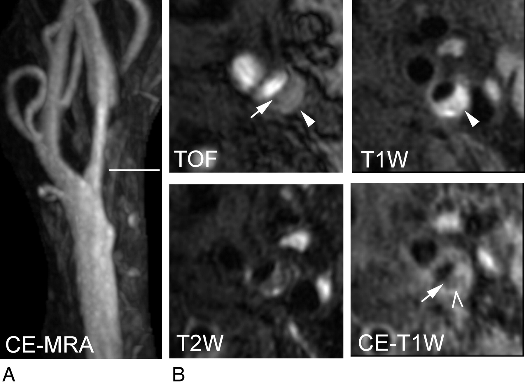
- Fig 1.
Coronally acquired MRA and transverse images of a complicated carotid plaque of the left carotid artery from a 75-year-old man with right-sided weakness. A, Maximum intensity projection of CE-MRA demonstrates a 55% smooth stenosis at the left internal carotid artery. The horizontal line indicates the level of the transverse carotid plaque images (shown in B). B, Disrupted dark band (arrow) on the TOF angiogram and discontinuation of the high-intensity band on CE-T1WI indicate a thin fibrous cap. High intensity on TOF and precontrast T1WI indicate regions of hemorrhage (arrowhead). The low-intensity area on the CE-T1WI indicates a lipid-rich necrotic core area occupying 31% of the wall area (chevron). Notice that the hemorrhage seen on TOF and T1WI almost completely fills the lipid-rich necrotic core as seen on the CE-T1WI. Symptomatic plaques tend to have a hemorrhagic lipid-rich necrotic core with a thin or ruptured fibrous cap.
- Fig 2.
Transverse carotid plaque images and coronally acquired CE-MRA of the left carotid artery from an asymptomatic 61-year-old woman. A, Maximum intensity projection of the CE-MRA demonstrates a 74% stenosis at the left internal carotid artery. The horizontal line indicates the level of the transverse carotid plaque images (shown in B). B, Transverse image of a TOF angiogram demonstrates a smooth luminal surface and a dark juxtaluminal band indicating an intact thick fibrous cap. The thick fibrous cap is easier to appreciate as a high-intensity band (arrow) on the CE-T1WI and the T2WI. An isointense area on TOF images and T1WI, an iso- to low-intensity area on the T2WI, and a low-intensity area on the CE-T1WI image indicate a lipid-rich necrotic core without hemorrhage occupying 29% of the wall area (arrowhead). Notice that the lipid-rich necrotic core is easiest to appreciate on the CE-T1WI. Asymptomatic plaques tend to have a smaller lipid-rich necrotic core without hemorrhage as well as a thick fibrous cap.
- Fig 3.
Strength of the association ROC between carotid plaque features and ipsilateral symptoms for 50 patients with mild/moderate carotid stenosis measured by CE-MRA.


Tables
Parameter T1WI With or Without Contrast Enhancement T2WI TOF Imaging CE-MRA Acquisition mode 2D 2D 3D 3D Acquisition sequence Fast spin-echo Fast spin-echo Spoiled gradient and flow compensation Enhanced gradient recalled-echo Blood suppression technique Quadruple inversion recovery Multisection double inversion recovery Saturation veins None TE (ms) 11 52 3.5 1.7 TR (ms) 800 4000 23 5.5 TI (ms) 520 250 n/a n/a Echo-train length 10 12 n/a n/a Excitation flip angle (degrees) 90 90 20 30 No. of signals acquired 1 2 1 FOVa 160 × 130 mm 160 × 120 mm 160 × 160 mm 150 mm Matrix size 256 × 256 256 × 256 288 × 256 256 × 256 No. of sections 18 18 48 40 Section thickness (mm) 2 2 1 0.8 Coverage (mm) 36 36 44 32 mm Imaging time (min:sec) 5:49 4:32 4:46 0:59 a The FOV of 140 × 140 cm was also used in some patients.
Variable Symptomatica (n = 13) Asymptomatica (n = 77) PValueb Age (yr) 68.7 ± 10.9 71.4 ± 8.3 .311 Male sex (%) 53.8 (7/13) 58.4 (45/77) .757 Hyperlipidemia (%) 69.2 (9/13) 70.1 (54/77) .948 Hypertension (%) 61.5 (8/13) 71.4 (55/77) .472 History of coronary artery disease (%) 30.8 (4/13) 41.6 (32/77) .463 History of peripheral vascular disease (%) 30.8 (4/13) 36.4 (28/77) .697 History of diabetes mellitus (%) 30.8 (4/13) 19.5 (15/77) .361 Current statin use (%) 76.9 (10/13) 81.8 (63/77) .677 Current smoker (%) 69.2 (9/13) 83.1 (64/77) .258 a Mean ± SD. Other values represent percentages; numbers in parentheses are used to calculate the percentages.
b P values by unpaired t test for age and by χ2 test or Fisher exact test for the other variables.
- Table 3:
Relationship between in vivo 3T carotid plaque components and recent ipsilateral thromboembolic symptoms for 50 patients with mild/moderate carotid stenosis measured by contrast-enhanced MR angiography
Variable Symptomatic (n = 6) Asymptomatic (n = 44) OR (95% CI) PValue AUC Plaque burden % wall volume 61.0 ± 5.8 56.1 ± 9.2 1.93 (0.70–5.31)a .204 0.689 % stenosis on MRA 50.8 ± 6.9 55.5 ± 9.7 0.61 (0.25–1.46)a .265 0.339 Ulceration on MRA 50% 36% 1.75 (0.32–9.72) .661 0.568 Prevalence plaque components Fibrous cap thin/ruptured 100% 39% n/a (2.13-infinity)b .006c 0.807 Lipid-rich necrotic core 100% 43% n/a (1.77-infinity)b .022c 0.784 Hemorrhage 67% 25% 6.00 (0.96–37.4) .055 0.708 Calcifications 67% 84% 0.38 (0.06–2.48) .311 0.413 % Volume of plaque components Lipid-rich necrotic core 18.5 ± 12.2 6.6 ± 11.3 1.90 (1.03–3.51)a .040 0.837 Hemorrhage 6.9 ± 6.6 1.9 ± 5.0 3.22 (0.97–10.71)a .056 0.750 Calcifications 2.9 ± 3.7 4.7 ± 5.9 0.44 (0.05–4.02)a .470 0.405 a OR and 95% CI for a 10% increase.
b Lower limit CI estimated by exact logistic regression analysis. “n/a” was used because OR cannot be calculated in the presence of zero cells in a 2 × 2 table.
c Fisher exact test.
- Table 4:
Relationship between in vivo 3T carotid plaque components and recent ipsilateral thromboembolic symptoms for 40 patients with severe carotid stenosis measured by contrast-enhanced MR angiography
Variable Symptomatic (n = 7) Asymptomatic (n = 33) OR (95% CI) PValue AUC Plaque burden % wall volume 63.7 ± 7.6 60.7 ± 8.8 1.52 (0.58–4.02)a .395 0.602 % stenosis on MRA 86.3 ± 6.5 81.7 ± 6.9 2.72 (0.77–9.65)a .121 0.669 Ulceration on MRA 86% 36% 10.50 (1.13–97.91) .039 0.747 Prevalence of plaque components Fibrous cap thin/ruptured 57% 52% 1.26 (0.24–6.50) .787 0.528 Lipid-rich necrotic core 57% 64% 0.76 (0.15–3.99) .748 0.481 Hemorrhage 43% 36% 1.31 (0.25–6.88) .748 0.532 Calcifications 86% 73% 2.25 (0.24–21.38) .480 0.565 Volume of plaque components Lipid-rich necrotic core 9.7 ± 11.8 10.6 ± 13.4 0.94 (0.49–1.82)a .863 0.481 Hemorrhage 2.8 ± 3.7 4.1 ± 7.4 0.73 (0.18–2.96)a 0.661 0.519 Calcifications 6.6 ± 5.5 5.0 ± 6.1 1.51 (0.43–5.31)a 0.525 0.608 a ORs for a 10% increase.
In this issue


Jump to section
Related Articles
Cited By...
- Carotid Intraplaque-Hemorrhage Volume and Its Association with Cerebrovascular Events
- Carotid Plaque CTA Analysis in Symptomatic Subjects with Bilateral Intraplaque Hemorrhage: A Preliminary Analysis
- Carotid Artery Wall Imaging: Perspective and Guidelines from the ASNR Vessel Wall Imaging Study Group and Expert Consensus Recommendations of the American Society of Neuroradiology
- Imaging Carotid Atherosclerosis Plaque Ulceration: Comparison of Advanced Imaging Modalities and Recent Developments
- Prediction of High-Risk Plaque Development and Plaque Progression With the Carotid Atherosclerosis Score
- Intracranial Stenting of Subacute Symptomatic Atherosclerotic Occlusion Versus Stenosis