Advertisement

Main menu

  • Home
  • Content
    • Current Issue
    • Accepted Manuscripts
    • Article Preview
    • Past Issue Archive
    • Video Articles
    • AJNR Case Collection
    • Case of the Week Archive
    • Case of the Month Archive
    • Classic Case Archive
  • Special Collections
    • AJNR Awards
    • Low-Field MRI
    • Alzheimer Disease
    • ASNR Foundation Special Collection
    • Photon-Counting CT
    • View All
  • Multimedia
    • AJNR Podcasts
    • AJNR SCANtastic
    • Trainee Corner
    • MRI Safety Corner
    • Imaging Protocols
  • For Authors
    • Submit a Manuscript
    • Submit a Video Article
    • Submit an eLetter to the Editor/Response
    • Manuscript Submission Guidelines
    • Statistical Tips
    • Fast Publishing of Accepted Manuscripts
    • Graphical Abstract Preparation
    • Imaging Protocol Submission
    • Author Policies
  • About Us
    • About AJNR
    • Editorial Board
    • Editorial Board Alumni
  • More
    • Become a Reviewer/Academy of Reviewers
    • Subscribers
    • Permissions
    • Alerts
    • Feedback
    • Advertisers
    • ASNR Home

User menu

  • Alerts
  • Log in

Search

  • Advanced search
American Journal of Neuroradiology

American Journal of Neuroradiology

ASHNR American Society of Functional Neuroradiology ASHNR American Society of Pediatric Neuroradiology ASSR
  • Alerts
  • Log in

Advanced Search

  • Home
  • Content
    • Current Issue
    • Accepted Manuscripts
    • Article Preview
    • Past Issue Archive
    • Video Articles
    • AJNR Case Collection
    • Case of the Week Archive
    • Case of the Month Archive
    • Classic Case Archive
  • Special Collections
    • AJNR Awards
    • Low-Field MRI
    • Alzheimer Disease
    • ASNR Foundation Special Collection
    • Photon-Counting CT
    • View All
  • Multimedia
    • AJNR Podcasts
    • AJNR SCANtastic
    • Trainee Corner
    • MRI Safety Corner
    • Imaging Protocols
  • For Authors
    • Submit a Manuscript
    • Submit a Video Article
    • Submit an eLetter to the Editor/Response
    • Manuscript Submission Guidelines
    • Statistical Tips
    • Fast Publishing of Accepted Manuscripts
    • Graphical Abstract Preparation
    • Imaging Protocol Submission
    • Author Policies
  • About Us
    • About AJNR
    • Editorial Board
    • Editorial Board Alumni
  • More
    • Become a Reviewer/Academy of Reviewers
    • Subscribers
    • Permissions
    • Alerts
    • Feedback
    • Advertisers
    • ASNR Home
  • Follow AJNR on Twitter
  • Visit AJNR on Facebook
  • Follow AJNR on Instagram
  • Join AJNR on LinkedIn
  • RSS Feeds

AJNR Awards, New Junior Editors, and more. Read the latest AJNR updates

Research ArticleBrain
Open Access

Accuracy and Reproducibility of Phase-Contrast MR Imaging Measurements for CSF Flow

A.L. Wentland, O. Wieben, F.R. Korosec and V.M. Haughton
American Journal of Neuroradiology August 2010, 31 (7) 1331-1336; DOI: https://doi.org/10.3174/ajnr.A2039
A.L. Wentland
  • Find this author on Google Scholar
  • Find this author on PubMed
  • Search for this author on this site
O. Wieben
  • Find this author on Google Scholar
  • Find this author on PubMed
  • Search for this author on this site
F.R. Korosec
  • Find this author on Google Scholar
  • Find this author on PubMed
  • Search for this author on this site
V.M. Haughton
  • Find this author on Google Scholar
  • Find this author on PubMed
  • Search for this author on this site
  • Article
  • Figures & Data
  • Info & Metrics
  • Responses
  • References
  • PDF
Loading

Article Figures & Data

Figures

  • Tables
  • Fig 1.
    • Download figure
    • Open in new tab
    • Download powerpoint
    Fig 1.

    Diagram of the setup used in phantom studies for evaluating CPC and RPC. Tubing was connected to a computer-controlled flow pump. The tubing was looped around a cubic MR imaging QA phantom and situated in the horizontal plane of the scanner along the z-axis.

  • Fig 2.
    • Download figure
    • Open in new tab
    • Download powerpoint
    Fig 2.

    Average measurements of constant velocities obtained with the RPC (A) and CPC (B) acquisitions compared with actual velocities. The solid line represents unity. Error bars are SDs.
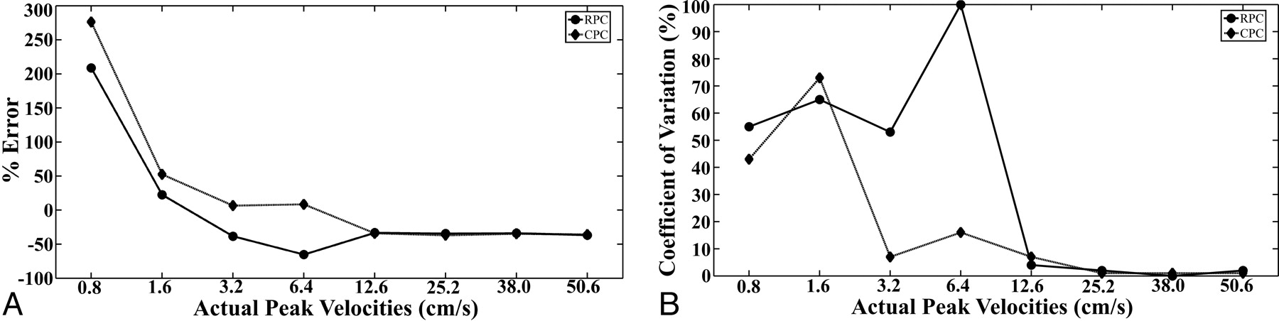
  • Fig 3.
    • Download figure
    • Open in new tab
    • Download powerpoint
    Fig 3.

    Error (A) and coefficient of variation (B) of peak velocity measurements in a constant flow phantom calculated from the RPC and CPC acquisitions. Actual peak velocities ranged from 0.8 to 50.6 cm/s.

  • Fig 4.
    • Download figure
    • Open in new tab
    • Download powerpoint
    Fig 4.

    Velocity (± SD) calculated from the CPC (A) and RPC (B) acquisition data plotted as a function of time for a sinusoidal flow rate of 1 mL/s at 60 beats per minute. The RPC data were acquired with 64 projections and 2 vps. The CPC data were acquired with 128 phase-encoding values and a ½ FOV. Three scans were obtained with each acquisition and error bars are shown. For each set of data, a sinusoidal curve of the form a + b × sin[2π(t − t0)] was fitted.

  • Fig 5.
    • Download figure
    • Open in new tab
    • Download powerpoint
    Fig 5.

    Phase images of CSF acquired at the level of the foramen magnum in 1 healthy volunteer with the CPC (TR/TE = 23.0/6.0 ms) (A) and RPC (TR/TE = 14.8/9.2 ms) (B) acquisitions. Arrows point to the area of the subarachnoid space at the level of the foramen magnum.

  • Fig 6.
    • Download figure
    • Open in new tab
    • Download powerpoint
    Fig 6.

    CSF flow over the R-R interval in the foramen magnum of 1 healthy volunteer as measured with the RPC and CPC acquisitions.

Tables

  • Figures
    • View popup
    Table 1:

    Average and peak velocities at the zenith and nadir of a sinusoidal waveform measured in a phantom with the RPC and CPC acquisitions

    RPCaCPCaAbsolute Difference
    Average velocity at zenith (cm/s)6.5 ± 0.16.4 ± 1.20.1
    Average velocity at nadir (cm/s)1.8 ± 0.72.0 ± 1.10.2
    Peak velocity at zenith (cm/s)11.5 ± 0.610.2 ± 0.91.3
    Peak velocity at nadir (cm/s)4.3 ± 0.45.0 ± 0.60.7
    Average0.6 ± 0.6
    • a Mean values ± SDs.

    • View popup
    Table 2:

    Peak systolic and diastolic CSF velocities in 5 human volunteers at the level of the foramen magnum defined in sagittal images by the tip of the clivus anteriorly and the occipital bone posteriorly

    RPCCPCAbsolute Difference
    Systolic peak velocity (cm/s)
    Volunteer 12.22.40.2
    Volunteer 24.34.00.3
    Volunteer 33.73.60.1
    Volunteer 42.12.20.1
    Volunteer 52.02.20.2
    Averagea2.9 ± 1.12.9 ± 0.90.2 ± 0.1
    Diastolic peak velocity (cm/s)
    Volunteer 11.01.10.1
    Volunteer 20.91.00.1
    Volunteer 31.61.40.2
    Volunteer 40.40.80.4
    Volunteer 50.80.80.0
    Averagea0.9 ± 0.41.0 ± 0.30.2 ± 0.2
    • a Mean values ± SDs.

PreviousNext
Back to top

In this issue

American Journal of Neuroradiology: 31 (7)
American Journal of Neuroradiology
Vol. 31, Issue 7
1 Aug 2010
  • Table of Contents
  • Index by author
Advertisement
Print
Download PDF
Email Article

Thank you for your interest in spreading the word on American Journal of Neuroradiology.

NOTE: We only request your email address so that the person you are recommending the page to knows that you wanted them to see it, and that it is not junk mail. We do not capture any email address.

Enter multiple addresses on separate lines or separate them with commas.
Accuracy and Reproducibility of Phase-Contrast MR Imaging Measurements for CSF Flow
(Your Name) has sent you a message from American Journal of Neuroradiology
(Your Name) thought you would like to see the American Journal of Neuroradiology web site.
CAPTCHA
This question is for testing whether or not you are a human visitor and to prevent automated spam submissions.
Cite this article
A.L. Wentland, O. Wieben, F.R. Korosec, V.M. Haughton
Accuracy and Reproducibility of Phase-Contrast MR Imaging Measurements for CSF Flow
American Journal of Neuroradiology Aug 2010, 31 (7) 1331-1336; DOI: 10.3174/ajnr.A2039

Citation Manager Formats

  • BibTeX
  • Bookends
  • EasyBib
  • EndNote (tagged)
  • EndNote 8 (xml)
  • Medlars
  • Mendeley
  • Papers
  • RefWorks Tagged
  • Ref Manager
  • RIS
  • Zotero
0 Responses
Respond to this article
Share
Bookmark this article
Accuracy and Reproducibility of Phase-Contrast MR Imaging Measurements for CSF Flow
A.L. Wentland, O. Wieben, F.R. Korosec, V.M. Haughton
American Journal of Neuroradiology Aug 2010, 31 (7) 1331-1336; DOI: 10.3174/ajnr.A2039
del.icio.us logo Twitter logo Facebook logo Mendeley logo
  • Tweet Widget
  • Facebook Like
  • Google Plus One
Purchase

Jump to section

  • Article
    • Abstract
    • Abbreviations
    • Materials and Methods
    • Results
    • Discussion
    • Conclusions
    • Acknowledgments
    • Footnotes
    • References
  • Figures & Data
  • Info & Metrics
  • Responses
  • References
  • PDF

Related Articles

  • No related articles found.
  • PubMed
  • Google Scholar

Cited By...

  • Human brain solute transport quantified by glymphatic MRI-informed biophysics during sleep and sleep deprivation
  • Patient-Specific 3D Simulation of Cyclic CSF Flow at the Craniocervical Region
  • CSF Flow through the Upper Cervical Spinal Canal in Chiari I Malformation
  • Crossref
  • Google Scholar

This article has not yet been cited by articles in journals that are participating in Crossref Cited-by Linking.

More in this TOC Section

  • White Matter Alterations in the Brains of Patients with Active, Remitted, and Cured Cushing Syndrome: A DTI Study
  • Qualitative and Quantitative Analysis of MR Imaging Findings in Patients with Middle Cerebral Artery Stroke Implanted with Mesenchymal Stem Cells
  • Fast Contrast-Enhanced 4D MRA and 4D Flow MRI Using Constrained Reconstruction (HYPRFlow): Potential Applications for Brain Arteriovenous Malformations
Show more Brain

Similar Articles

Advertisement

Indexed Content

  • Current Issue
  • Accepted Manuscripts
  • Article Preview
  • Past Issues
  • Editorials
  • Editor's Choice
  • Fellows' Journal Club
  • Letters to the Editor
  • Video Articles

Cases

  • Case Collection
  • Archive - Case of the Week
  • Archive - Case of the Month
  • Archive - Classic Case

More from AJNR

  • Trainee Corner
  • Imaging Protocols
  • MRI Safety Corner
  • Book Reviews

Multimedia

  • AJNR Podcasts
  • AJNR Scantastics

Resources

  • Turnaround Time
  • Submit a Manuscript
  • Submit a Video Article
  • Submit an eLetter to the Editor/Response
  • Manuscript Submission Guidelines
  • Statistical Tips
  • Fast Publishing of Accepted Manuscripts
  • Graphical Abstract Preparation
  • Imaging Protocol Submission
  • Evidence-Based Medicine Level Guide
  • Publishing Checklists
  • Author Policies
  • Become a Reviewer/Academy of Reviewers
  • News and Updates

About Us

  • About AJNR
  • Editorial Board
  • Editorial Board Alumni
  • Alerts
  • Permissions
  • Not an AJNR Subscriber? Join Now
  • Advertise with Us
  • Librarian Resources
  • Feedback
  • Terms and Conditions
  • AJNR Editorial Board Alumni

American Society of Neuroradiology

  • Not an ASNR Member? Join Now

© 2025 by the American Society of Neuroradiology All rights, including for text and data mining, AI training, and similar technologies, are reserved.
Print ISSN: 0195-6108 Online ISSN: 1936-959X

Powered by HighWire