Advertisement

Main menu

  • Home
  • Content
    • Current Issue
    • Accepted Manuscripts
    • Article Preview
    • Past Issue Archive
    • Video Articles
    • AJNR Case Collection
    • Case of the Week Archive
    • Case of the Month Archive
    • Classic Case Archive
  • Special Collections
    • AJNR Awards
    • Low-Field MRI
    • Alzheimer Disease
    • ASNR Foundation Special Collection
    • Photon-Counting CT
    • View All
  • Multimedia
    • AJNR Podcasts
    • AJNR SCANtastic
    • Trainee Corner
    • MRI Safety Corner
    • Imaging Protocols
  • For Authors
    • Submit a Manuscript
    • Submit a Video Article
    • Submit an eLetter to the Editor/Response
    • Manuscript Submission Guidelines
    • Statistical Tips
    • Fast Publishing of Accepted Manuscripts
    • Graphical Abstract Preparation
    • Imaging Protocol Submission
    • Author Policies
  • About Us
    • About AJNR
    • Editorial Board
    • Editorial Board Alumni
  • More
    • Become a Reviewer/Academy of Reviewers
    • Subscribers
    • Permissions
    • Alerts
    • Feedback
    • Advertisers
    • ASNR Home

User menu

  • Alerts
  • Log in

Search

  • Advanced search
American Journal of Neuroradiology

American Journal of Neuroradiology

ASHNR American Society of Functional Neuroradiology ASHNR American Society of Pediatric Neuroradiology ASSR
  • Alerts
  • Log in

Advanced Search

  • Home
  • Content
    • Current Issue
    • Accepted Manuscripts
    • Article Preview
    • Past Issue Archive
    • Video Articles
    • AJNR Case Collection
    • Case of the Week Archive
    • Case of the Month Archive
    • Classic Case Archive
  • Special Collections
    • AJNR Awards
    • Low-Field MRI
    • Alzheimer Disease
    • ASNR Foundation Special Collection
    • Photon-Counting CT
    • View All
  • Multimedia
    • AJNR Podcasts
    • AJNR SCANtastic
    • Trainee Corner
    • MRI Safety Corner
    • Imaging Protocols
  • For Authors
    • Submit a Manuscript
    • Submit a Video Article
    • Submit an eLetter to the Editor/Response
    • Manuscript Submission Guidelines
    • Statistical Tips
    • Fast Publishing of Accepted Manuscripts
    • Graphical Abstract Preparation
    • Imaging Protocol Submission
    • Author Policies
  • About Us
    • About AJNR
    • Editorial Board
    • Editorial Board Alumni
  • More
    • Become a Reviewer/Academy of Reviewers
    • Subscribers
    • Permissions
    • Alerts
    • Feedback
    • Advertisers
    • ASNR Home
  • Follow AJNR on Twitter
  • Visit AJNR on Facebook
  • Follow AJNR on Instagram
  • Join AJNR on LinkedIn
  • RSS Feeds

AJNR Awards, New Junior Editors, and more. Read the latest AJNR updates

Research ArticleHead and NeckF

Imaging Characteristics of Primary Laryngeal Lymphoma

N.A. Siddiqui, B.F. Branstetter, B.E. Hamilton, L.E. Ginsberg, C.M. Glastonbury, H.R. Harnsberger, E.L. Barnes and E.N. Myers
American Journal of Neuroradiology August 2010, 31 (7) 1261-1265; DOI: https://doi.org/10.3174/ajnr.A2085
N.A. Siddiqui
  • Find this author on Google Scholar
  • Find this author on PubMed
  • Search for this author on this site
B.F. Branstetter IV
  • Find this author on Google Scholar
  • Find this author on PubMed
  • Search for this author on this site
B.E. Hamilton
  • Find this author on Google Scholar
  • Find this author on PubMed
  • Search for this author on this site
L.E. Ginsberg
  • Find this author on Google Scholar
  • Find this author on PubMed
  • Search for this author on this site
C.M. Glastonbury
  • Find this author on Google Scholar
  • Find this author on PubMed
  • Search for this author on this site
H.R. Harnsberger
  • Find this author on Google Scholar
  • Find this author on PubMed
  • Search for this author on this site
E.L. Barnes
  • Find this author on Google Scholar
  • Find this author on PubMed
  • Search for this author on this site
E.N. Myers
  • Find this author on Google Scholar
  • Find this author on PubMed
  • Search for this author on this site
  • Article
  • Figures & Data
  • Info & Metrics
  • Responses
  • References
  • PDF
Loading

Article Figures & Data

Figures

  • Fig 1.
    • Download figure
    • Open in new tab
    • Download powerpoint
    Fig 1.

    A 65-year-old woman with laryngeal lymphoma. A, Coronal T1-weighted MR image demonstrates an isointense supraglottic soft-tissue nodule within the right false vocal cord (asterisk). B, Coronal T1-weighted contrast-enhanced MR image shows enhancement of the nodular lesion (asterisk) and the adjacent mucosa of the supraglottic larynx (arrow), which was also found to be positive for lymphoma.

  • Fig 2.
    • Download figure
    • Open in new tab
    • Download powerpoint
    Fig 2.

    A 65-year-old woman with laryngeal lymphoma. Axial CT scan demonstrates a uniformly enhancing mass involving the left false vocal cord (asterisk). Note the thin layer of fat separating this submucosal lesion from the mucosa (arrow).
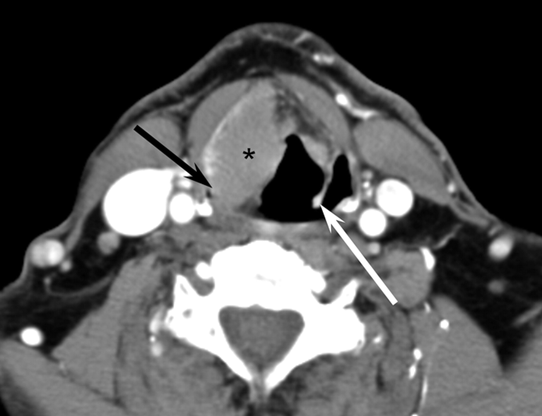
  • Fig 3.
    • Download figure
    • Open in new tab
    • Download powerpoint
    Fig 3.

    A 53-year-old man with laryngeal lymphoma. Axial CT scan demonstrates moderate uniform enhancement of a right false vocal cord mass (asterisk) with involvement of both the right aryepiglottic fold and the right pyriform sinus (black arrow). Note the normal configuration of the left aryepiglottic fold (white arrow), which is indistinguishable on the right.
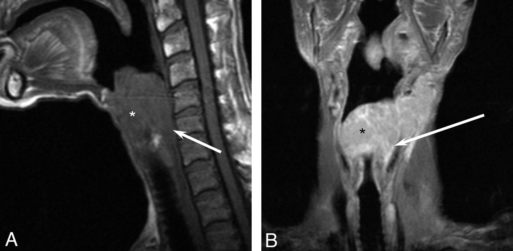
  • Fig 4.
    • Download figure
    • Open in new tab
    • Download powerpoint
    Fig 4.

    A 64-year-old man with laryngeal lymphoma. A, Sagittal T1-weighted MR image demonstrates a large isointense supraglottic laryngeal tumor (asterisk ) with posterior extension to the hypopharynx (arrow ). B, Coronal T1-weighted contrast-enhanced MR image shows homogeneous enhancement of the large supraglottic lesion (asterisk ) with posterolateral extension into the hypopharynx (arrow ).

  • Fig 5.
    • Download figure
    • Open in new tab
    • Download powerpoint
    Fig 5.

    A 65-year-old woman with laryngeal lymphoma. Axial PET-CT image demonstrates avid FDG uptake in a focal right false vocal cord lesion.

  • Fig 6.
    • Download figure
    • Open in new tab
    • Download powerpoint
    Fig 6.

    A 64-year-old man with laryngeal lymphoma (same patient as in Fig 4). A, Axial T1-weighted MR image demonstrates an isointense large supraglottic mass (asterisk ) extending into the hypopharynx (arrows ). B, Axial T2-weighted MR image shows the mass to be hyperintense (asterisk ). It is again seen extending into the hypopharynx (arrows ). C, Axial T1-weighted contrast-enhanced MR image demonstrates homogeneous enhancement (asterisk ) of the mass, which is again seen to be extending into the hypopharynx (arrows). Note the bulky bilateral lymphadenopathy.

PreviousNext
Back to top

In this issue

American Journal of Neuroradiology: 31 (7)
American Journal of Neuroradiology
Vol. 31, Issue 7
1 Aug 2010
  • Table of Contents
  • Index by author
Advertisement
Print
Download PDF
Email Article

Thank you for your interest in spreading the word on American Journal of Neuroradiology.

NOTE: We only request your email address so that the person you are recommending the page to knows that you wanted them to see it, and that it is not junk mail. We do not capture any email address.

Enter multiple addresses on separate lines or separate them with commas.
Imaging Characteristics of Primary Laryngeal Lymphoma
(Your Name) has sent you a message from American Journal of Neuroradiology
(Your Name) thought you would like to see the American Journal of Neuroradiology web site.
CAPTCHA
This question is for testing whether or not you are a human visitor and to prevent automated spam submissions.
Cite this article
N.A. Siddiqui, B.F. Branstetter, B.E. Hamilton, L.E. Ginsberg, C.M. Glastonbury, H.R. Harnsberger, E.L. Barnes, E.N. Myers
Imaging Characteristics of Primary Laryngeal Lymphoma
American Journal of Neuroradiology Aug 2010, 31 (7) 1261-1265; DOI: 10.3174/ajnr.A2085

Citation Manager Formats

  • BibTeX
  • Bookends
  • EasyBib
  • EndNote (tagged)
  • EndNote 8 (xml)
  • Medlars
  • Mendeley
  • Papers
  • RefWorks Tagged
  • Ref Manager
  • RIS
  • Zotero
0 Responses
Respond to this article
Share
Bookmark this article
Imaging Characteristics of Primary Laryngeal Lymphoma
N.A. Siddiqui, B.F. Branstetter, B.E. Hamilton, L.E. Ginsberg, C.M. Glastonbury, H.R. Harnsberger, E.L. Barnes, E.N. Myers
American Journal of Neuroradiology Aug 2010, 31 (7) 1261-1265; DOI: 10.3174/ajnr.A2085
del.icio.us logo Twitter logo Facebook logo Mendeley logo
  • Tweet Widget
  • Facebook Like
  • Google Plus One
Purchase

Jump to section

  • Article
    • Abstract
    • Abbreviations
    • Materials and Methods
    • Results
    • Discussion
    • Conclusions
    • Footnotes
    • References
  • Figures & Data
  • Info & Metrics
  • Responses
  • References
  • PDF

Related Articles

  • No related articles found.
  • PubMed
  • Google Scholar

Cited By...

  • Primary Composite Lymphoma of the Larynx, Composed of Diffuse Large B-Cell Lymphoma and Peripheral T-Cell Lymphoma, Not Otherwise Specified, Presenting as Left Subglottic Tracheal Fistula, Esophageal Diverticulum, and Neck Abscess
  • Crossref
  • Google Scholar

This article has not yet been cited by articles in journals that are participating in Crossref Cited-by Linking.

More in this TOC Section

  • Correlation of Apparent Diffusion Coefficient at 3T with Prognostic Parameters of Retinoblastoma
  • Parathyroid Lesions: Characterization with Dual-Phase Arterial and Venous Enhanced CT of the Neck
  • MR Diagnosis of Facial Neuritis: Diagnostic Performance of Contrast-Enhanced 3D-FLAIR Technique Compared with Contrast-Enhanced 3D-T1-Fast-Field Echo with Fat Suppression
Show more HEAD AND NECK

Similar Articles

Advertisement

Indexed Content

  • Current Issue
  • Accepted Manuscripts
  • Article Preview
  • Past Issues
  • Editorials
  • Editor's Choice
  • Fellows' Journal Club
  • Letters to the Editor
  • Video Articles

Cases

  • Case Collection
  • Archive - Case of the Week
  • Archive - Case of the Month
  • Archive - Classic Case

More from AJNR

  • Trainee Corner
  • Imaging Protocols
  • MRI Safety Corner
  • Book Reviews

Multimedia

  • AJNR Podcasts
  • AJNR Scantastics

Resources

  • Turnaround Time
  • Submit a Manuscript
  • Submit a Video Article
  • Submit an eLetter to the Editor/Response
  • Manuscript Submission Guidelines
  • Statistical Tips
  • Fast Publishing of Accepted Manuscripts
  • Graphical Abstract Preparation
  • Imaging Protocol Submission
  • Evidence-Based Medicine Level Guide
  • Publishing Checklists
  • Author Policies
  • Become a Reviewer/Academy of Reviewers
  • News and Updates

About Us

  • About AJNR
  • Editorial Board
  • Editorial Board Alumni
  • Alerts
  • Permissions
  • Not an AJNR Subscriber? Join Now
  • Advertise with Us
  • Librarian Resources
  • Feedback
  • Terms and Conditions
  • AJNR Editorial Board Alumni

American Society of Neuroradiology

  • Not an ASNR Member? Join Now

© 2025 by the American Society of Neuroradiology All rights, including for text and data mining, AI training, and similar technologies, are reserved.
Print ISSN: 0195-6108 Online ISSN: 1936-959X

Powered by HighWire