
Article Figures & Data
Figures
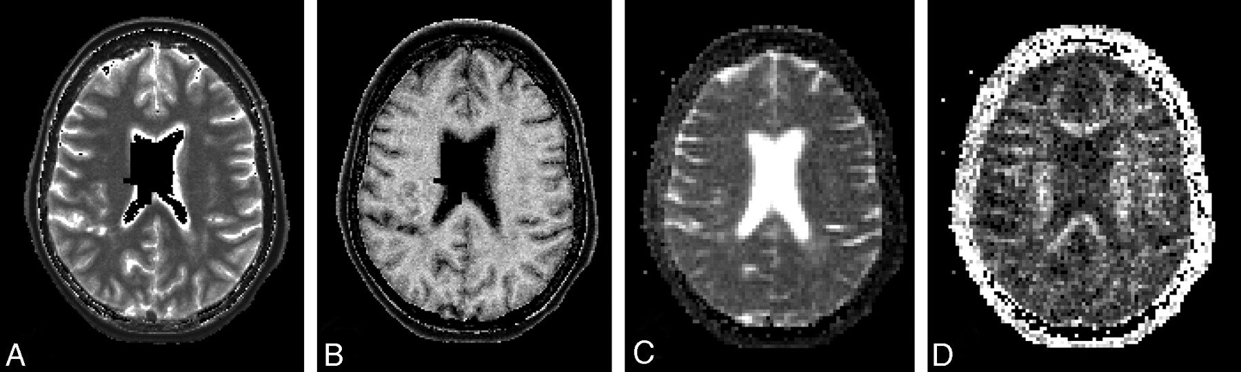
- Fig 1.
Sample of maps of quantitative MR imaging measures for 1 axial section in a female patient with PPMS (59.5 years of age; disease duration, 8.3 years; EDSS score, 4.0; total focal WM lesion volume on T2, 14.9 mL). Images show maps of the T1 (A), MTR (B), ADC (C), and FA (D). The empty (black) voxels located centrally in the maps of T1 and MTR are voxels excluded from analyses because of insufficient accuracy in determining the local effective B1 field strength in and close to the CSF. Details of methods are provided in the text.
- Fig 2.
Early-echo (Pd) images from a T2-weighted 2D dual-echo fast spin-echo sequence used to identify and outline focal WM lesions, DAWM, and NAWM. Images shown are from a male patient with SPMS (45.7 years of age; disease duration, 11.1 years; EDSS score, 4.0; total focal WM lesion volume on T2, 21.9 mL). The top row shows the images of 5 consecutive sections used for placing ROIs in this patient. The bottom row shows the same images but with the ROIs overlaid. The ROIs placed in each section are left frontal focal WM lesion (blue), left frontal DAWM (green) (A); right parieto-occipital DAWM (blue) (B); right frontal DAWM (red) (C); right frontal focal WM lesion (green), right parieto-occipital focal WM lesion (yellow), left parieto-occipital focal WM lesion (pink), and left parieto-occipital DAWM (yellow) (D); and right frontal NAWM (blue), left frontal NAWM (green), right parieto-occipital NAWM (pink), and left parieto-occipital NAWM (yellow) (E).
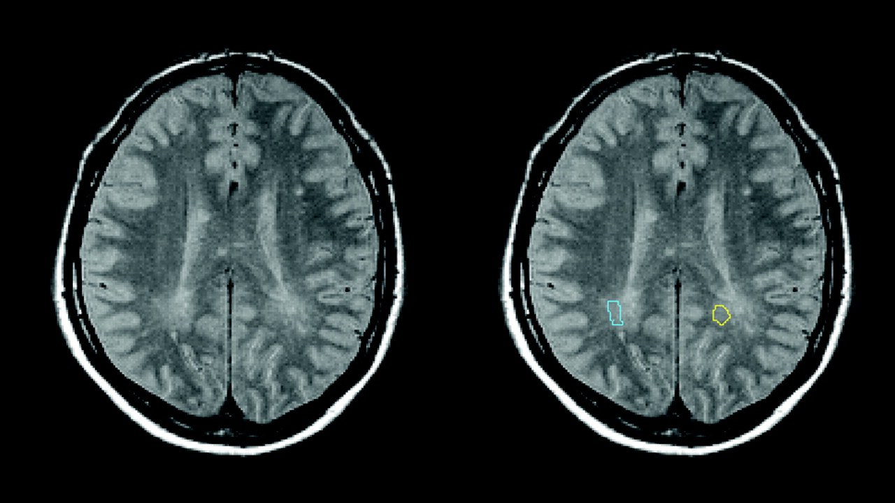
- Fig 3.
Further illustration of the radiologic definition of DAWM and placement of ROIs. Images shown are from a male patient with PPMS (52.3 years of age; disease duration, 2.3 years; EDSS score, 3.0; total focal WM lesion volume on T2, 6.9 mL). The left part of the figure shows the early-echo (Pd) image of 1 section from the T2-weighted 2D dual-echo fast spin-echo sequence. Images are in radiologic convention: The right side of the body is on the left side of the image. The right part of the figure shows the same image but with the parieto-occipital DAWM ROIs used in this study as a color overlay. The right parieto-occipital DAWM ROI is shown in light blue; the left parieto-occipital DAWM ROI is shown in yellow.
- Fig 4.
Boxplots showing median, range, and 25th and 75th percentiles for T1 (A), MTR (B), ADC (C), and FA (D). Each boxplot shows data split according to patient group (either PP or SPMS) as well as according to tissue type (NAWM, DAWM, or focal WM lesions). The plots clearly demonstrate that the values for the quantitative MR imaging measures observed in DAWM are intermediate to those observed in NAWM and focal WM lesions.


Tables
PPMS SPMS P Value for Comparison: PPMS vs SPMS No. of patients 7 10 Sex: M/F 3/4 3/7 .6 Age (yr) (mean ± SD) 59.2 ± 5.9 43.8 ± 10.3 .002 Disease duration (yr): Median (IQR) 8.3 (3.8–16.4) 15.9 (8.7–23.7) .3 EDSS score: median (IQR) 4.5 (3.0–4.5) 5.5 (4.0−6.75) .2 Supratentorial focal WM lesion volume (mL): median (IQR) 3.6 (1.4–13.8) 15.5 (3.9–23.6) .07 a P values for the comparison between patients with PPMS and SPMS are derived from the Mann-Whitney U test, except for sex, for which Pearson χ2 was used.
- Table 2:
Median number (volume) of analyzed voxels per patient in each tissue type for each quantitative MR imaging measurea
Tissue Type Technique ADC/FA MTR T1 Pd NAWM 117.5 (2.8 mL) 624 (2.5 mL) 660 (2.6 mL) 660 (2.6 mL) DAWM 53 (1.3 mL) 327 (1.3 mL) 283 (1.1 mL) 200 (0.8 mL) Focal WM lesions 19.5 (0.5 mL) 117 (0.5 mL) 123.5 (0.5 mL) 80.5 (0.3 mL) a Median values are given for the entire group of patients in this study. A more detailed subdivision by anatomic region and disease type is provided in Table 3. The size of each region of interest was defined as the total number of voxels included in the ROI, after warping it to the corresponding quantitative MR imaging maps as described in the text. The corresponding ROI volume was calculated by multiplying the number of voxels by the appropriate voxel volume, which was 4 mm3 for the T1 and MTR maps, and 24 mm3 for the ADC and FA maps. The column headed “Pd” gives the values for the original ROIs as drawn on the Pd-weighted images.
Disease Type, Region NAWM DAWM Lesions Median IQR Median IQR Median IQR PPMS Frontal 314.0 247.5–338.5 89.0 79.5–120.0 25.0 17.0–56.0 Parieto-occipital 318.0 305.0–379.5 128.0 111.0–156.0 32.0 26.0–52.0 Combined 593.0 528.0–713.0 212.0 190.5–265.5 57.0 17.0–80.0 SPMS Frontal 394.0 238.0–452.0 99.0 69.0–121.0 40.5 32.0–87.0 Parieto-occipital 390.0 319.0–501.0 94.5 76.0–114.0 56.0 36.0–61.0 Combined 703.0 629.0–921.0 193.0 141.0–238.0 95.0 71.0–144.0 a The “Combined” ROI for each patient is the combination of the frontal and parieto-occipital ROIs for that tissue type (NAWM, DAWM, or lesions).
NAWM DAWM Lesions Significant Pair-wise Comparisons between Tissue Types Significant Comparisons between Disease Types ADC (μm2 s−1) PP 805 ± 53 842 ± 48 1042 ± 216 Lesions vs NAWM, P < .05 – SP 798 ± 40 903 ± 77 1201 ± 131 DAWM vs NAWM, P < .001; DAWM vs lesions, P < .001; lesions vs NAWM, P < .001 FA PP 0.383 ± 0.058 0.350 ± 0.047 0.318 ± 0.110 DAWM vs NAWM P < .01; DAWM vs lesions, P < .05; lesions vs NAWM, P < .001 – SP 0.365 ± 0.033 0.322 ± 0.030 0.242 ± 0.054 DAWM vs NAWM, P < .001; DAWM vs lesions, P < .001; lesions vs NAWM, P < .001 MTR (%) PP 32.9 ± 0.6 31.1 ± 1.0 28.2 ± 3.6 DAWM vs NAWM, P < .01; lesions vs NAWM, P < .05 DAWM SP vs PP, P < .001; lesions SP vs PP, P < .01 SP 32.8 ± 1.2 28.8 ± 0.8 21.3 ± 3.3 DAWM vs NAWM, P < .001; DAWM vs lesions, P < .001; lesions vs NAWM, P < .001 T1 (ms) PP 759 ± 27 823 ± 38 1040 ± 185 DAWM vs NAWM, P < .05 DAWM SP vs PP, P < .001; lesions SP vs PP, P < .05 SP 815 ± 47 958 ± 67 1419 ± 298 DAWM vs NAWM, P < .001; DAWM vs lesions, P < .001; lesions vs NAWM, P < .001 a Means and SDs of ADC, FA, MTR, and T1 in NAWM, DAWM, and lesions by clinical group (PP or SPMS). Bonferroni-corrected P values derived from general linear mixed model analysis are indicated for statistically significant pair-wise differences between tissue types and disease types. Details of statistical analyses are provided in the text.


