
Article Figures & Data
Figures
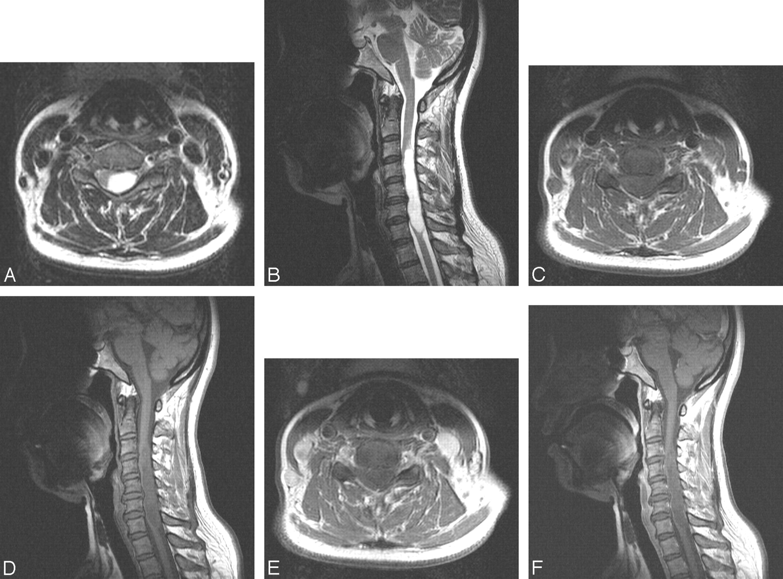
- Fig 1.
A 50-year-old woman with nonenhancing WHO grade II diffuse astrocytoma. Her symptoms were progressive left-handed weakness, hypoesthesia, and lower right-legged paresthesia starting 5 months earlier. A and B, Axial and sagittal T2-weighted MR images show a well-demarcated hyperintense intramedullary mass at the cervical spinal cord. The mass is slightly eccentric to the left side from the spinal cord center on the axial image. There is no peritumoral edema, periapical cap, or hemorrhage. C and D, The mass is hypointense on axial and sagittal T1-weighted images. E and F, Contrast enhanced T1-weighted images show that the mass is not enhanced at all.
- Fig 2.
A 47-year-old woman with a nonenhancing WHO grade III anaplastic astrocytoma. Her chief symptoms were a left upper extremity tingling sense and weakness, which had gradually extended from the distal to the proximal portion. Thereafter, the same symptoms occurred in both lower extremities. A and B, Axial and sagittal T2-weighted MR images show an ill-defined slightly hyperintense mass at cervical spinal cord. C, The mass is isointense to the spinal cord on sagittal T1-weighted images. D and E, Contrast-enhanced axial and sagittal T1-weighted images show a nonenhanced mass.
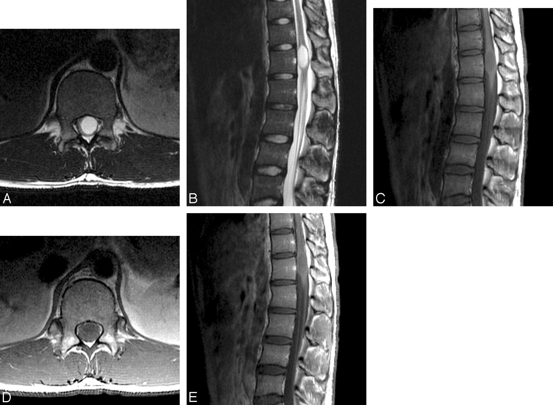
- Fig 3.
A 19-year-old man with a nonenhancing WHO grade III anaplastic astrocytoma. His chief symptoms were left lower extremity weakness, hypoesthesia, and lower back pain starting 6 months earlier. A and B, Axial and sagittal T2-weighted MR images show a well-demarcated hyperintense mass at the center of the conus medullaris. C, The mass is hypointense on the sagittal T1-weighted image. D and E, Contrast-enhanced axial and sagittal T1-weighted images show that the mass is not enhanced at all.


Tables
Enhancement Pattern WHO Grade Total (n = 19) (%) I (n = 3) (%) II (n = 13) (%) III (n = 3) (%) None 0 (0) 4 (31) 2 (67) 6 (32) Focal nodular 1 (33) 4 (31) 0 (0) 5 (26) Patchy 1 (33) 2 (15) 0 (0) 3 (16) Inhomogeneous diffuse 1 (33) 3 (23) 1 (33) 5 (26) Homogeneous diffuse 0 (0) 0 (0) 0 (0) 0 (0) T1 Signal Cases (%) Hypointense 11 (58) Isointense 7 (37) Hyperintense 1 (5) Axial Location Central 13 (68) Eccentric 6 (32) Other Findings Edema 7 (37) Intratumoral cyst 4 (21) Peritumoral cyst 3 (16) T2 Signal Cases (%) Hyperintense 18 (95) Isointense 1 (5) Longitudinal Location Cervical 10 (53) Thoracic 5 (26) Cervical/thoracic 2 (11) Thoracic/lumbar 1 (5) Cervical/thoracic/lumbar 1 (5) Other Findings Syrinx 0 (0) Hemorrhage 2 (11) Study Year Total No. No. Enhanced (%) Comment White et al.7 2007 35 10 (29) Includes 2 nonenhancing anaplastic astrocytomas Lin and Zhang13 2004 4 2 (50) Patel et al.11 1998 15 1 (7) Breger et al.12 1989 3 0 (0) Parizel et al.10 1989 7 0 (0) Includes 1 pilocytic and 6 low-grade astrocytomas Dillon et al.9 1989 6 0 (0) Includes 6 recurrent astrocytomas Sze et al.8 1988 6 1 (17) Includes 4 primary and 2 recurrent astrocytomas Total 76 14 (18)
In this issue


Jump to section
Related Articles
Cited By...
- No citing articles found.