Advertisement

Main menu

  • Home
  • Content
    • Current Issue
    • Accepted Manuscripts
    • Article Preview
    • Past Issue Archive
    • Video Articles
    • AJNR Case Collection
    • Case of the Week Archive
    • Case of the Month Archive
    • Classic Case Archive
  • Special Collections
    • AJNR Awards
    • Low-Field MRI
    • Alzheimer Disease
    • ASNR Foundation Special Collection
    • Photon-Counting CT
    • View All
  • Multimedia
    • AJNR Podcasts
    • AJNR SCANtastic
    • Trainee Corner
    • MRI Safety Corner
    • Imaging Protocols
  • For Authors
    • Submit a Manuscript
    • Submit a Video Article
    • Submit an eLetter to the Editor/Response
    • Manuscript Submission Guidelines
    • Statistical Tips
    • Fast Publishing of Accepted Manuscripts
    • Graphical Abstract Preparation
    • Imaging Protocol Submission
    • Author Policies
  • About Us
    • About AJNR
    • Editorial Board
    • Editorial Board Alumni
  • More
    • Become a Reviewer/Academy of Reviewers
    • Subscribers
    • Permissions
    • Alerts
    • Feedback
    • Advertisers
    • ASNR Home

User menu

  • Alerts
  • Log in

Search

  • Advanced search
American Journal of Neuroradiology

American Journal of Neuroradiology

ASHNR American Society of Functional Neuroradiology ASHNR American Society of Pediatric Neuroradiology ASSR
  • Alerts
  • Log in

Advanced Search

  • Home
  • Content
    • Current Issue
    • Accepted Manuscripts
    • Article Preview
    • Past Issue Archive
    • Video Articles
    • AJNR Case Collection
    • Case of the Week Archive
    • Case of the Month Archive
    • Classic Case Archive
  • Special Collections
    • AJNR Awards
    • Low-Field MRI
    • Alzheimer Disease
    • ASNR Foundation Special Collection
    • Photon-Counting CT
    • View All
  • Multimedia
    • AJNR Podcasts
    • AJNR SCANtastic
    • Trainee Corner
    • MRI Safety Corner
    • Imaging Protocols
  • For Authors
    • Submit a Manuscript
    • Submit a Video Article
    • Submit an eLetter to the Editor/Response
    • Manuscript Submission Guidelines
    • Statistical Tips
    • Fast Publishing of Accepted Manuscripts
    • Graphical Abstract Preparation
    • Imaging Protocol Submission
    • Author Policies
  • About Us
    • About AJNR
    • Editorial Board
    • Editorial Board Alumni
  • More
    • Become a Reviewer/Academy of Reviewers
    • Subscribers
    • Permissions
    • Alerts
    • Feedback
    • Advertisers
    • ASNR Home
  • Follow AJNR on Twitter
  • Visit AJNR on Facebook
  • Follow AJNR on Instagram
  • Join AJNR on LinkedIn
  • RSS Feeds

AJNR Awards, New Junior Editors, and more. Read the latest AJNR updates

Research ArticlePediatric Neuroimaging

Corroboration of Normal and Abnormal Fetal Cerebral Lamination on Postmortem MR Imaging with Postmortem Examination

E. Widjaja, S. Geibprasert, S. Zarei Mahmoodabadi, N.E. Brown and P. Shannon
American Journal of Neuroradiology November 2010, 31 (10) 1987-1993; DOI: https://doi.org/10.3174/ajnr.A2193
E. Widjaja
  • Find this author on Google Scholar
  • Find this author on PubMed
  • Search for this author on this site
S. Geibprasert
  • Find this author on Google Scholar
  • Find this author on PubMed
  • Search for this author on this site
S. Zarei Mahmoodabadi
  • Find this author on Google Scholar
  • Find this author on PubMed
  • Search for this author on this site
N.E. Brown
  • Find this author on Google Scholar
  • Find this author on PubMed
  • Search for this author on this site
P. Shannon
  • Find this author on Google Scholar
  • Find this author on PubMed
  • Search for this author on this site
  • Article
  • Figures & Data
  • Info & Metrics
  • Responses
  • References
  • PDF
Loading

Article Figures & Data

Figures

  • Tables
  • Fig 1.
    • Download figure
    • Open in new tab
    • Download powerpoint
    Fig 1.

    A 22-week fetus with normal cerebral lamination. A, On a T1-weighted image, the germinal matrix is of high T1 signal intensity, the intermediate zone is of slightly lower T1 signal intensity relative to the subplate layer, and the cortex is of high T1 signal intensity. B, On a T2-weighted image, the germinal matrix is of low T2 signal intensity, the intermediate zone is of lower T2 signal intensity relative to the subplate layer, and the cortex is of lower T2 signal intensity compared with subplate layer.

  • Fig 2.
    • Download figure
    • Open in new tab
    • Download powerpoint
    Fig 2.

    A 22-week fetus with neuroaxonal dystrophy. A, Coronal T1-weighted image shows disruption of normal cerebral lamination, with loss of distinction between the cortex and subplate layer (asterisk), and the high T1 signal intensity of the germinal matrix is not visualized. B, Coronal T2-weighted image shows the presence of normal distinct cerebral lamination. The abnormal cerebral lamination is detected on the T1-weighted image (A) but not on the T2-weighted image (B). The sulcation and gyration of the cerebral hemispheres are abnormal. C and D, Gross pathology section of the cerebrum (C) and whole mount histology (D) stain (hematoxylin-eosin, 1× magnification) show the presence of attenuated lamination in the cerebral mantle. The temporal horns are dilated with a thinned temporal cortex (black arrow).
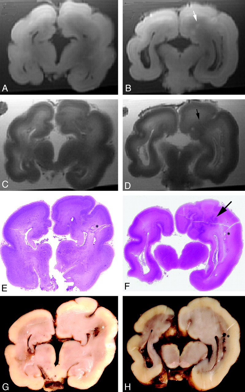
  • Fig 3.
    • Download figure
    • Open in new tab
    • Download powerpoint
    Fig 3.

    A 29-week fetus with neuronal migration disorder. A −D, Coronal T1- (A and B) and T2-weighted (C and D) images show disruption of the normal cerebral lamination, and the cortical layer appears thickened. There is gray matter heterotopia within the left cerebral mantle (small arrow). The sulcation and gyration are abnormal, and there is also agenesis of the corpus callosum. E−H, Whole mount (E and F) stain (hematoxylin-eosin, 1× magnification) and gross pathology (G and H) sections of the brain illustrate agenesis of the corpus callosum and caudate nuclei as well as focal infarcts as indicated by asterisks. There are absence of the normal cerebral lamination and a focal area of deranged cortical architecture within the cerebral mantle (black arrow).

  • Fig 4.
    • Download figure
    • Open in new tab
    • Download powerpoint
    Fig 4.

    A 22-week fetus with neuronal migration disorder. Coronal T1- (A) and T2-weighted (B) images demonstrate an attenuated cortex, subplate layer, and intermediate zone on the medial aspect of the left frontal lobe, associated with focal deepening of the sulcus at the same site (arrowhead). C, Gross pathology section of the cerebrum demonstrates a deep sulcus on the medial aspect of the left frontal lobe. D, Histology stain (hematoxylin-eosin, 50× magnification) shows dysplastic hypocellular cortex noted at the site of an abnormally deep sulcus and the overlying arachnoid contains a cyst lined by a single layer of choroid plexus−type epithelium (arrow). There is also agenesis of the corpus callosum.

  • Fig 5.
    • Download figure
    • Open in new tab
    • Download powerpoint
    Fig 5.

    A, 25-week fetus with hypoxic-ischemic injury. Coronal T1- (A) and T2-weighted (B) images demonstrate patchy areas of increased T1 and reduced T2 signal intensity (small arrows) within the subplate layer and intermediate zone. The sulcation and gyration pattern is normal. C, Histology stain (hematoxylin-eosin) demonstrates normal cerebral lamination that is age-appropriate. D, Microscopy (630× magnification) demonstrates karyorrhectic neurons (large arrow) in brain stem nuclei, likely due to hypoxic-ischemic injury.

Tables

  • Figures
    • View popup
    Table 1:

    Agreement between T1- and T2-weighted postmortem MRI assessment of germinal matrix, intermediate zone, subplate layer, cortex, and overall cerebral lamination and histology

    T1-Weighted ImagesT2-Weighted Images
    AbnormalNormalAbnormalNormal
    Germinal matrix20/2529/3019/2530/30
    Intermediate zone23/2626/2918/2628/29
    Subplate layer22/2627/2918/2628/29
    Cortex21/2629/2915/2629/29
    Overall cerebral lamination25/2626/2919/2628/29
    • View popup
    Table 2:

    Sensitivity, specificity, and positive and negative predictive values of the germinal matrix, intermediate zone, subplate layer, and cortex on T1-weighted postmortem MRI

    SensitivitySpecificityPPVNPV
    T1 germinal matrix80.00%96.67%95.24%85.29%
    (CI, 58.70%–92.39%)(CI, 80.95%–99.83%)(CI, 74.13%–99.75%)(CI, 68.17%–94.46%)
    T1 intermediate zone88.46%89.66%88.00%89.66%
    (CI, 68.72%–96.97%)(CI, 71.50%–97.29%)(CI, 68.72%–96.97%)(CI, 71.50%–97.29%)
    T1 subplate layer84.62%93.10%91.67%87.10%
    (CI, 64.27%–94.95%)(CI, 75.79%–98.80%)(CI, 71.53%–98.54%)(CI, 69.24%–95.78%)
    T1 cortex80.77%100%100%85.29%
    (CI, 60.02%–92.69%)(CI, 85.44%–100%)(CI, 80.76%–100%)(CI, 68.17%–94.46%)
    T1 overall96.15%89.66%89.29%96.29%
    (CI, 78.42%–99.80%)(CI, 71.50%–97.29%)(CI, 70.63%–97.19%)(CI, 79.11%–99.80%)
    • View popup
    Table 3:

    Sensitivity, specificity, and positive and negative predictive values of the germinal matrix, intermediate zone, subplate layer, and cortex on T2-weighted postmortem MRI

    SensitivitySpecificityPPVNPV
    T2 germinal matrix76.00%100%100%83.33%
    (CI, 54.48%–89.84%)(CI, 85.87%–100%)(CI, 79.08%–100%)(CI, 66.53%–93.04%)
    T2 intermediate zone69.23%96.55%94.74%77.78%
    (CI, 48.10%–4.91%)(CI, 80.37%–99.82%)(CI, 71.89%–99.72%)(CI, 60.41%–89.27%)
    T2 subplate layer69.23%96.55%94.74%77.78%
    (CI, 48.10%–84.91%)(CI, 80.37%–88.82%)(CI, 71.89%–99.72%)(CI, 60.41%–89.27%)
    T2 cortex57.69%100%100%72.50%
    (CI, 37.19%–76.03%)(CI, 85.44%–100%)(CI, 74.65%–100%)(CI, 55.86%–84.86%)
    T2 overall73.08%96.55%95.00%80.00%
    (CI, 51.95%–87.65%)(CI, 80.37%–99.82%)(CI, 73.06%–99.74%)(CI, 62.54%–90.94%)
PreviousNext
Back to top

In this issue

American Journal of Neuroradiology: 31 (10)
American Journal of Neuroradiology
Vol. 31, Issue 10
1 Nov 2010
  • Table of Contents
  • Index by author
Advertisement
Print
Download PDF
Email Article

Thank you for your interest in spreading the word on American Journal of Neuroradiology.

NOTE: We only request your email address so that the person you are recommending the page to knows that you wanted them to see it, and that it is not junk mail. We do not capture any email address.

Enter multiple addresses on separate lines or separate them with commas.
Corroboration of Normal and Abnormal Fetal Cerebral Lamination on Postmortem MR Imaging with Postmortem Examination
(Your Name) has sent you a message from American Journal of Neuroradiology
(Your Name) thought you would like to see the American Journal of Neuroradiology web site.
CAPTCHA
This question is for testing whether or not you are a human visitor and to prevent automated spam submissions.
Cite this article
E. Widjaja, S. Geibprasert, S. Zarei Mahmoodabadi, N.E. Brown, P. Shannon
Corroboration of Normal and Abnormal Fetal Cerebral Lamination on Postmortem MR Imaging with Postmortem Examination
American Journal of Neuroradiology Nov 2010, 31 (10) 1987-1993; DOI: 10.3174/ajnr.A2193

Citation Manager Formats

  • BibTeX
  • Bookends
  • EasyBib
  • EndNote (tagged)
  • EndNote 8 (xml)
  • Medlars
  • Mendeley
  • Papers
  • RefWorks Tagged
  • Ref Manager
  • RIS
  • Zotero
0 Responses
Respond to this article
Share
Bookmark this article
Corroboration of Normal and Abnormal Fetal Cerebral Lamination on Postmortem MR Imaging with Postmortem Examination
E. Widjaja, S. Geibprasert, S. Zarei Mahmoodabadi, N.E. Brown, P. Shannon
American Journal of Neuroradiology Nov 2010, 31 (10) 1987-1993; DOI: 10.3174/ajnr.A2193
del.icio.us logo Twitter logo Facebook logo Mendeley logo
  • Tweet Widget
  • Facebook Like
  • Google Plus One
Purchase

Jump to section

  • Article
    • Abstract
    • Abbreviations
    • Materials and Methods
    • Results
    • Discussion
    • Conclusions
    • Footnotes
    • References
  • Figures & Data
  • Info & Metrics
  • Responses
  • References
  • PDF

Related Articles

  • No related articles found.
  • PubMed
  • Google Scholar

Cited By...

  • No citing articles found.
  • Crossref
  • Google Scholar

This article has not yet been cited by articles in journals that are participating in Crossref Cited-by Linking.

More in this TOC Section

  • Frontal Paraventricular Cysts
  • Sodium MRI in Pediatric Brain Tumors
  • FRACTURE MR in Congenital Vertebral Anomalies
Show more Pediatric Neuroimaging

Similar Articles

Advertisement

Indexed Content

  • Current Issue
  • Accepted Manuscripts
  • Article Preview
  • Past Issues
  • Editorials
  • Editor's Choice
  • Fellows' Journal Club
  • Letters to the Editor
  • Video Articles

Cases

  • Case Collection
  • Archive - Case of the Week
  • Archive - Case of the Month
  • Archive - Classic Case

More from AJNR

  • Trainee Corner
  • Imaging Protocols
  • MRI Safety Corner
  • Book Reviews

Multimedia

  • AJNR Podcasts
  • AJNR Scantastics

Resources

  • Turnaround Time
  • Submit a Manuscript
  • Submit a Video Article
  • Submit an eLetter to the Editor/Response
  • Manuscript Submission Guidelines
  • Statistical Tips
  • Fast Publishing of Accepted Manuscripts
  • Graphical Abstract Preparation
  • Imaging Protocol Submission
  • Evidence-Based Medicine Level Guide
  • Publishing Checklists
  • Author Policies
  • Become a Reviewer/Academy of Reviewers
  • News and Updates

About Us

  • About AJNR
  • Editorial Board
  • Editorial Board Alumni
  • Alerts
  • Permissions
  • Not an AJNR Subscriber? Join Now
  • Advertise with Us
  • Librarian Resources
  • Feedback
  • Terms and Conditions
  • AJNR Editorial Board Alumni

American Society of Neuroradiology

  • Not an ASNR Member? Join Now

© 2025 by the American Society of Neuroradiology All rights, including for text and data mining, AI training, and similar technologies, are reserved.
Print ISSN: 0195-6108 Online ISSN: 1936-959X

Powered by HighWire