
Article Figures & Data
Figures
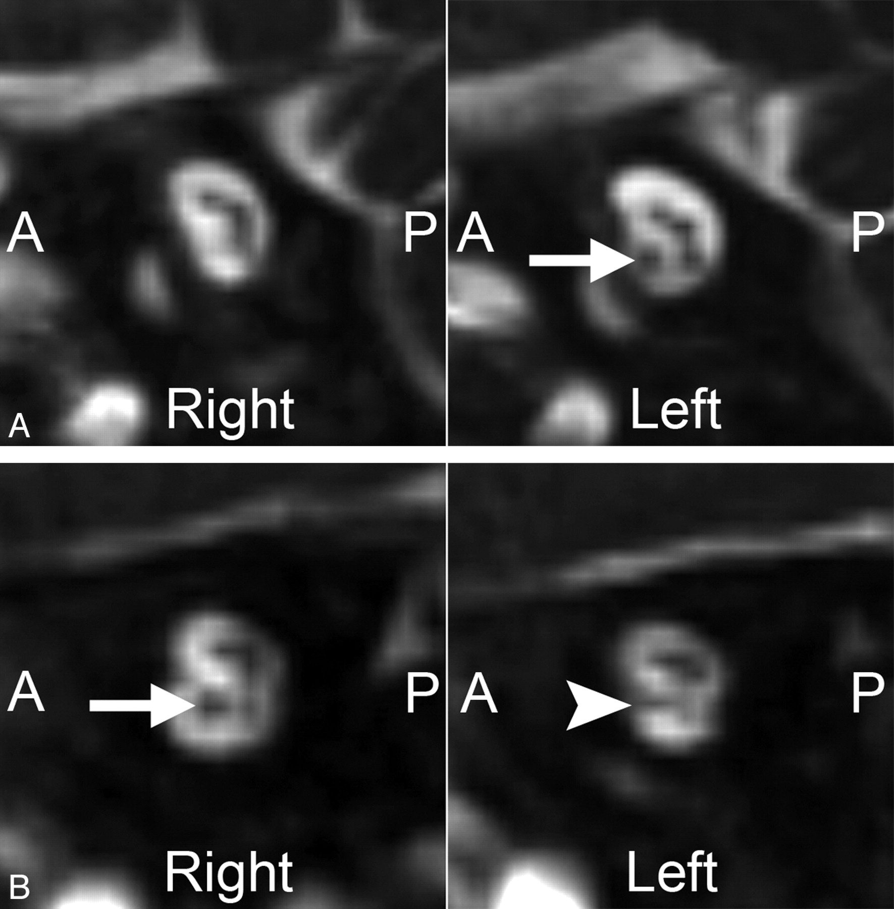
- Fig 1.
Two cases of unilateral CND. Magnified oblique sagittal CISS images through the IACs in 2 different patients with ANSD. A, An 8-month-old boy with complete absence of the right cochlear nerve. Compare these findings with the normal-appearing cochlear nerve on the left (arrow). B, A 10-month-old girl with a hypoplastic left cochlear nerve. The left cochlear nerve (arrowhead) is present but is noticeably smaller than the normal-appearing right cochlear nerve (arrow).
- Fig 2.
Inner ear and brain abnormalities in a 2-year-old girl with ANSD and bilateral CND. A, Axial CISS images through the bilateral inner ears and IACs. The left IAC is stenotic, particularly at the level of the porus acousticus (arrow), while the right IAC is normal in caliber. Both cochleae are isolated and dysplastic, with truncated apical turns, and the right modiolus is deficient. B, Midsagittal T1-weighted image demonstrates callosal agenesis, pontine hypoplasia, and inferior vermian hypoplasia. There is a prominent ventral cleft at the level of the pontomedullary junction. C, Axial T2-weighted image through the lateral ventricles demonstrates ventriculomegaly and diffuse pachygyria with relatively few sulci.
- Fig 3.
Inner ear and brain abnormalities in patients with bilateral CND and pontine segmental cap dysplasia. A, Axial CISS images through the inner ears in a 1-year-old girl. Both cochleae demonstrate deficient modioli and incomplete partitioning. The BCNCs are also small and possibly atretic. B, Sagittal midline T1-weighted image in a 2-year-old boy demonstrates characteristic pontine hypoplasia and an abnormal exophytic masslike band along the dorsal superior surface of the pons (white arrow). The vermis is also hypoplastic inferiorly. C, Axial T2-weighted image at the level of the MCPs in the same patient as A. The pons and MCPs are small, and there is a transversely oriented band along the dorsal pons, separated from the remainder of the pons by a hypointense horizontal cleft (black arrowheads).
- Fig 4.
Vestibular and SCC abnormalities in a patient with bilateral CND. Axial CISS images in a 1-year-old girl with CHARGE syndrome. No semicircular canals are present, and both vestibules are enlarged and dysplastic. The right BCNC appears narrowed and the left BCNC is atretic.


Tables
Type of Brain Abnormality Forebrain Developmental Mid- or Hindbrain Developmental CSF-Related WM Callosal dysgenesis Pontine hypoplasia Ventriculomegaly Patchy foci of abnormal WM signal (nonspecific) Cortical malformations (pachygyria) Dandy-Walker malformation Enlarged extra-axial CSF spaces Delayed myelination Gray matter heterotopias Cerebellar hypoplasia Arachnoid cysts PVL Porencephalic cysts Vermian hypoplasia Septo-optic dysplasia Pontine tegmental cap dysplasia Type of Temporal Bone Abnormality Cochlear Nerve Status Normal (n = 69) Unilateral CND (n = 19) Bilateral CND (n = 15) None 68 (98.6%)1,2 2 (10.5%)1 1 (6.7%)2 Any labyrinthine abnormality 1 (1.4%)3 3 (15.8%)4 11 (73.3%)3,4 Cochlear malformations 1 (1.4%)5 2 (10.5%)6 8 (53.3%)5,6 Vestibular malformations 0 (0.0%)7 2 (10.5%) 5 (33.3%)7 SCC malformations 0 (0.0%)8 2 (10.5%) 4 (26.7%)8 Large endolymphatic duct/sac 1 (1.4%) 0 (0.0%) 0 (0.0%) IAC stenosis/atresia 0 (0.0%)9,10 5 (26.3%)9 7 (46.7%)10 BCNC stenosis/atresia 0 (0.0%)11,12 15 (78.9%)11 13 (86.7%)12 -
a Superscripts denote significant group pair differences: 1–5, 7–12, P ≤ .001; 6, P =.010.
-
Type of Intracranial Abnormality Cochlear Nerve Status Normal (n = 69) Unilateral CND (n = 19) Bilateral CND (n = 15) None 44 (63.8%) 16 (84.2%) 6 (40.0%) Any intracranial abnormality 25 (36.2%) 3 (15.8%)1 9 (60.0%)1 Forebrain malformations 3 (4.3%) 0 (0.0%) 4 (26.7%) Mid-/hindbrain malformations 7 (10.1%)2 0 (0.0%)3 6 (40.0%)2,3 CSF abnormalities 12 (17.4%) 3 (11.7%) 4 (26.7%) Abnormalities of WM 16 (23.2%) 0 (0.0%) 2 (13.3%) -
a Superscripts denote significant group pair differences: 1, P = .012; 2, P = .010; 3, P = .004.
-


