Advertisement

Main menu

  • Home
  • Content
    • Current Issue
    • Accepted Manuscripts
    • Article Preview
    • Past Issue Archive
    • Video Articles
    • AJNR Case Collection
    • Case of the Week Archive
    • Case of the Month Archive
    • Classic Case Archive
  • Special Collections
    • AJNR Awards
    • Low-Field MRI
    • Alzheimer Disease
    • ASNR Foundation Special Collection
    • Photon-Counting CT
    • View All
  • Multimedia
    • AJNR Podcasts
    • AJNR SCANtastic
    • Trainee Corner
    • MRI Safety Corner
    • Imaging Protocols
  • For Authors
    • Submit a Manuscript
    • Submit a Video Article
    • Submit an eLetter to the Editor/Response
    • Manuscript Submission Guidelines
    • Statistical Tips
    • Fast Publishing of Accepted Manuscripts
    • Graphical Abstract Preparation
    • Imaging Protocol Submission
    • Author Policies
  • About Us
    • About AJNR
    • Editorial Board
    • Editorial Board Alumni
  • More
    • Become a Reviewer/Academy of Reviewers
    • Subscribers
    • Permissions
    • Alerts
    • Feedback
    • Advertisers
    • ASNR Home

User menu

  • Alerts
  • Log in

Search

  • Advanced search
American Journal of Neuroradiology

American Journal of Neuroradiology

ASHNR American Society of Functional Neuroradiology ASHNR American Society of Pediatric Neuroradiology ASSR
  • Alerts
  • Log in

Advanced Search

  • Home
  • Content
    • Current Issue
    • Accepted Manuscripts
    • Article Preview
    • Past Issue Archive
    • Video Articles
    • AJNR Case Collection
    • Case of the Week Archive
    • Case of the Month Archive
    • Classic Case Archive
  • Special Collections
    • AJNR Awards
    • Low-Field MRI
    • Alzheimer Disease
    • ASNR Foundation Special Collection
    • Photon-Counting CT
    • View All
  • Multimedia
    • AJNR Podcasts
    • AJNR SCANtastic
    • Trainee Corner
    • MRI Safety Corner
    • Imaging Protocols
  • For Authors
    • Submit a Manuscript
    • Submit a Video Article
    • Submit an eLetter to the Editor/Response
    • Manuscript Submission Guidelines
    • Statistical Tips
    • Fast Publishing of Accepted Manuscripts
    • Graphical Abstract Preparation
    • Imaging Protocol Submission
    • Author Policies
  • About Us
    • About AJNR
    • Editorial Board
    • Editorial Board Alumni
  • More
    • Become a Reviewer/Academy of Reviewers
    • Subscribers
    • Permissions
    • Alerts
    • Feedback
    • Advertisers
    • ASNR Home
  • Follow AJNR on Twitter
  • Visit AJNR on Facebook
  • Follow AJNR on Instagram
  • Join AJNR on LinkedIn
  • RSS Feeds

AJNR Awards, New Junior Editors, and more. Read the latest AJNR updates

Research ArticleBrain
Open Access

Cranial Dural Arteriovenous Fistula: Diagnosis and Classification with Time-Resolved MR Angiography at 3T

R.I. Farb, R. Agid, R.A. Willinsky, D.M. Johnstone and K.G. terBrugge
American Journal of Neuroradiology September 2009, 30 (8) 1546-1551; DOI: https://doi.org/10.3174/ajnr.A1646
R.I. Farb
  • Find this author on Google Scholar
  • Find this author on PubMed
  • Search for this author on this site
R. Agid
  • Find this author on Google Scholar
  • Find this author on PubMed
  • Search for this author on this site
R.A. Willinsky
  • Find this author on Google Scholar
  • Find this author on PubMed
  • Search for this author on this site
D.M. Johnstone
  • Find this author on Google Scholar
  • Find this author on PubMed
  • Search for this author on this site
K.G. terBrugge
  • Find this author on Google Scholar
  • Find this author on PubMed
  • Search for this author on this site
  • Article
  • Figures & Data
  • Info & Metrics
  • Responses
  • References
  • PDF
Loading

Article Figures & Data

Figures

  • Fig 1.
    • Download figure
    • Open in new tab
    • Download powerpoint
    Fig 1.

    Case 15. A type I DAVF in a 46-year-old woman with left conjunctival erythema. Submentovertex (A–D) and sagittal (E–H) corresponding MIPs of consecutive whole-head volumes with an effective frame rate of 2 s. Frame A is arbitrarily taken as time zero. I, DSA lateral view of injection into the left internal carotid artery. Rapid arterial phase filling of the left superior (arrows) and inferior (arrowheads) ophthalmic vein as well as filling of the left angular vein of the face (curved arrows) is seen because of a left carotid-cavernous fistula. There is no evidence of CVR. Note the faint amount of signal intensity overlying the superior sagittal and sigmoid sinuses on F (open arrows). This is artifactual signal intensity because of the modified k-space collection scheme of the TRICKS technique (ie, the sharing of peripheral k-space data, some of which is collected well into the venous phase), which results in a small amount of normal venous “contamination” in the final images. This contamination is easily differentiated from pathologic early venous filling because it is not evident in the earlier frames (A and E), nor does it match the intensity of the arteries or the truly abnormal early venous filling (until later frames coincident with normal venous filling as in H).

  • Fig 2.
    • Download figure
    • Open in new tab
    • Download powerpoint
    Fig 2.

    Case 31. A type II DAVF in a 71-year-old man with a progressive neurologic deficit. Submentovertex (A–C), sagittal (D–F), and anteroposterior (G; frame as C and F) MIPs of whole-head trMRA volumes. H, DSA lateral view of an injection into the left common carotid artery. A DAVF located at the torcula with early arterial phase filling of the superior sagittal sinus (arrowheads), straight sinus (open arrow), and vein of Galen as well as rapid filling of multiple tortuous superficial cortical veins over both cerebral convexities (arrows). Note the differential rate of filling of the superior sagittal sinus and refluxing cortical veins vs the later normal venous phase filling of the left sigmoid sinus (dashed arrow), which fills via the brain.
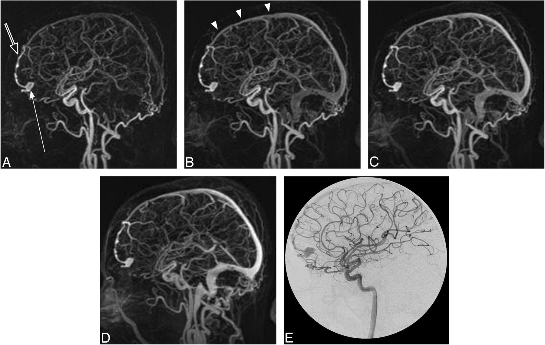
  • Fig 3.
    • Download figure
    • Open in new tab
    • Download powerpoint
    Fig 3.

    Case 5. A type III DAVF in a 60-year-old man presenting with an intracranial hemorrhage. A–D, trMRA sagittal MIPs (2-s frame rate) and (E) DSA lateral view from an injection into the left internal carotid artery. There is a DAVF located over the left aspect of the cribriform plate with rapid early arterial phase filling of a subarachnoid venous pouch (arrow), which drains via an abnormal leptomeningeal vein (open arrow) to the superior sagittal sinus (arrowheads).

  • Fig 4.
    • Download figure
    • Open in new tab
    • Download powerpoint
    Fig 4.

    Case 23, a patient with treated DAVF with residual Borden type I shunt. Anteroposterior (A–E) and submentovertex (F–J) MIPs of consecutive whole-head volumes with an effective frame rate of 2 s. K and L are DSA anteroposterior and lateral views of an injection into the left external carotid artery. A persistent DAVF is located at the left sigmoid sinus, with residual shunt into the left transverse sinus and across the torcula to the right transverse sinus and down the right jugular vein (arrows). The left jugular vein is occluded. Endovascular coils from previous treatment can be seen overlying the left transverse sinus (open arrow) on DSA. Note the normal (later) filling of the superior sagittal sinus at 6 s in Figs D and I (curved arrow). In this case, reader 1 incorrectly suspected CVR.

  • Fig 5.
    • Download figure
    • Open in new tab
    • Download powerpoint
    Fig 5.

    Case 13. A type I, low-flow, residual carotid-cavernous fistula in a 55-year-old man after previous embolization. This represents a false-negative result in which one of the readers did not correctly identify the presence of a fistula. Submentovertex (A–C) and anteroposterior (D) MIPs of trMRA whole-head volumes. DSA (E) frontal and lateral (F) views of a left internal carotid injection. A small amount of early filling of the left cavernous sinus (arrow) with truncated outflow to the superior ophthalmic vein (open arrow) is seen on both modalities.

PreviousNext
Back to top

In this issue

American Journal of Neuroradiology: 30 (8)
American Journal of Neuroradiology
Vol. 30, Issue 8
1 Sep 2009
  • Table of Contents
  • Index by author
Advertisement
Print
Download PDF
Email Article

Thank you for your interest in spreading the word on American Journal of Neuroradiology.

NOTE: We only request your email address so that the person you are recommending the page to knows that you wanted them to see it, and that it is not junk mail. We do not capture any email address.

Enter multiple addresses on separate lines or separate them with commas.
Cranial Dural Arteriovenous Fistula: Diagnosis and Classification with Time-Resolved MR Angiography at 3T
(Your Name) has sent you a message from American Journal of Neuroradiology
(Your Name) thought you would like to see the American Journal of Neuroradiology web site.
CAPTCHA
This question is for testing whether or not you are a human visitor and to prevent automated spam submissions.
Cite this article
R.I. Farb, R. Agid, R.A. Willinsky, D.M. Johnstone, K.G. terBrugge
Cranial Dural Arteriovenous Fistula: Diagnosis and Classification with Time-Resolved MR Angiography at 3T
American Journal of Neuroradiology Sep 2009, 30 (8) 1546-1551; DOI: 10.3174/ajnr.A1646

Citation Manager Formats

  • BibTeX
  • Bookends
  • EasyBib
  • EndNote (tagged)
  • EndNote 8 (xml)
  • Medlars
  • Mendeley
  • Papers
  • RefWorks Tagged
  • Ref Manager
  • RIS
  • Zotero
0 Responses
Respond to this article
Share
Bookmark this article
Cranial Dural Arteriovenous Fistula: Diagnosis and Classification with Time-Resolved MR Angiography at 3T
R.I. Farb, R. Agid, R.A. Willinsky, D.M. Johnstone, K.G. terBrugge
American Journal of Neuroradiology Sep 2009, 30 (8) 1546-1551; DOI: 10.3174/ajnr.A1646
del.icio.us logo Twitter logo Facebook logo Mendeley logo
  • Tweet Widget
  • Facebook Like
  • Google Plus One
Purchase

Jump to section

  • Article
    • Abstract
    • Materials and Methods
    • Results
    • Discussion
    • Conclusions
    • References
  • Figures & Data
  • Info & Metrics
  • Responses
  • References
  • PDF

Related Articles

  • No related articles found.
  • PubMed
  • Google Scholar

Cited By...

  • Vessel-Selective 4D-MRA Using Superselective Pseudocontinuous Arterial Spin-Labeling with Keyhole and View-Sharing for Visualizing Intracranial Dural AVFs
  • Endovascular Management of Intracranial Dural AVFs: Principles
  • Assessment of 4D MR Angiography at 3T Compared with DSA for the Follow-up of Embolized Brain Dural Arteriovenous Fistula: A Dual-Center Study
  • Arterial Spin-Labeling Improves Detection of Intracranial Dural Arteriovenous Fistulas with MRI
  • Standard and Guidelines: Intracranial Dural Arteriovenous Shunts
  • Contrast-Enhanced Time-Resolved MRA for Follow-Up of Intracranial Aneurysms Treated with the Pipeline Embolization Device
  • Clinical and Angiographic Characteristics of Multiple Dural Arteriovenous Shunts
  • Intracranial Dural Arteriovenous Fistulas: Classification, Imaging Findings, and Treatment
  • Noninvasive Evaluation of Cerebral Arteriovenous Malformations by 4D-MRA for Preoperative Planning and Postoperative Follow-Up in 56 Patients: Comparison with DSA and Intraoperative Findings
  • Simultaneous Arteriovenous Shunting and Venous Congestion Identification in Dural Arteriovenous Fistulas Using Susceptibility-Weighted Imaging: Initial Experience
  • Identification of Venous Signal on Arterial Spin Labeling Improves Diagnosis of Dural Arteriovenous Fistulas and Small Arteriovenous Malformations
  • CT Angiography as a Screening Tool for Dural Arteriovenous Fistula in Patients with Pulsatile Tinnitus: Feasibility and Test Characteristics
  • Detection and Classification of Cranial Dural Arteriovenous Fistulas Using 4D-CT Angiography: Initial Experience
  • Intracranial Dural Arteriovenous Fistula with Retrograde Cortical Venous Drainage: Use of Susceptibility-Weighted Imaging in Combination with Dynamic Susceptibility Contrast Imaging
  • Spontaneous Angiographic Conversion of Intracranial Dural Arteriovenous Shunt: Long-Term Follow-Up in Nontreated Patients
  • Crossref
  • Google Scholar

This article has not yet been cited by articles in journals that are participating in Crossref Cited-by Linking.

More in this TOC Section

  • Evaluating the Effects of White Matter Multiple Sclerosis Lesions on the Volume Estimation of 6 Brain Tissue Segmentation Methods
  • Quiet PROPELLER MRI Techniques Match the Quality of Conventional PROPELLER Brain Imaging Techniques
  • Predictors of Reperfusion in Patients with Acute Ischemic Stroke
Show more Brain

Similar Articles

Advertisement

Indexed Content

  • Current Issue
  • Accepted Manuscripts
  • Article Preview
  • Past Issues
  • Editorials
  • Editor's Choice
  • Fellows' Journal Club
  • Letters to the Editor
  • Video Articles

Cases

  • Case Collection
  • Archive - Case of the Week
  • Archive - Case of the Month
  • Archive - Classic Case

More from AJNR

  • Trainee Corner
  • Imaging Protocols
  • MRI Safety Corner
  • Book Reviews

Multimedia

  • AJNR Podcasts
  • AJNR Scantastics

Resources

  • Turnaround Time
  • Submit a Manuscript
  • Submit a Video Article
  • Submit an eLetter to the Editor/Response
  • Manuscript Submission Guidelines
  • Statistical Tips
  • Fast Publishing of Accepted Manuscripts
  • Graphical Abstract Preparation
  • Imaging Protocol Submission
  • Evidence-Based Medicine Level Guide
  • Publishing Checklists
  • Author Policies
  • Become a Reviewer/Academy of Reviewers
  • News and Updates

About Us

  • About AJNR
  • Editorial Board
  • Editorial Board Alumni
  • Alerts
  • Permissions
  • Not an AJNR Subscriber? Join Now
  • Advertise with Us
  • Librarian Resources
  • Feedback
  • Terms and Conditions
  • AJNR Editorial Board Alumni

American Society of Neuroradiology

  • Not an ASNR Member? Join Now

© 2025 by the American Society of Neuroradiology All rights, including for text and data mining, AI training, and similar technologies, are reserved.
Print ISSN: 0195-6108 Online ISSN: 1936-959X

Powered by HighWire